These are the cranial and caudal anatomical landmarks for a lateral thoracic radiograph.
What are thoracic inlet and diaphragm?
These are the proximal and distal landmarks for a properly positioned radius/ulna radiograph.
What are the elbow and the carpus?
What is wrong with this patient? 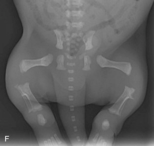
What is nothing? It is a puppy.
This radiograph is too light, too dark or properly exposed.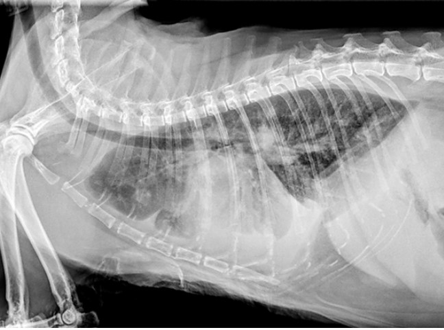
What is properly exposed?
Standard PPE for x-ray exposure
What is a lead gown, thyroid shield, dosimeter badge +/- lead gloves and goggles?
These are the cranial and caudal anatomical landmarks for a VD abdominal radiograph.
What are the entire liver to the wings of the ilium?
These are the proximal and distal landmarks for a properly positioned humoral radiograph.
What are the elbow and the distal aspect of the scapula?
These are the proximal and distal landmarks for a tibia/fibular radiograph.
What are the stifle and tarsus?
This radiopaque substance can be seen in this radiograph.
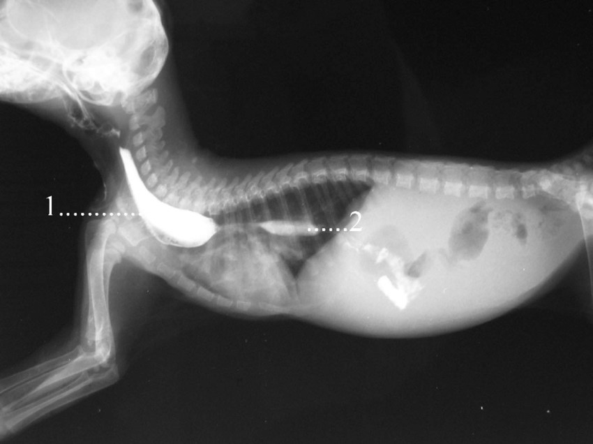
What is barium?
A moving x-ray
What is fluoroscopy?
This phase of breathing is the best to obtain thoracic radiographs.
What is inhalation?
This is a properly or poorly positioned elbow radiograph. 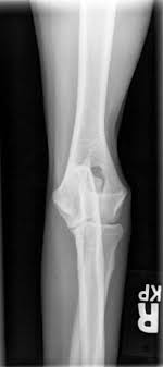
What is poorly and should be retaken?
This radiographs is too light, too dark or properly exposed? 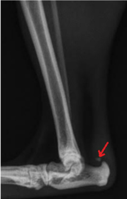
What is overexposed?
This is the best order of positioning to highlight GI foreign bodies.
What is LL, VD, RL?
These tissues are at the highest risk when exposed to x-ray radiation.
Hands, eyes, reproductive organs, rapidly dividing cells, thyroid
These structures are highlighted on this radiographs. 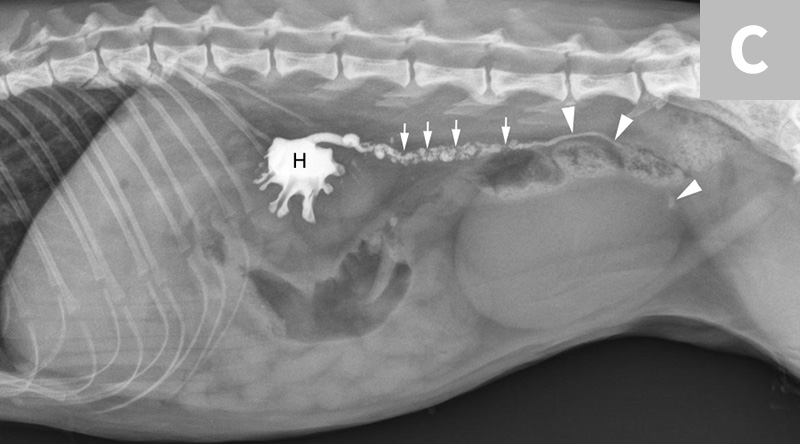
What are the kidney, ureter and ureteral stones?
This radiograph of the shoulder joint is properly positioned or should be repeated. 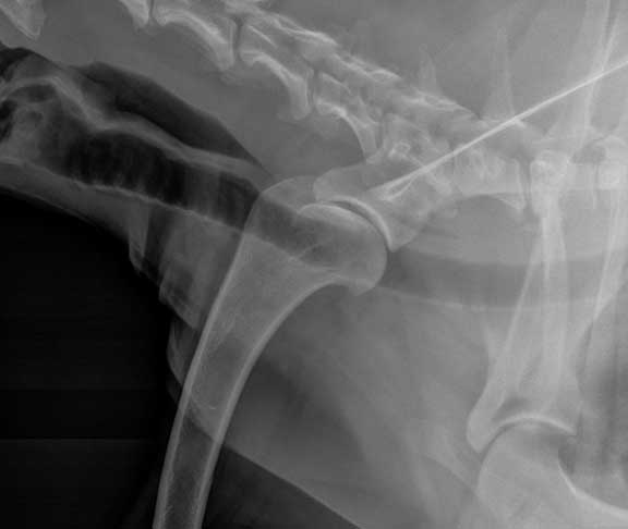
What is poorly positioned and should be retaken?
This positioning is called a _____-______ pelvis view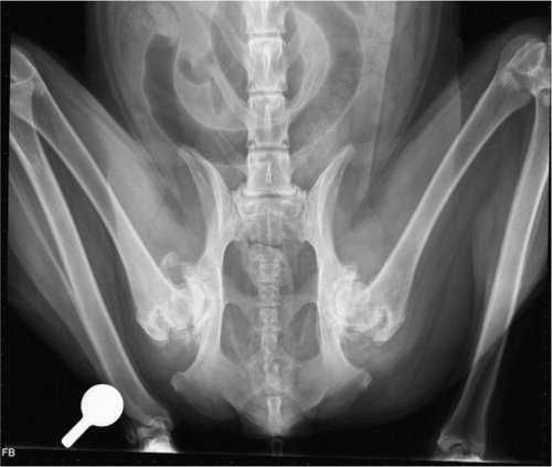
What is a frog-leg pelvis radiograph?
The number of cervical vertebrae in all but three mammals
What is seven?
ALARA stands for
What is As Low As Reasonably Achievable?
This organ's placement in the abdomen will help you determine which side is right vs left. 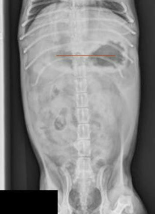
The stomach is normally located on the left in a VD radiograph.
Your DVM asks for a stress view of the carpus. To obtain this radiograph your patient will be in sternal or lateral recumbency.
What is lateral recumbency?
These are the anatomical landmarks for a properly positioned VD pelvis.
What are the entire pelvis to just below the stifles?
The condition seen here.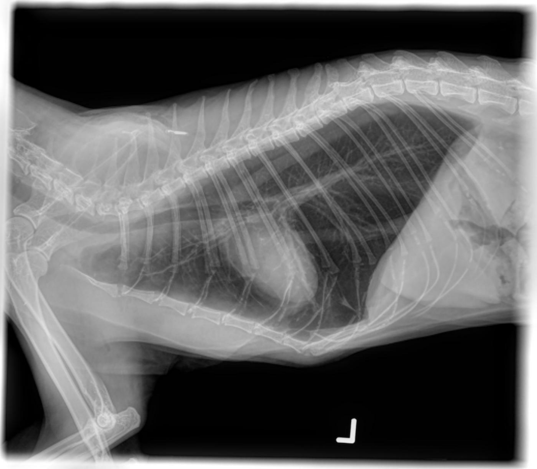
What is pneumothorax?
Patients get the most exposure to x-ray radiation from the direct beam. Personnel get the most exposure from __________?
What is scatter radiation?