Polypssss
Hysteroscopy
Submucosal fibroids
Hysteroscopic uh-oh's
Hysteroscope set up
100
Definition of a polyp and risk of malignancy.
Hyperplastic growth of the endometrium
Post menopausal 5%, pre menopausal 1%
Size has been inconsistently shown to be associated with malignancy!!
100
3 types of distention media.
Sorbitol, mannitol, dextran, normal saline.
100
Based on FIGO classification, these types of fibroids are amenable to hysteroscopic resection. (also must define location of each fibroid)
Type 0-3!!

100
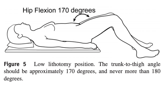
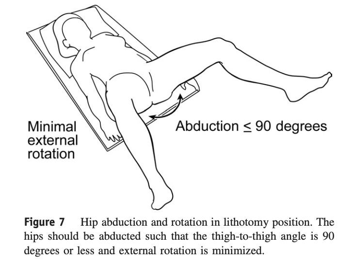
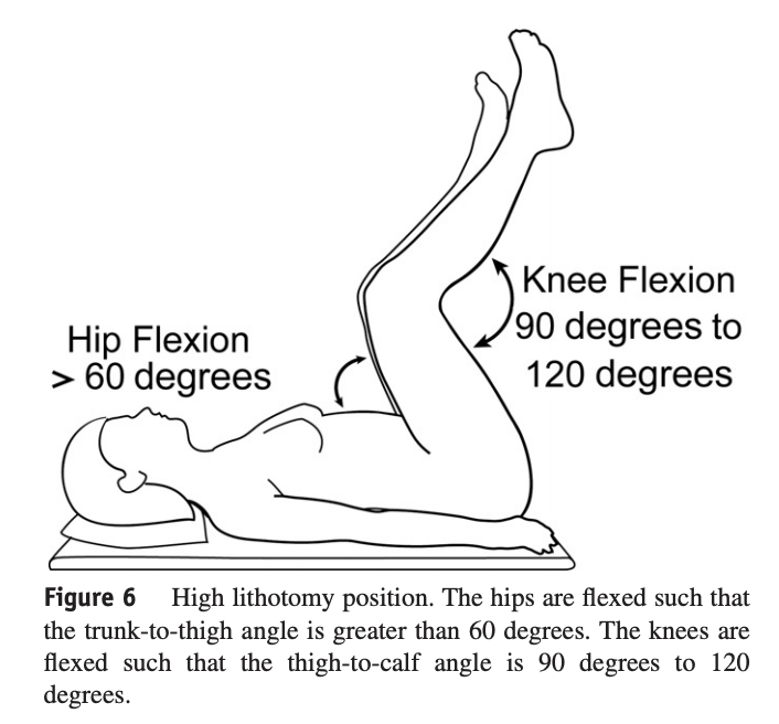
Describe the correct positioning during lithotomy for the following:
1) hip flexion
2) knee flexion
3) hip abduction
4) hip rotation
Four important elements of correct lithotomy positioning are: (1) hip flexion of 60 degrees to 170 degrees; (2) knee flexion of 90 degrees to 120 degrees; (3) hip abduction of 90 degrees or less; and (4) minimal external hip rotation
100
The 6 parts of Myosure hysteroscope that you hold in your hand.
What are...
1. Camera head
2. Scope
3. Light cable/source
4. Inflow (distention media)
5. Outflow (suction)
6. Myosure (cutting) device
200
Indications for polyp removal. (Name 4 clinical scenarios)
Anyone who is post menopausal
Anyone who is premenopausal if it causes AUB, infertility, risk factors for EH, recurrent, >1.5 cm, multiple polyps, prolapsed
200
3 factors for systemic fluid loss/deficit during hysteroscopy.
1. Intrauterine pressure: the higher the pressure, the greater the degree of systemic absorption.
2. Length of surgery.
3. Disruption of venous sinuses within endometrium and myometrium.
200
Three fibroid characteristics that increase risk of incomplete resection.
>5 cm in diameter
>50 percent extension into the myometrium (FIGO type 2-3)
Multiple fibroids
200
True or false: hysteroscopy does not require preoperative antibiotics.
FALSE: history of PID or active hydrosalpinx are indications per ACOG.
If the procedure demonstrates dilated fallopian tubes, 100 mg of doxycycline may be given twice daily for five days. In women with a history of pelvic infection, doxycycline can be administered before the procedure and continued if dilated fallopian tubes are found.
200
The Myosure device is inserted into this part of hysteroscope. The hysteroscopic needle is inserted into this part of the hysteroscope.
The myosure device is inserted into the outflow WITHOUT the cannula.
The hysteroscopic needle is inserted into the outflow WITH the cannula in place.
300
Methods for polyp diagnosis. (name 3 methods)
Endometrial biopsy
thickening of endometrial stripe on ultrasound
SIS
300
The Myosure and Truclear uses ______ energy for resection while the resectoscope uses _______ energy for resection.
Mechanical energy vs bipolar energy
300
Reliable imaging used to evaluate fibroid candidacy for hysteroscopic resection. (name 3)
SIS--once uterus is >12 weeks hard to maintain uterine distention and see the endometrium, also may have too many fibroids to determine via US
MRI--$$$ but good at delineating size, #, location
Office hysteroscopy--gold standard, not always available
300
Maximum fluid deficit and factors that affect what the number is set as.
2500 mL is the maximum allowable systemic absorption in a hysteroscopic procedure
Individuals of small stature, or those compromised by comorbidities such as heart failure, ESRD, would not tolerate high volumes.
300
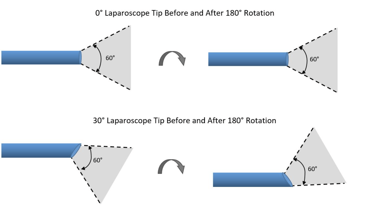
Angle (degree of scope) of hysteroscopes.
0 degree to 70 degrees.
We use 0 degree scopes at HBH!
400
Spontaneous regression rate of polyps in patients.
50% if premenopausal and SMALL polyps.
400
If you are using the resectoscope, you must use this type of media distention.
Normal saline if bipolar, hypotonic if monopolar
400
Rate of complete resection for type 2 fibroids?
60-80%
High need for two step procedure!!
400
The fourth year resident starts the hysteroscopy by dilating a postmenopausal cervix. Upon insertion of the scope, it is difficult to see but the attending decides to increase the intrauterine pressure. While doing so, the myosure starts to ding loudly and a high fluid deficit is note. A bloody hole is noted along the left lateral portion of hte lower uterine segment. What are next steps? What could have been done to prevent this?
1. Check vitals with anesthesia
2. Lateral perforation--> laparoscopy. 3 factors to determine if lap is indicated type of instrument (blunt vs sharp), area of perforation (lat vs fundal), HD stability of pt (determines degree of bleeding)
Prevention: preoperative misoprostol, pediatric dilators, omniscope
400
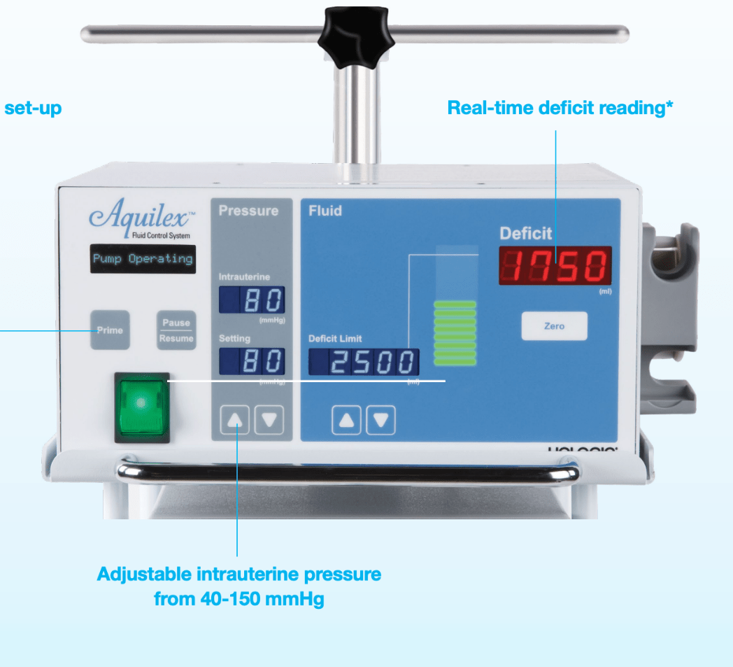
Number of readings on the Myosure machine and definitions.
Set Intrauterine pressure
Actual intrauterine pressure
Set fluid deficit
Actual fluid deficit
500
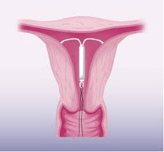
This drug can prevent recurrence of polyps.
PROGESTERONE IUD!!!
500
Medication (including dosing) you can give to decrease systemic fluid absorption and two methods of administration.
Transcervical dilute vasopressin (decreases fluid absorption by up to ~50% in some studies with hysteroscopic myomectomy)
1. Inject into the uterus at cervicovaginal junction
2. Injection via direct visualization into the uterus
0.1-0.4 units per mL dilution! (ie: 20 units in 100 cc of normal saline, 4 units in 10 cc of lidocaine)
500
Risk of synechiae after hysteroscopic resection.
20% or higher if using bipolar device!
500
While performing a hysteroscopy the CRNA notes that there is fluid pooling up towards the patient's head. The circulator does some math and realizes the Myosure actually was not calculating the fluid deficit. Based on what is on the floor, the deficit is about 4000cc in this healthy 30 year old patient. What would you do next?
1. Confirm fluid deficit
2. Ask CRNA about vitals, difficulty ventilating
3. In PACU obtain BMP, CBC, complete respiratory exam.
4. If normal, monitor, fluid restrict.
5. If abnormal, consult medicine for hyponatremia, hypervolemia!!!
500
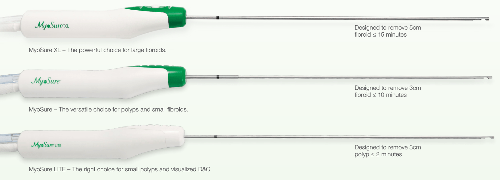
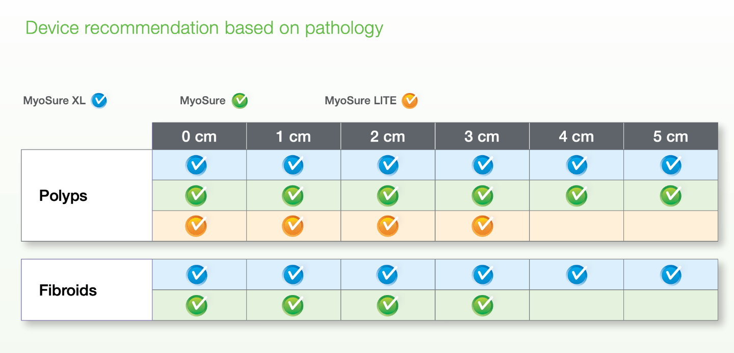
Differences in size and type of pathology between 3 Myosure devices.