Digestive
Respiratory System
Urinary System
Repro Female
Repro Male
100
These specific structures are found in one part of the digestive system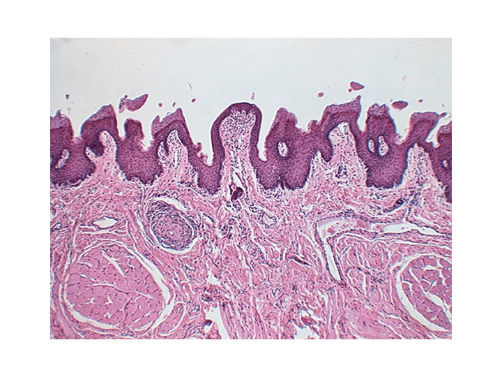
What is: surface of the tongue
100
A: From what organ is the image made
B: Name the structures deep to Tunica Mucosa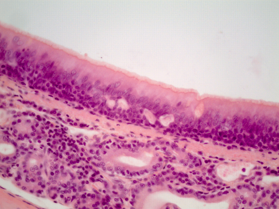
A: Trachea
B: Tracheal glands (submucosal glands)
100
A: Name the epithelial tissue at the tip of the pointer.
B: From what organ is this slide made?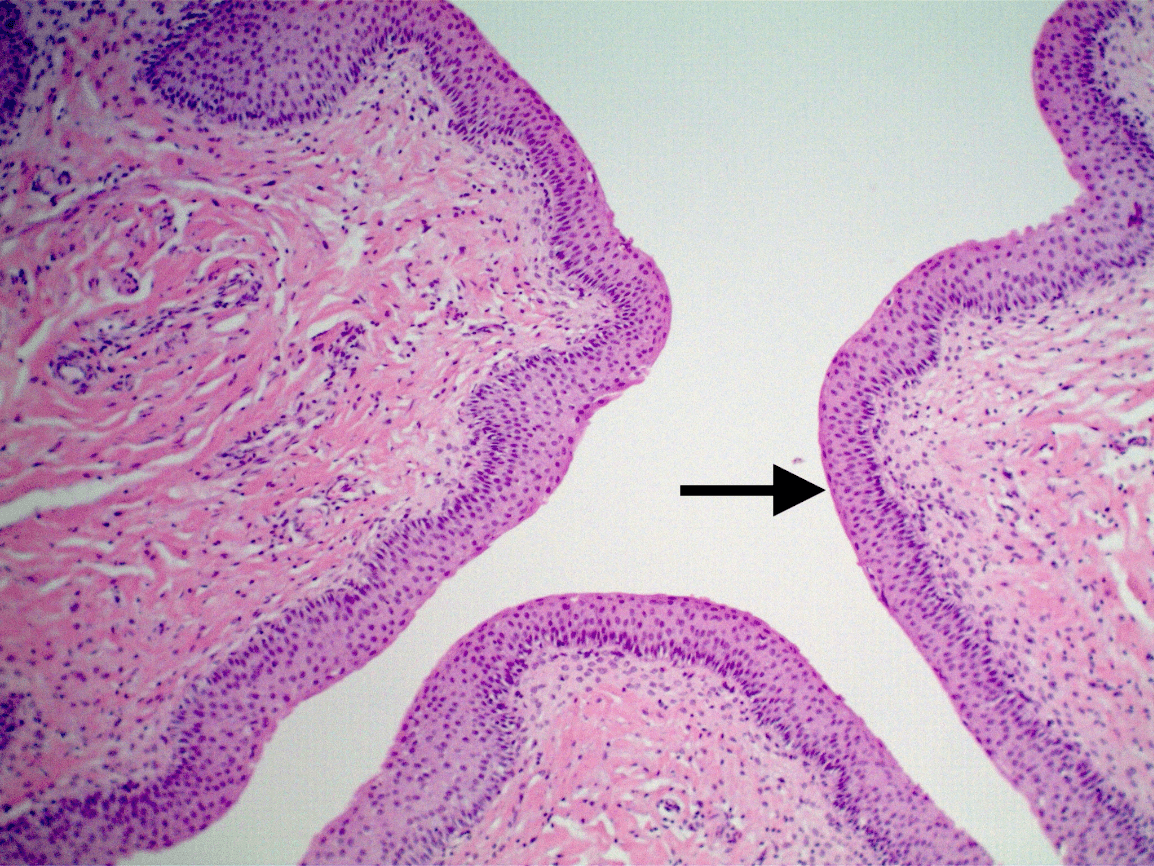
Transitional ET
B: Urinary bladder.
100
Name the organ from which this slide is made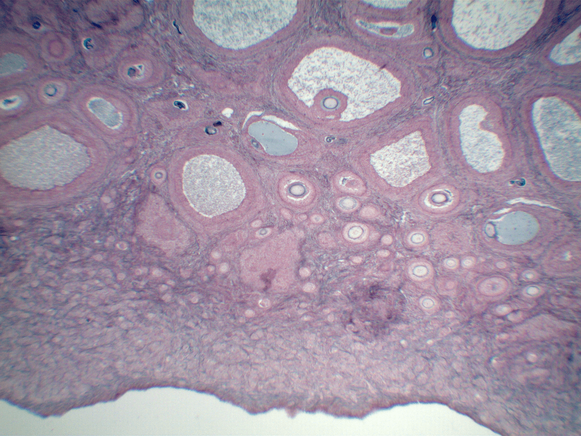
Ovary
100
Name the organ from which this slide is taken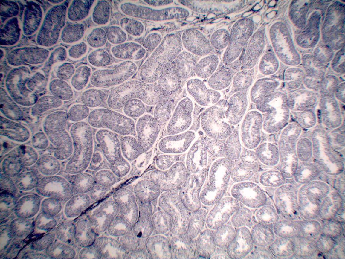
Testis.
200
Name of this structure specifically ?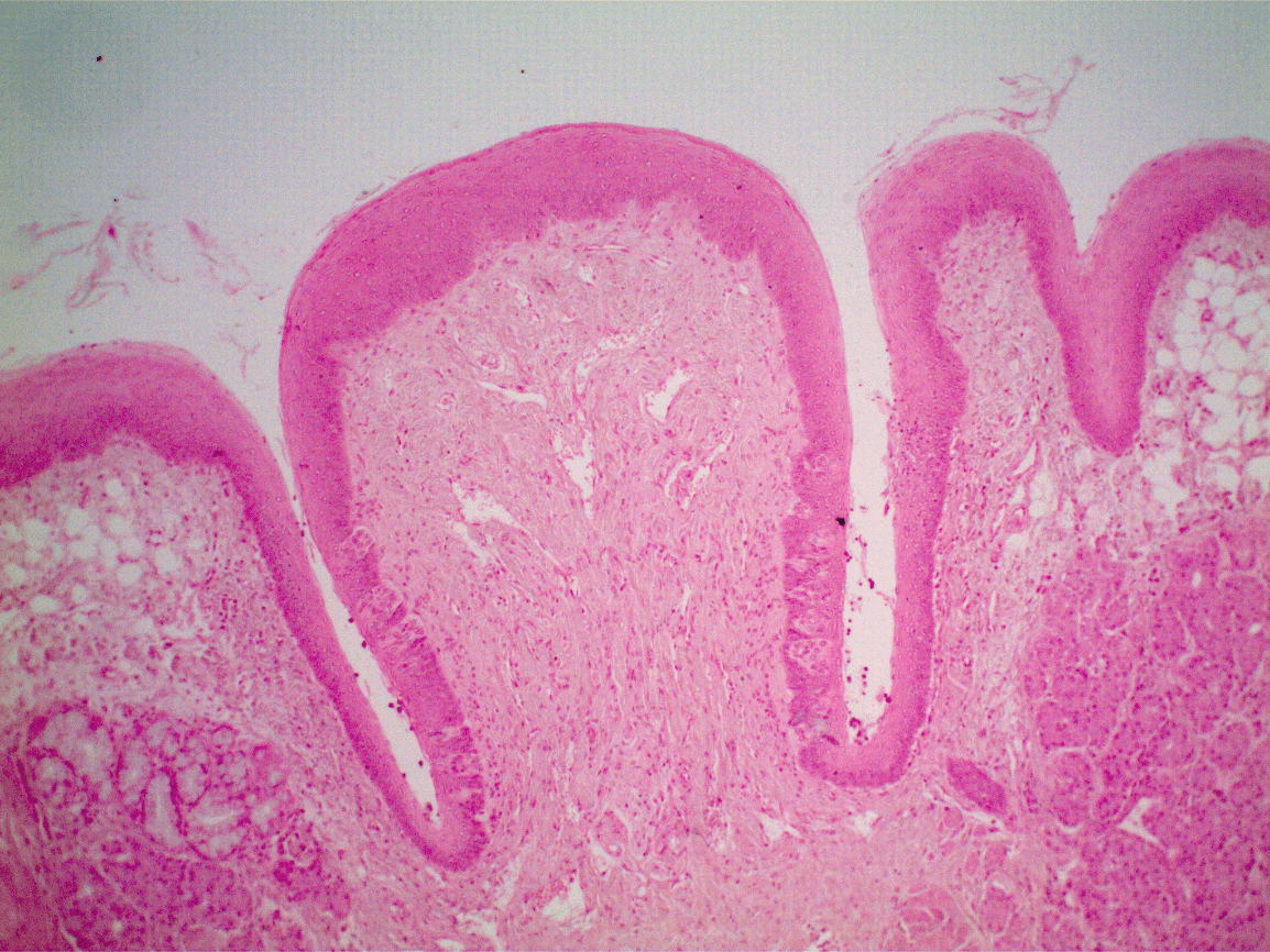
What is: circumvallate papilla
200
Name the structure in the center of the field of view
Trachealis muscle
200
Name the structure at the tip of the pointer. Be specific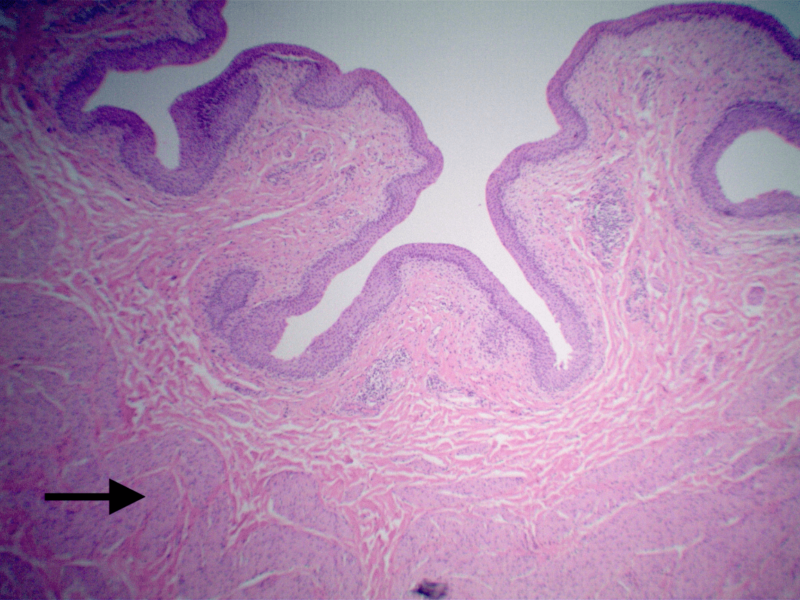
Detrusor muscle
200
Name the structure
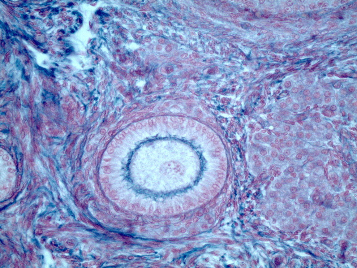
What is: primary follicle
200
Name the structures that fill the field of view
Seminiferous tubules
300
This image can be made from this specific organ
What is: Stomach
300
Name the thick walled structures in the center of the field of view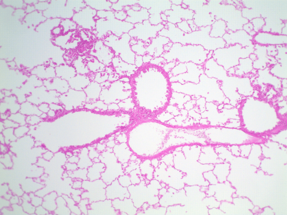
Bronchioles
300
Name the organ from which this slide is made

Ureter
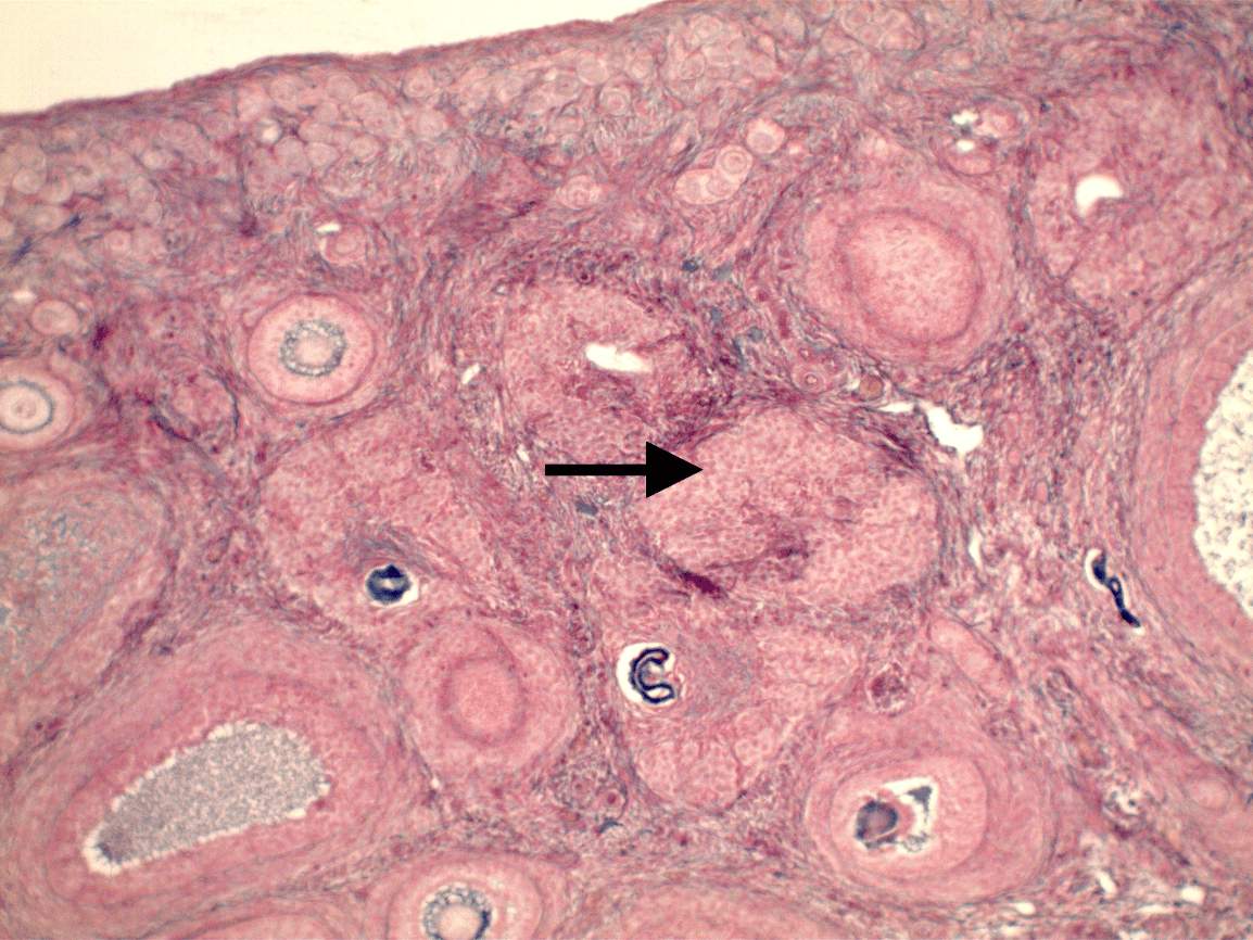
300
Name the structure at the tip of the pointer
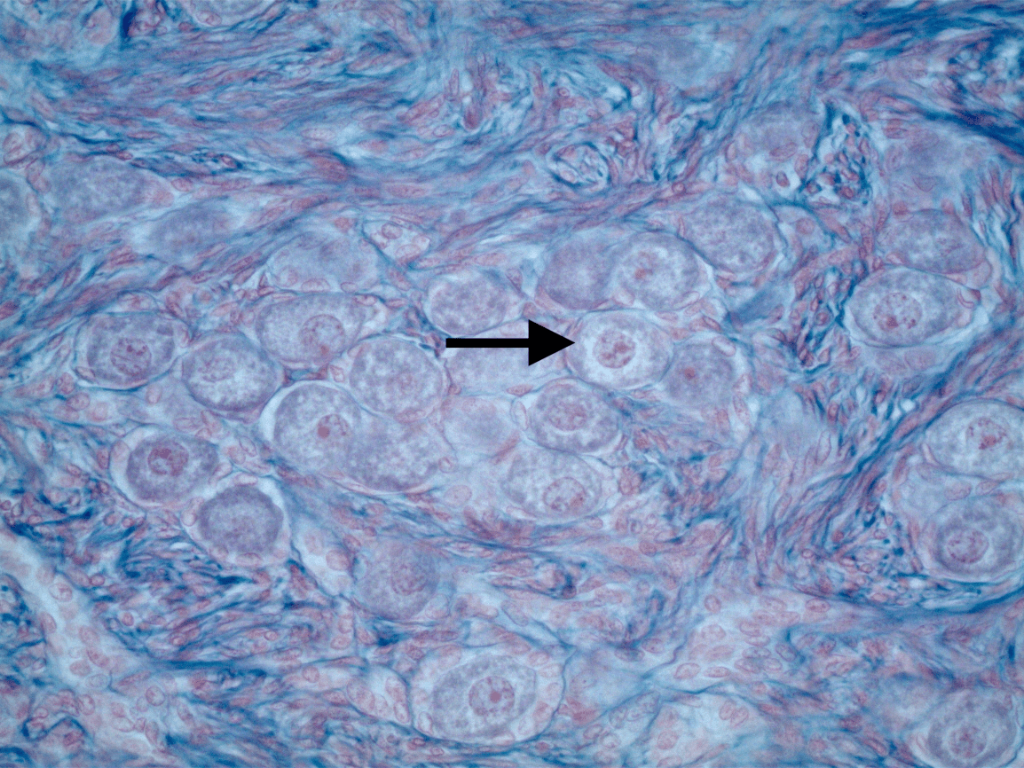
Primordial follicle
300
Name the structure that fills the field of view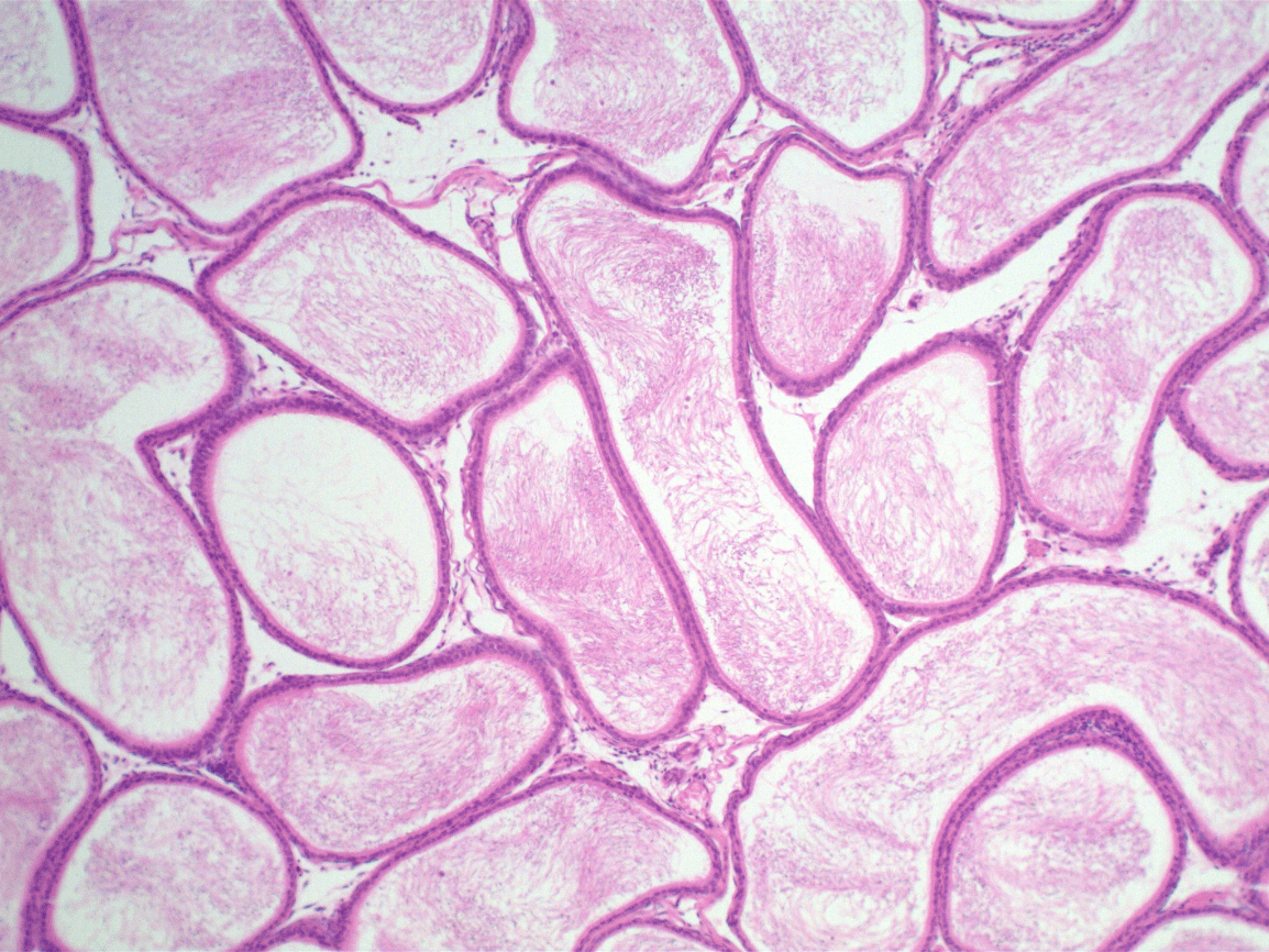
Epididymis
400
A. Name the white space at the bottom left
B. Name the rest of the white spaces

A. Central Vein
B. Sinusoids
400
Name the cell at the tip of the pointer.
Give its function.
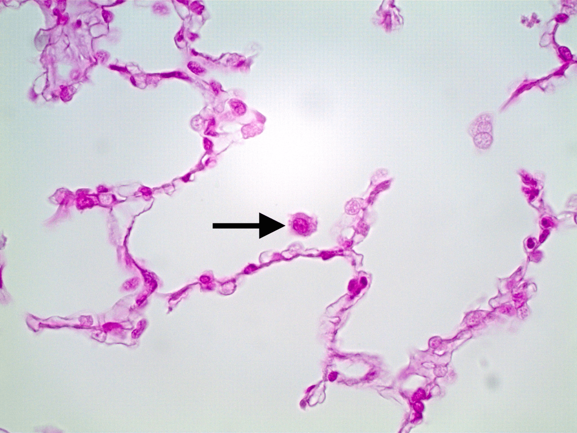
What is: alveolar macrophage
Phagocytic protection for the respiratory alveolus
400
A. Name the large tubes.
B. Name the small tubes
A. Collecting Ducts
B. Thin (Descending) Limb of Nephron Loop
400
A: Name the organ from which this slide is made
B: Name the specific layer indicated by the red line.
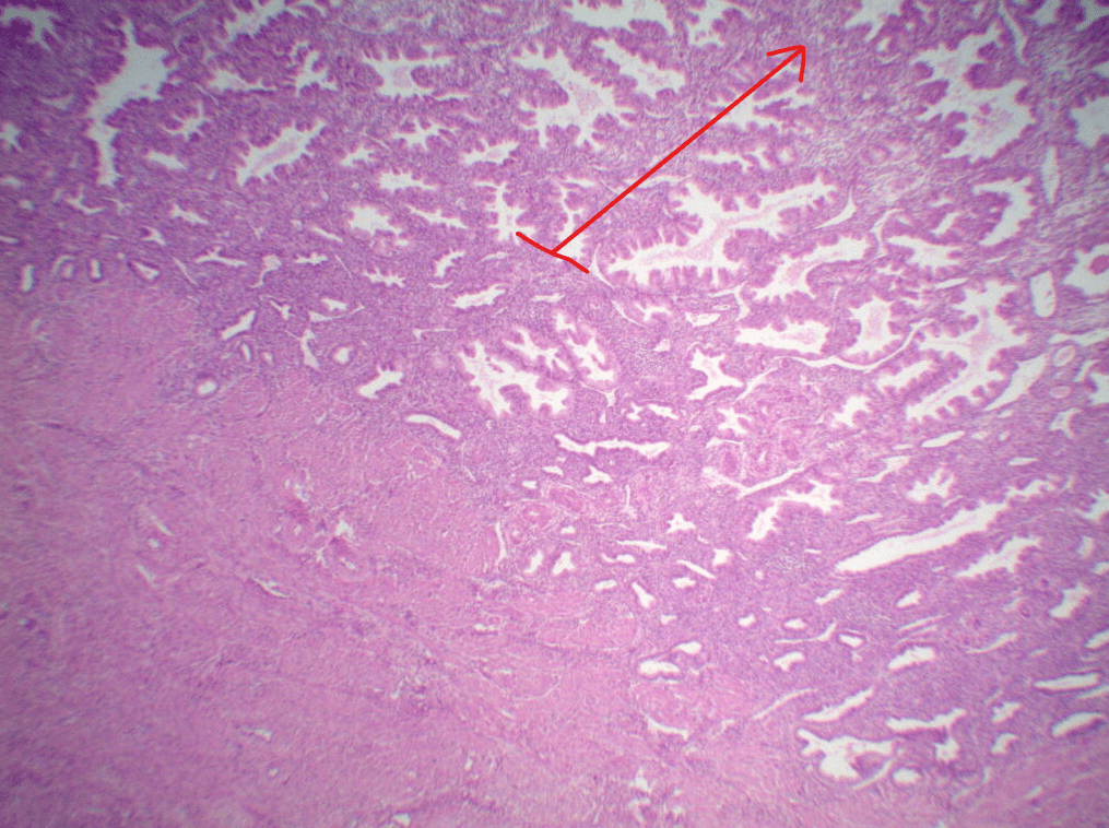
A: Uterus
B: Stratum functionals
400
Identify the epithelial tissue in this structure
Pseudostratified columnar ET with stereocilia
500
Name the two cells indicated by the pointers and what is secreted by each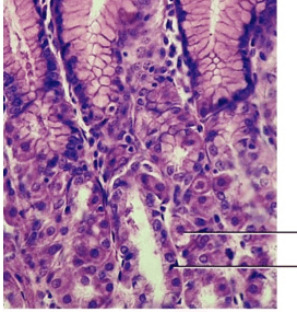
What are: Parietal cell secreting HCl, and chief cell secreting Pepsinogen
500
Name the structure surrounding the black line.
Alveolar duct.
500
A. Name the boxed cells
B. Give their function
A. Macula Densa
B. Monitor salt content in Pre-Urine Filtrate in Distal Convoluted Tubule
500
Name the specific structure at the tip of the pointer
Corpus luteum
500
A. Name the cell in the red box
B. Name the cell in the yellow circle
A. Sustentacular Cell (Sertoli Cell)
B. Interstitial Cell (Leydig Cell)