Basic Neuroanatomy
EDX
Stroke 1.0
Stroke 2.0
100
Which nerve predominately innervates the muscles of the deep posterior compartment of the leg?
Correct answer- Tibial
Per PMR recap -
The deep posterior leg compartment contains the tibialis posterior, flexor digitorum longus, and flexor hallucis longus, which are all innervated by the tibial nerve.
Explanation:
The deep posterior leg compartment contains the tibialis posterior, flexor digitorum longus, and flexor hallucis longus, which are all innervated by the tibial nerve.
100
During an exam, perhaps much like this one, when a question stem mentions EMG findings of painless myokymia in C5-C6/upper trunk muscles, you will astutely recall that the most likely etiology of these findings is which of the following?
A Radiation plexopathy
B Active denervation
C Myotonia congenita
D Pancoast tumor
Correct Answer- A Radiation plexopathy
From PMR recap - Radiation plexopathy is classically the cause of myokymia found on EMG, which classically affects C5-C6/upper trunk muscles, and is usually a painless finding. Lower trunk plexopathy with pain is concerning for pancoast (lung) tumor compressing the lower trunk of the brachial plexus. Myotonia congenita will demonstrate myotonic discharges (divebomber sound). Active denervation will demonstrate fibrillations and positive sharp waves. Examiners thoroughly enjoy asking you about radiation plexopathy and myokymia, so be sure to know this topic well.
100
A 75-year-old man with a recent anterior communicating artery aneurysm, treated by neurosurgical clipping is admitted to the inpatient rehabilitation unit for poststroke care. Deep vein thrombosis prophylaxis should include:
(a) heparin 5,000 units BID. (b) warfarin doses based on INR values. (c) pneumatic compression stocking (ankle or calf). (d) continuous passive motion devices.
Correct Answer- (C) pneumatic compression stocking (ankle or calf)
AAPMR SAE 2001 explanation-
Venous thromboembolic events can occur in as many as 75% of untreated patients with afterstroke. Prophylaxis is typically pharmacologic or manual venous compression. In patients with documented intracerebral bleeding, anticoagulation is not recommended, and alternating pneumatic compression derives are best used.
100
A patient suffers a stroke and he is transferred to the stroke rehab unit after medical stabilization. He is seen by physical, occupational, and speech therapies. He has significant aphasia during the initial H&P encounter. You contemplate melodic intonation therapy as a means to help this patient; which type of aphasia below could melodic intonation therapy benefit?
A Transcortical sensory aphasia
B Broca aphasa
C Wernicke aphasia
D Conduction aphasia
Correct answer- B Broca aphasa
PMR recap-
Melodic intonation therapy can be useful for patients with nonfluent aphasia, including Broca’s aphasia. The other answer choices are fluent aphasias. Melodic intonation therapy recruits the right hemisphere (speech is typically a left hemisphere activity) by incorporating melodies or rhythms into communication.
Explanation:
Melodic intonation therapy can be useful for patients with nonfluent aphasia, including Broca’s aphasia. The other answer choices are fluent aphasias. Melodic intonation therapy recruits the right hemisphere (speech is typically a left hemisphere activity) by incorporating melodies or rhythms into communication.
200
During a blink reflex study, the input and output are via which cranial nerves, respectively?
A 5, 7
B 7, 7
C 7, 5
D 5, 5
Correct Answer (A) 5, 7
Per PMR recap- The blink reflex study tests CN V and CN VII in the brainstem and peripherally. CN V has two nuclei being tested (Vm and Vs). The Vm nucleus lies within the pons. It accepts CN V input from the face and conducts it to the ipsilateral CN VII nucleus, causing an ipsilateral blink (R1 response). It also simultaneously conducts the impulse to the medulla where the Vs nucleus lies. Vs accepts the impulse from Vm and sends the impulse to the bilateral CN VII nuclei, which then causes a bilateral blink via the orbicularis oculi muscles (R2 response).
Explanation:
The blink reflex study tests CN V and CN VII in the brainstem and peripherally. CN V has two nuclei being tested (Vm and Vs). The Vm nucleus lies within the pons. It accepts CN V input from the face and conducts it to the ipsilateral CN VII nucleus, causing an ipsilateral blink (R1 response). It also simultaneously conducts the impulse to the medulla where the Vs nucleus lies. Vs accepts the impulse from Vm and sends the impulse to the bilateral CN VII nuclei, which then causes a bilateral blink via the orbicularis oculi muscles (R2 response).
200
You are performing an EMG/NCS on a patient. The sensory nerve action potential (SNAP) to digit 2 and compound motor action potential (CMAP) to the abductor pollicis brevis are normal. The SNAP to digit 5 reveals prolonged latency. The CMAP to the abductor digiti minimi is normal. Needle EMG reveals no abnormalities. Which of the following is the most likely diagnosis?
A Medial cord plexopathy
B Severe ulnar neuropathy
C Mild ulnar neuropathy
D Normal study
Correct Answer- C Mild ulnar neuropathy
Per PMR recap- Mild ulnar neuropathy is most consistent with an otherwise normal study with isolated prolonged ulnar SNAP - indicating ulnar nerve changes somewhere, most likely between and including the elbow and the wrist. All values would have to be normal for this to be recorded as a normal study. Medial cordopathy would have to be better supported, possibly including an abnormal medial antebrachial cutaneous SNAP. Finally, severe ulnar neuropathy would show active denervation in ulnar nerve muscles, not shown here.
200
A 65 year-old female is brought to the ED due to sudden-onset right hemiplegia. On exam you discover she has impaired sensation to light touch on her right side. Her extraocular motility and visual acuity are normal bilaterally. When asked to stick out her tongue, her tongue deviates toward the left side. What is the most likely diagnosis?
A Right Weber Syndrome
B Left Weber Syndrome
C Right medial medullary syndrome
D Left medial medullary syndrome
Correct answer is D Left medial medullary syndrome
Per PMR recap - This patient has suffered a stroke of her left medial medulla. In the medial medulla lies the corticospinal tract, medial lemniscus, and CN 12 nucleus. Thus, damage to these structures will cause, respectively, contralateral hemiparesis/plegia, contralateral impaired light touch, and ipsilateral CN 12 palsy (tongue deviates toward the side of the lesion). Weber syndrome presents with contralateral hemiparesis with ipsilateral CN 3 palsy.
Explanation:
This patient has suffered a stroke of her left medial medulla. In the medial medulla lies the corticospinal tract, medial lemniscus, and CN 12 nucleus. Thus, damage to these structures will cause, respectively, contralateral hemiparesis/plegia, contralateral impaired light touch, and ipsilateral CN 12 palsy (tongue deviates toward the side of the lesion). Weber syndrome presents with contralateral hemiparesis with ipsilateral CN 3 palsy.
300
Which peripheral nerve stimulates the bladder to facilitate urine storage?
A Direct innervation from the S2-S4 nerve roots
B Pudendal
C Hypogastric
D Pelvic
Correct Answer (C) Hypogastric
Per PMR recap-Remember, the hypogastric (“hippogastric”) nerve makes your bladder as big as a hippo! Thus, it facilitates urine storage with all that extra space.
Explanation:
Remember, the hypogastric (“hippogastric”) nerve makes your bladder as big as a hippo! Thus, it facilitates urine storage with all that extra space.
300
You are treating a 48-year-old man who has had two lumbar laminectomies for what you suspect is a recurrent right L5 radiculopathy. You perform an electromyogram to confirm the diagnosis, and it reveals 2+ positive waves and fibrillations with decreased recruitment in the right anterior tibialis. The patient informs you that he can only tolerate the examination of one more muscle. Of the following you would choose?
(a) extensor hallucis longus. (b) L5 paraspinals. (c) vastus medialis. (d) flexor digitorum longus.
Correct Answer- (d) flexor digitorum longus.
Per AAPMR SAE 2001- The history is suggestive of an L5 radiculopathy. Given the previous laminectomies, examining a single level of paraspinals would provide limited information. Although you cannot form any firm conclusions based on such a limited examination, study of the flexor digitorum longus will provide findings outside the peroneal distribution and could lend support to the clinical diagnosis.
400
For patients who undergo a posterior approach to total hip arthroplasty, hip precautions are routinely continued for?
A.12 weeks.
B.4 months.
C.6 weeks.
D. 6 months.
Correct answer- C. 6 weeks
AAPMR - Total hip precautions include avoiding flexion of more than 90 degrees, adduction past midline, and internal rotation past neutral. Total hip precautions for a posterior approach total hip arthroplasty is prescribed for 6 weeks.
400
A 45-year-old colon cancer patient presents with new urinary incontinence and dull pain radiating into the right buttock. Physical examination fails to reveal evidence of lower extremity motor or sensory deficits. Of the following, the MOST appropriate next step in the patient's evaluation would include
(a) pelvic computed tomography with contrast. (b) lower extremity electromyogram. (c) positron-emission tomography scan. (d) urodynamics studies.
Correct Answer- (a) pelvic computed tomography with contrast.
Per AAPMR SAE 2001-
The sacral plexus is usually involved by tumor from the colon, prostate, bladder, or uterus.
Presenting symptoms usually begin as a dull, aching, midline pain, which may radiate into the
5
buttocks. The pain may be associated with numbness in the perianal region. Numbness and
aresthesias may extend to involve the buttock and posterior aspect of the thigh. Bowel and bladder
function are often compromised. Computed tomography and magnetic resonance imaging scans of
the pelvis are excellent tools for detecting presacral masses and sacral destruction.
400
Weber (medial midbrain) syndrome is caused by an interruption of the medial midbrain’s blood supply (typically from the ipsilateral posterior cerebral artery - PCA), leading to paralysis of the medially located corticospinal tract fibers, which will cause a contralateral hemiparesis, as these fibers have yet to decussate (cross) in the medulla more inferiorly. As we are in the midbrain, medially we have the CN 3 nucleus, thus causing an ipsilateral CN 3 palsy. Medial medullary syndrome typically causes contralateral hemiparesis and ipsilateral tongue deviation towards the side of the lesion.
A Right Weber Syndrome
B Left Weber Syndrome
C Right medial medullary syndrome
D Left medial medullary syndrome
correct answer- A Right Weber Syndrome
PMR recap- Weber (medial midbrain) syndrome is caused by an interruption of the medial midbrain’s blood supply (typically from the ipsilateral posterior cerebral artery - PCA), leading to paralysis of the medially located corticospinal tract fibers, which will cause a contralateral hemiparesis, as these fibers have yet to decussate (cross) in the medulla more inferiorly. As we are in the midbrain, medially we have the CN 3 nucleus, thus causing an ipsilateral CN 3 palsy. Medial medullary syndrome typically causes contralateral hemiparesis and ipsilateral tongue deviation towards the side of the lesion.
500
You are examining a 70 year-old female with a history of epilepsy and diabetes mellitus who complains of atraumatic shoulder pain of sudden onset. Speed, Empty Can, and O’Brien tests are positive. Active range of motion is limited to 45 degrees of abduction. In attempting passive abduction of the shoulder, you note significant resistance as you slowly abduct the shoulder between 50 and 100 degrees, which causes the patient severe pain. Which of the following is the most likely diagnosis?
A Adhesive capsulitis
B Glenohumeral joint osteoarthritis
C Rotator cuff syndrome
D Biceps tendinopathy
Correct Answer- A Adhesive capsulitis
per PMR recap- This patient with a history of diabetes mellitus is at risk of frozen shoulder (adhesive capsulitis). Adhesive capsulitis is more common in patients with diabetes, stroke, or trauma. Patients with adhesive capsulitis often exhibit “panpositive” physical exam findings. A tight, inflamed shoulder capsule causes severe glenohumeral joint range of motion (ROM) restriction with severe pain during attempted ROM. There is often tenderness to palpation throughout the entire shoulder. A patient with adhesive capsulitis may also truly have rotator cuff or biceps tendon dysfunction/pain, however this will be masked by the severe pain in most planes of motion encountered in this diagnosis. A key thing to remember in these patients is during passive ROM the severe pain and tight resistance to ROM due to a tight, inflamed shoulder capsule. ROM is still generally possible, however this occurs against resistance, almost like a rubber band resisting the examiner. In contrast, osteoarthritis of the shoulder generally presents as a firm stop/block to passive ROM, rather than a gradual “rubber band” resistance.
500
During the electromyographic evaluation of a patient, you note discharges consistent with myotonia and small motor units in the distal muscles of the upper and lower extremities. The most likely diagnosis is
(a) paramyotonia congenita. (b) myotonic dystrophy. (c) myotonia congenita. (d) hyperkalemic periodic paralysis
correct answer- (b) myotonic dystrophy.
Per AAPMR SAE 2001-
Although myotonic discharges may be seen in all of these disorders, myotonic dystrophy is the only one with low-amplitude motor units in the distal muscles.
500
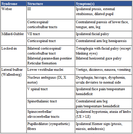
Which stroke syndrome has ipsilateral ptosis, external strabismus, a dilated pupil, and contralateral weakness of the lower face, tongue, arm, and leg?
A.Wallenberg, B.Weber, C. Millard-Gubler ,D. Locked-in
correct answer B.Weber
Per AAPMR -
Weber syndrome includes the symptoms of ipsilateral ptosis, external strabismus, a dilated pupil, and contralateral weakness of the lower face, tongue, arm, and leg. The chart below delineates each syndrome in detail.
500
A 53-year-old right-handed male presents with alexia without agraphia following a stroke. The most common blood vessel infarcted is:
A.Left posterior cerebral artery, B. Left posterior inferior cerebellar artery, C. Left anterior cerebral artery, D. Left retinal artery
Correct answer - A. Left posterior cerebral artery
Per AAPMR- PCA infarct in the left occipital lobe can result in alexia without agraphia. These patients are not able to read but can write. The infarct includes the left primary visual cortex, causing a right homonymous hemianopsia. It also includes the posterior corpus callosum, which disconnects the right primary visual cortex from the language network. Thus, the patient can see words in the left hemisphere but cannot transfer visual images to the language centers. Writing is not affected because there is no disconnection between the language network and motor cortex. Patients with this lesion may have some left neglect syndrome as well. Left posterior inferior cerebellar artery infarction results in lateral medullary syndrome. Left anterior cerebral artery infarction could result in -a number of symptoms such as motor weakness, sensory changes, or incontinence. Left retinal artery infarction results in sudden loss of eyesight in one eye.