Pelvic hurdles
Leepin' femurs
I kneed you
physical exam findings
100
Beneath plain film shows this pathology
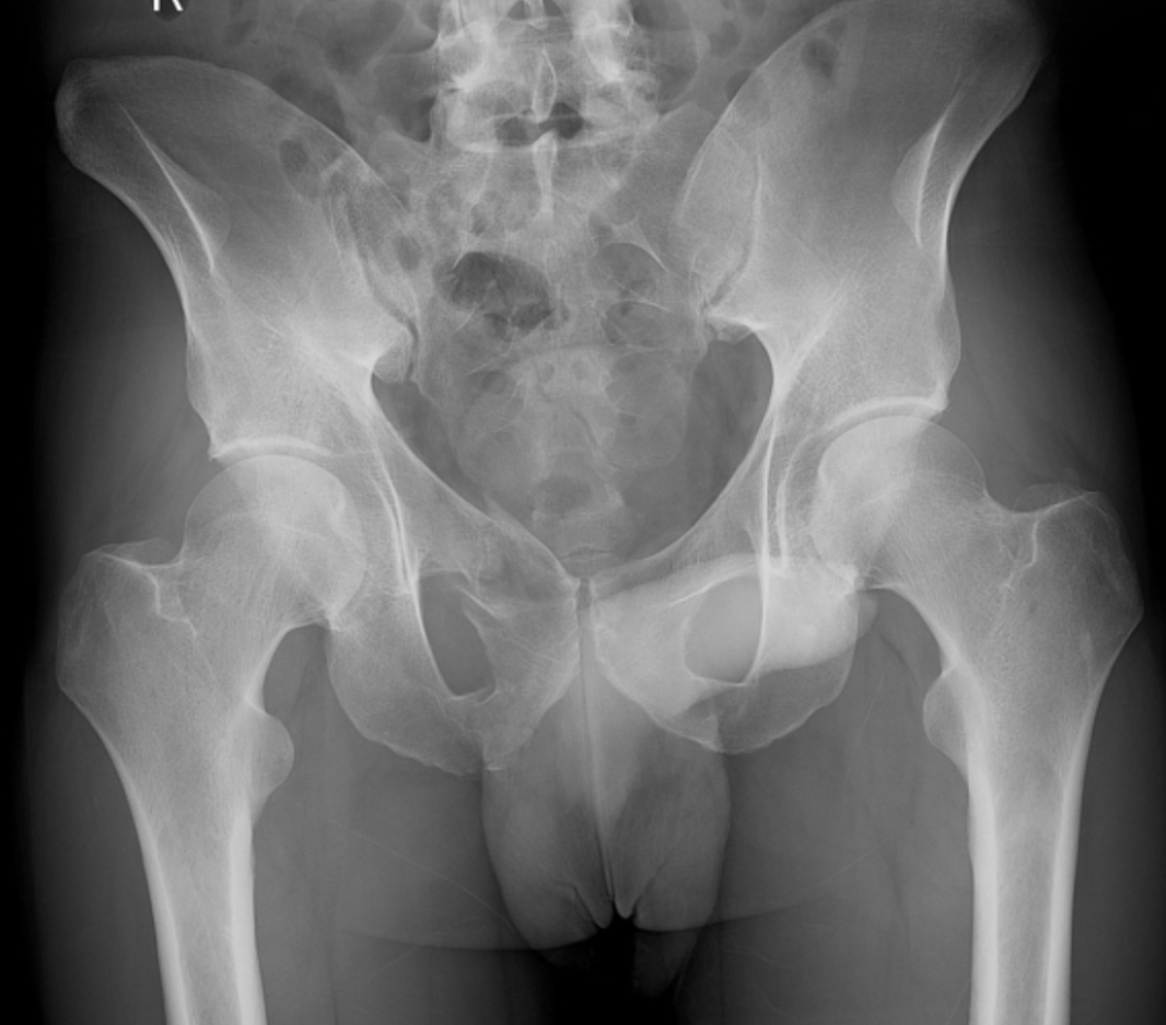
What is a right superior pubic rami fracture?
Learning point: Most common pelvic fracture in elderly falls. The ring rarely breaks in one place, so look hard for a posterior (sacral/SI) injury. Superior is more common than inferior, but minimally. 70% managed non-operatively
100
This is the treatment for what you see below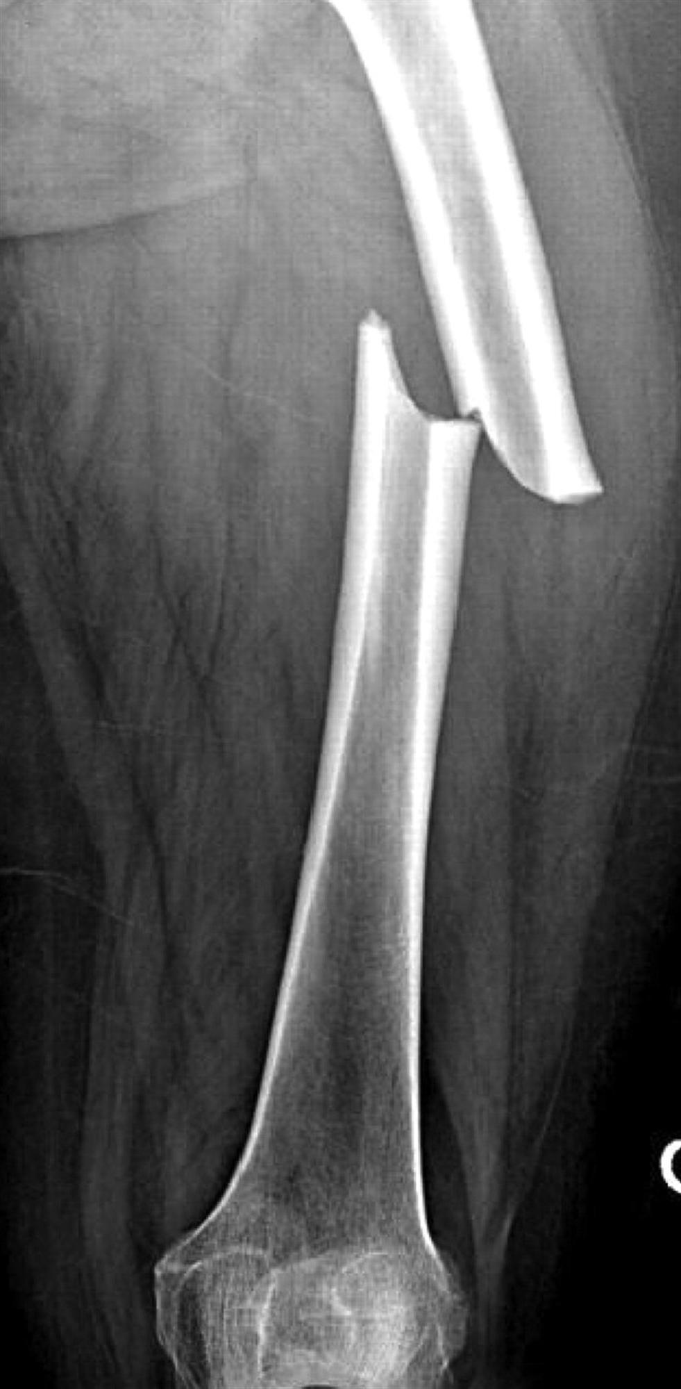
What is Buck's traction?
Learning point: Neutralizes deforming muscle forces. Ortho decides skin traction (the foam boot) vs skeletal (putting a temporary pin in the distal femur) based on need, timing, stability, etc. Think Hemodynamically unstable patient who cannot go to the OR for multiple days, other fractures (tib plateau).
100
If you see the plain film below, you need to evaluate for this injury
_moved.jpg)
What is a popliteal arterial laceration?
Learning point: popliteal arterial complications in 10% of tibia-femoral dislocations. After reduction, a patient with palpable symmetric pulses and ABI ≥0.9can be observed with serial exams; ABI <0.9 warrants CT angiography or vascular consultation. An ABI <0.9 has been shown to be predictive of arterial injury and reduces unnecessary CTA
100
This type of stress test
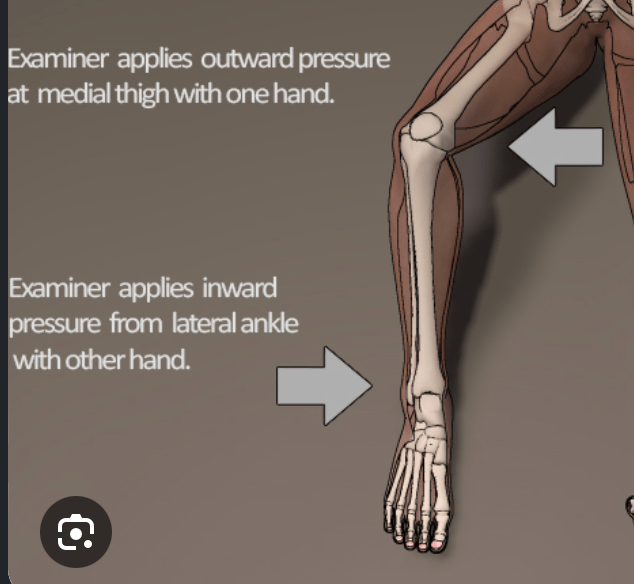
What is varus stress test?
Teaching point: testing for LCL sprain. gum vs rum
200
This is 3 and 4c (in order)
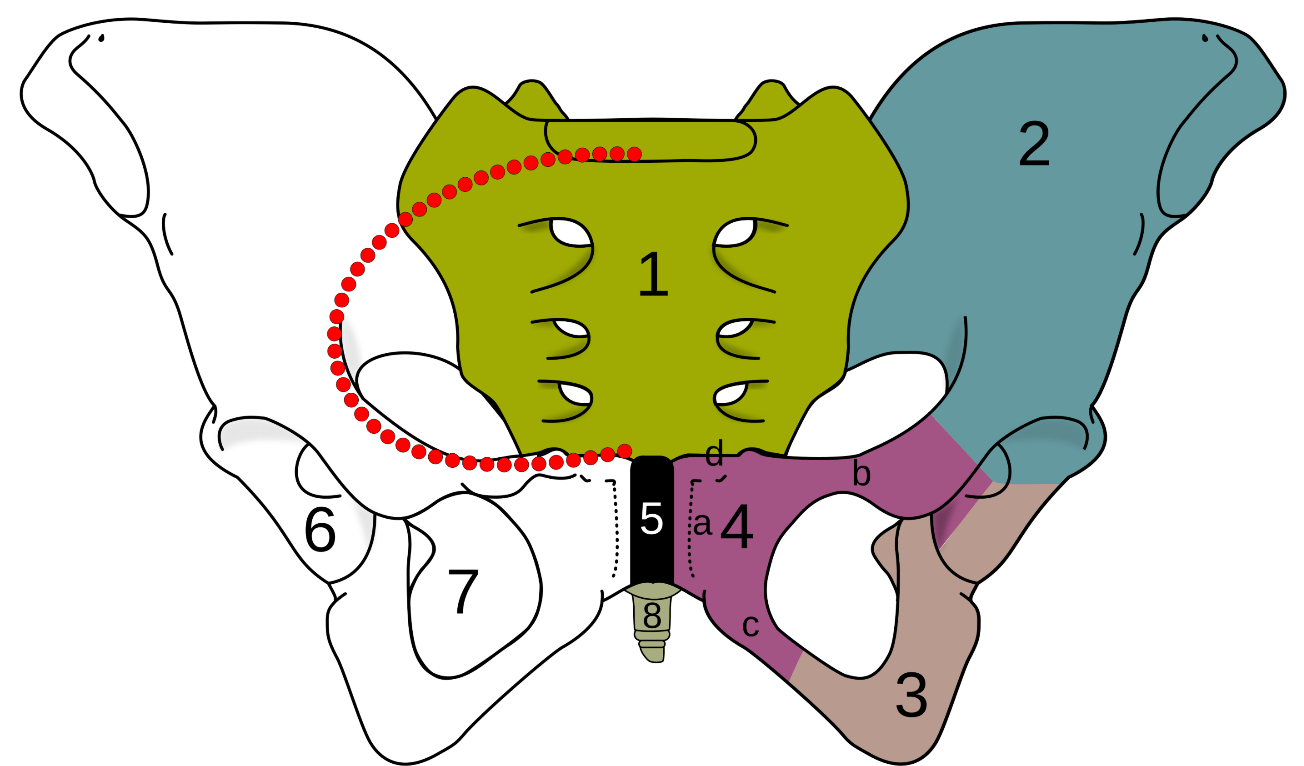
What are the ischium and inferior pubic rami?
Learning point: 1. Sacrum (os sacrum), 2. Ilium (os ilium), 3. Ischium (os ischii)
4. Pubic bone (os pubis) (4a. corpus, 4b. ramus superior, 4c.ramus inferior, 4d. tuberculum pubicum)
5. Pubic symphysis, 6. Acetabulum (of the hip joint), 7. Obturator foramen, 8. Coccyx/tailbone (os coccygis)
Dotted. Linea terminalis of the pelvic brim.
200
This is fractured
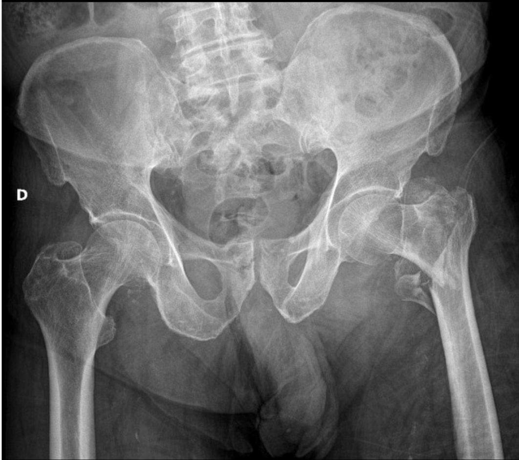
What is an L intertrochanteric fracture?
learning point: 4 types: femoral head, femoral neck, intertrochanteric and subtrochanteric.
200
The commonplace name for the fracture below
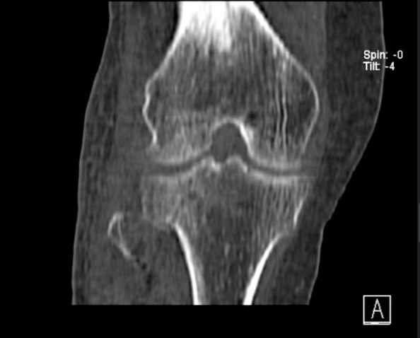
What is a bumper or fender fracture?
Learning point: tib plateau make up 1% of all adult fractures, Need urgent reduction and early MOBILIZATION. immobilization longer than 3 weeks with worse outcomes. ORIF is always gold standard. 5% require knee arthroplasty later in life.
Displaced intra-articular fractures, joint instability, malalignment, and high-energy/complexpatterns are the core indications for ORIF, while < 4mm displacement, nonop, same outcomes. Post-traumatic osteoarthritis is common regardless of treatment. Today, Improvement in outcomes compared to older data (regardless of surgery), but still average 100 day light work
200
This pathology reveals the below physical exam finding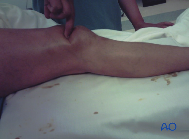
What is quadriceps tendon rupture?
Learning point: corticosteroids and fluoroquinolones are recognized precipitants. uncommon injury, men between 50-70 (still moving around, but not as strong as they used to be). Caused by rapid quad eccentric contraction (walking down stairs, slip, straighten quad to catch you)
300
The technical name for this type of "fracture"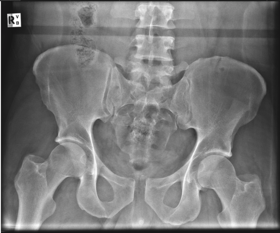
What is anterior-posterior compression injury?
Learning point: most bleeding will be venous (presacral and prevesical (paravesical) venous plexuses and the raw cancellous bone surfaces of the disrupted sacroiliac joint and posterior ring) but arterial will almost always be branches of the internal iliac (hypogastric) artery
300
If you see the below X-ray, you should do this physical exam pathway next

What is TEN-4-FACESp? (will accept skeletal survey and CT head)
Learning point: femoral shaft fractures in children are relatively uncommon and accompany a high energy force mechanism. All children with a femoral shaft fracture should undergo a thorough ten-4-facesp exam and get skeletal survey/ct head 3d recon if story is concerning etc
300
The name of the fracture seen below
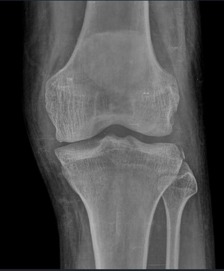
What is a segond fracture?
Learning point: Small avulsion off the lateral tibial rim. Highly associated with ACL tear and meniscal injury. A tiny fragment with big significance.
300
Performing these two maneuvers are part of the newborn physical exam
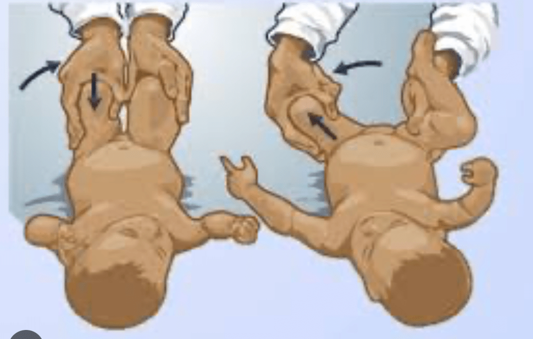
What are Barlow and Ortolani maneveurs?
Teaching point: checking for developmental dysplasia of the hip. Risk factors are breech, female, first born, restrictive swaddling. Treatment is Pavlik harness. untreated= 33% of hip replacements < 65 years old
400
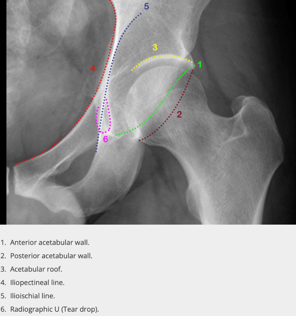
This type of fracture is seen below
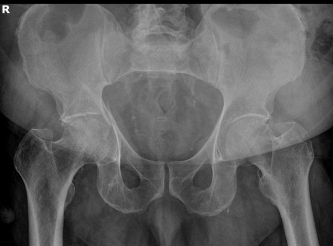
What is a R acetabular fracture?
Learning point: high energy trauma with axial loading of the femur, ie fall from height. Should evaluate the below anatomy of your pelvis to identify whether a acetabular fx is present
400
If you see this finding below, it is called this
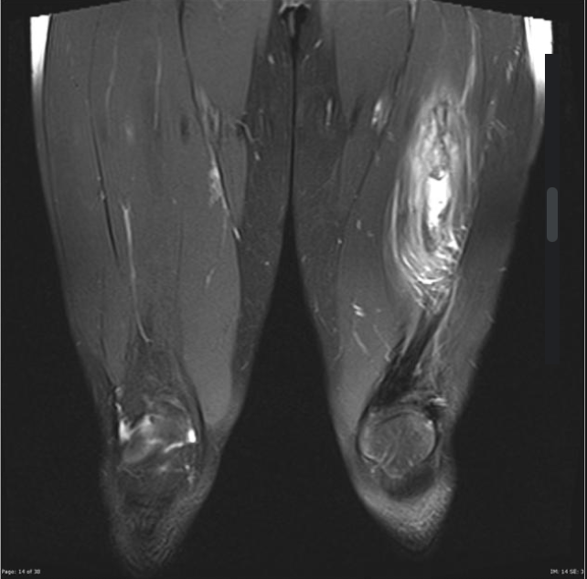
What is myositis ossificans?
Learning point: most common causes are trauma, prolonged immobilization/neurologic injury, burns. 3 stages:
1. calcium deposition often begins ~7–10 days after symptom onset, radiographically apparent for 6 weeks or longer. MRI shows extensive muscle edema at this point.
2. Intermediate stage (4–8 weeks): osteoblastic differentiation with immature osteoid maturing into bone at the periphery, producing the characteristic zonal pattern (cellular center, ossifying rim). Peripheral calcification generally becomes visible on radiograph/CT between 2 and 4 weeks
3. Mature stage (>8 weeks): a well-organized mass of mature lamellar bone with a distinctive peripheral shell — reliably distinguishes MO from osteosarcoma (which ossifies centrally). 6 weeks to several months
Surgical excision is reserved for mature lesions that cause persistent pain, muscle weakness, or a fixed loss of joint motion/function. delay until complete maturation due to recurrence concern. Otherwise, bisphonates and NSAIDS
400
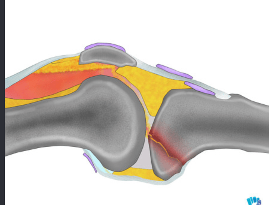
The tibial plateau fracture below also has this finding
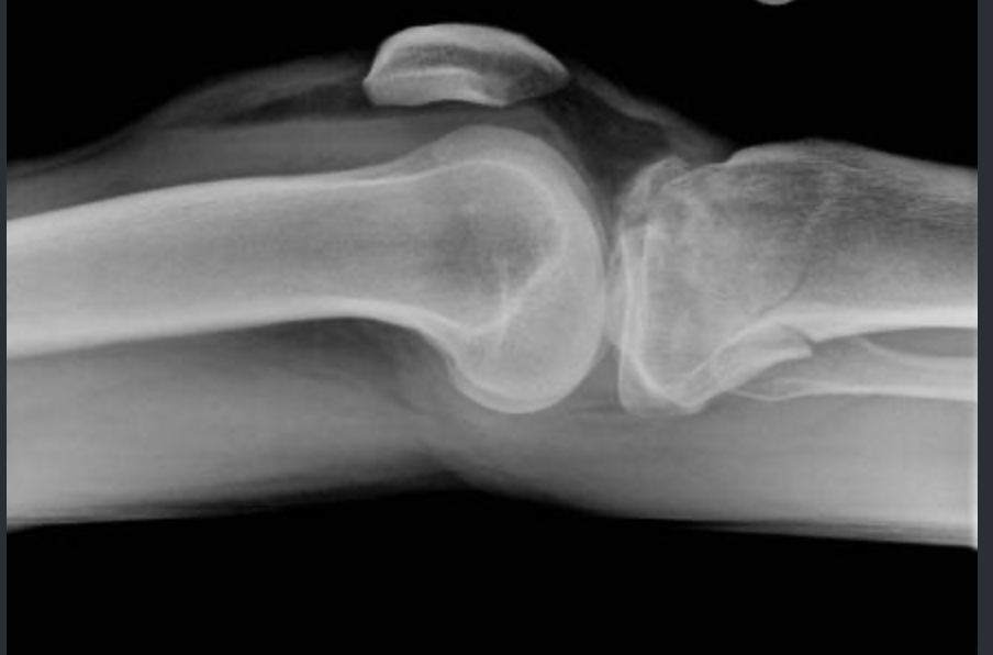
What is lipohemarthrosis?
Learning point: intra-articular fracture disrupts cortex and marrow, releasing marrow fat and blood. lateral radiography, horizontal x-ray beam captures the layered interface in the suprapatellar bursa
400
A positive short arc test is helpful for ruling in this pathology
What is septic arthritis of the knee?
Learning point: minimal passive ROM (<10 degrees) with axial traction should not be painful in cellulitis of the knee, osteoarthritis/rheumatoid arthritis. Also remember to not perform arthocentesis if obvious overlying cellulitis is present as can provide nidus into septic joint
500
The common name of this fracture (not named after anatomy)
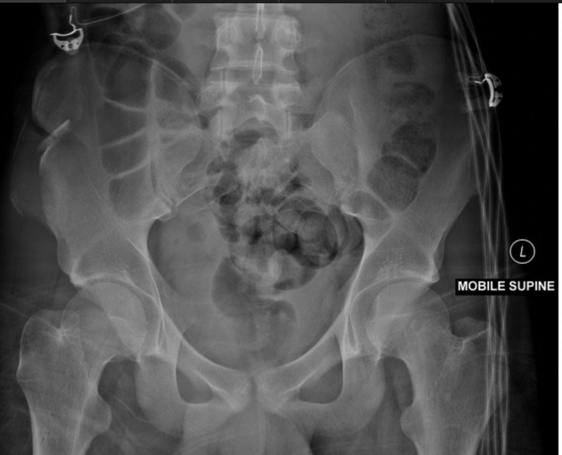
What is a Duverney fracture?
Learning point: direct blow to the ilium, with a resultant isolated iliac wing fracture
It is named after Joseph Guichard Duverney (1648-1730), a French surgeon who described it after a patient fell onto, and broke, an iron tire. The patient died days later. During dissection, a pus-filled fracture of the ilium was discovered 5.
500
Patient has a left intertrochanteric fracture, this is why they have it (not trauma)
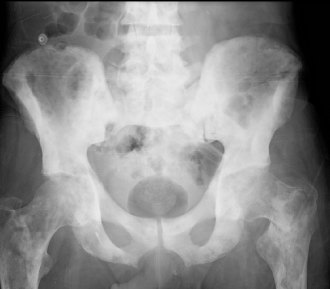
What is a pathologic fracture?
Learning point: pathologic fracture is any broken bone due to underlying disease that does not correlate with traumatic mechanism. Most common locations are subtrochanteric, humeral head, and vertebral body. Admittable criteria, need PTH, malignancy work-up etc. Should also get CT scan
500
The image below is indicative of this traumatic knee injury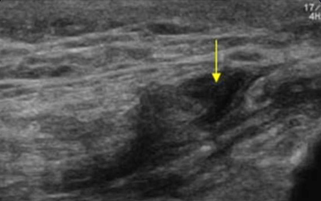
What is a quadriceps tendon tear?
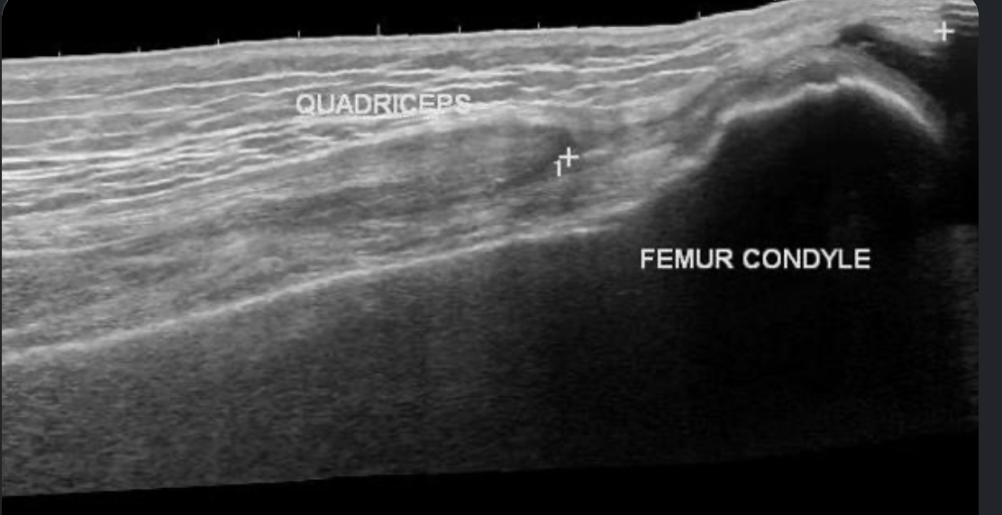
Learning point: ultrasound is often underused in the ED for MSK injuries. It is easy to evaluate the patella and tendon insertion sites as noted below. A hematoma with difficulty visualizing the insertion site is concern for a quad tendon tear. This injury does not require emergent surgery- immobilize in full extension and f/u OP
500
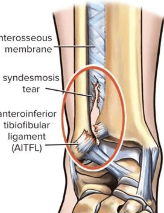
Orthopedics will stress ankle fractures afterwards to check for ankle mortise widening secondary to this anatomical injury
What is a syndesmotic injury?