Tissue propagation
Mechanics of ultrasound
Image generation
Echogenicity 1
Echogenicity 2
100
At what speed does sound propagate through soft tissue?
1540 m/sec
100
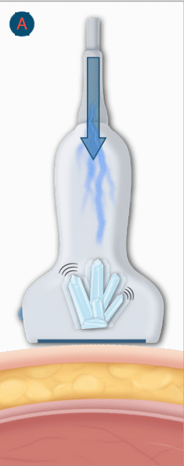
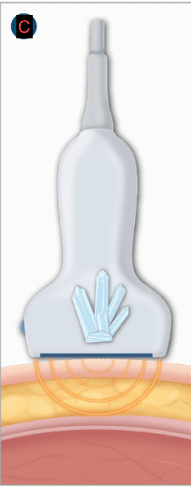
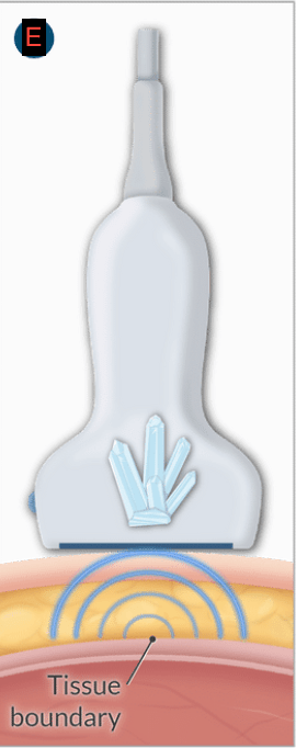
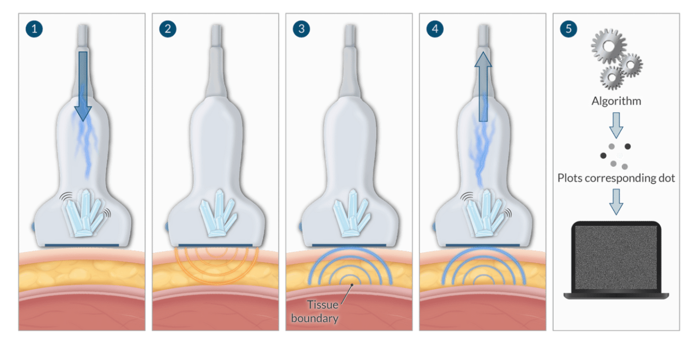
What happens when you apply an electrical current to a piezo electric material?
It vibrates and emits soundwaves
100
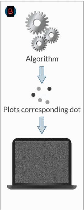
What determines the vertical placement of each dot that makes up the ultrasound image?
Time delay
100
Define anechoic
Tissue that does not produce echos
100
How will an anechoic image appear on the screen?
Black
200
At what speed does sound propagate through air?
343 m/sec
200
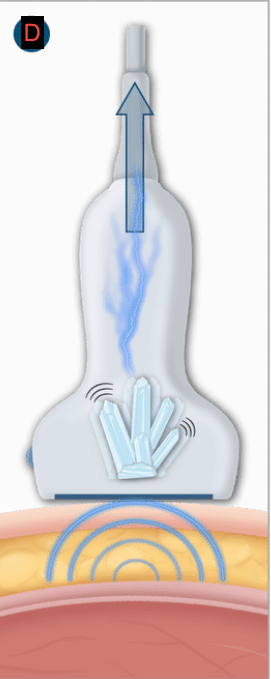
What happens when you subject a material to soundwaves?
The material will generate an electrical potential
200
What is time delay?
How long it takes for the echo to return to the transducer
200
Define hypoechoic
Tissue that produces some echos
200
How will a hypoechoic tissue appear on screen?
Grey
300
At what speed does sound propagate through bone?
3,000-5,000 m/sec
300
What piezoelectric material is used in an ultrasound probe
Lead zirconate titanate
300
What determines the horizontal placement of each dot?
It is determined by which particular crystal receives the returning echo
300
Define hyperechoic
Tissue that produces high amplitude echos
300
How will a hyperechoic tissue appear on the screen?
White
400
Will lower frequencies have shallower or deeper tissue penetration and what will the image quality be with a low frequency probe?
Tissue penetration will be deeper but the image quality will be poorer.
400
400
Which below is long access and which is short access?
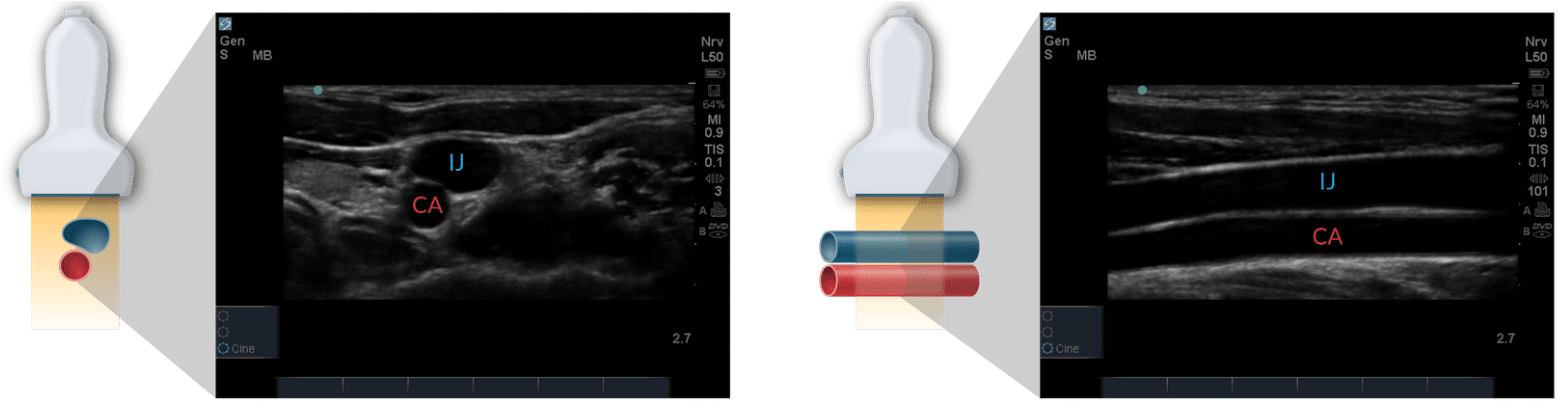
Short access Long access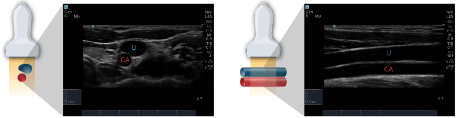
400
Peripheral nerves appear to be ____ while distal nerves appear to be _____
Hyperechoic, Anechoic
Peripheral nerves appear to be anechoic while distal nerves appear to be hyperecohic
400
What is an example of hyperechoic tissue?
Bone
500
Will higher frequencies have shallower or deeper tissue penetration and what will the image quality be with a high frequency probe?
The tissue penetration will be more shallow but produce a higher quality image.
500
Describe the doppler effect and a way in which it can be used
A positive doppler shift or red shift is observed when the source is moving toward the observer. A decreased wavelength and increased frequency is noted.
A negative doppler shift or blue shift is observed when the source is moving away from the observer. An increased wavelength and decreased frequency is noted.
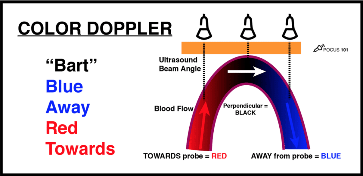
500
Describe the function of the gain control
The gain control adjusts the strength of the returning echoes displayed on the screen. It is important to note that adjusting the gain will not improve your anatomic view, it will only brighten up the view you have at that time.
500
When scanning vascular structures how do you differentiate between arterial vessels and venous vessels?
Venous vessels are compressible while arterial vessels pulsate.
500
What is an example of anechoic tissue?
Vascular structures, cysts or ascites