This type of tissue is found in the lining of the trachea.
What is pseudo-stratified ciliated columnar epithelium?
What is TLC = RV + VC
The _____ are the muscles that keep the atrioventricular valves closed.
What are papillary muscles?
In gas diffusion between a capillary and alveoli, how many cells does the gas diffuse through and what type of cells are they?
What is a pneumothorax?
The collapsing of a lung due to air entering the pleural cavity.
The four types of tissues.
What are:
Muscle
Connective
Nervous
Epithelium
During inspiration, the ribs ____ and the diaphragm _____ and ______.
Lift, contracts and flattens out
This type of connective tissue makes up the epiglottis and the outer ear.
What is elastic cartilage?
What is an aneurysm?
The "ballooning" of a weak spot in a blood vessel under pressure.
These two muscle types only have one nucleus?
What is Cardiac and Smooth?
External respiration is:
What is, the process of gas diffusion between the alveoli and the capillaries in the lungs?
Describe the flow of blood through the heart, include the oxygen status at each location.
- Oxygen poor blood is returned to the right atrium through the superior and inferior vena cava.
- Oxygen poor blood passes through the tricuspid atrioventricular valve into the right ventricle
- Oxygen poor blood passes through the pulmonary semilunar valve into the pulmonary truk
- Oxygen poor blood is carried from the pulmonary trunk to the pulmonary arteries to the lungs where the blood becomes oxygenated
- Oxygen rich blood is returned to the left atrium through the pulmonary veins
- Oxygen rich blood passes through the bicuspid atrioventricular valve into the left ventricle.
- Oxygen rich blood is forced out of the ventricle through the aortic semilunar valve into the aorta
- The blood is systemically circulated
The function of simple cuboidal epithelium.
What does M.I stand for, what is it known as colloquially, and what is it caused by?
What is, Myocardial Infarction, Heart attack, and a lack of oxygen to cardiac muscle caused by a coronary artery blockage.
The features of this muscle tissue include intercalated discs and bifurcation (branching) of the cells.
What is cardiac muscle?
This structure is the beginning of the lower respiratory pathway.
What is the trachea?
The three layers of the heart.
Pericardium:Serous membrane
Myocardium: Cardiac muscles
Endocardium: Epithelial lining of the cardiac chambers
What type of connective tissue is found in the inter-vertebral discs?
What is atherosclerosis and what is it caused by?
The function of this connective tissue is to provide protection and store energy
What is adipose connective tissue?
Name the three parts of the pleura and where they are found.
Visceral Pleura: Snug against lung tissues.
Pleural Cavity: Between the two pleural membranes, contains serous fluid.
Parietal Pleura: Lines the walls of the thoracic cavity.

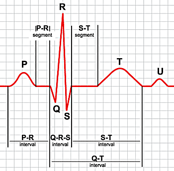
Describe what is happening during each of the following:
- P wave
- QRS Complex
- T wave
- P-R interval
- Q - T interval
What is:
P Wave - SA node fires causing atrial depolarization and contraction ( atrial systole).
QRS Complex - Depolarization and contraction of the ventricles (ventricular systole).
T Wave - Ventricular re-polarization and relaxation (return to diastole).
P - R interval - From atrial depolarization to ventricular depolarization.
Q-T interval - Total time of ventricular contraction and relaxation.
Which of the following is NOT a type of fibrous connective tissue?
- Dense
- Collagen
- Elastic
- Loose
What is Collagen?
The condition of hypohidrosis is the dis-functioning of your sweat glands. What type of gland is a sweat gland?
What is exocrine?