The Basics
Calculate this
Impact
Name that VHD
Patho
100
The leaky valve suffers from this.
Regurgitation/Insufficiency
100
Normal IVC measurement
<2.1 cm
100
Atrium can't hypertrophy so they do this instead.
Dilate
100
 The report description for the imaged aortic valve. Be Profesh!
Sclerotic AV
100
Potenially caused by rhematic fever, this is the shape of the anterior leaflet during diastole.
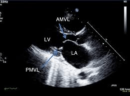
Hockey Stick
200
The kinked/narrowed valve suffers from this.
Stenosis
200
I add this value to RAP with <2.1 cm IVC diameter and no collapse.
8 mmHg
200
Sterling would agree, that increased pressure also does this.
Increases Afterload
200
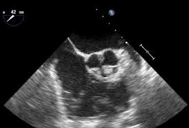
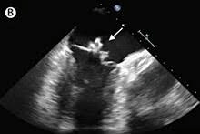
 That aortic valve, though.
Bicuspid Aortic Valve.
200
The A, B, C ..... shape caused by pressure
D shaped septum
300
Pathology affecting heart valves not as prevalent since modern medicine.
Rhematic Fever
300
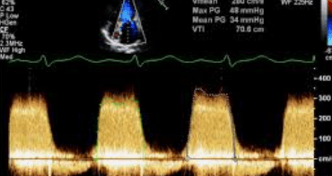
The formula that calculates pressure through the valve.
P=4v(squared)
300
Regurgitation increases this idea from Sterling.
Preload
300
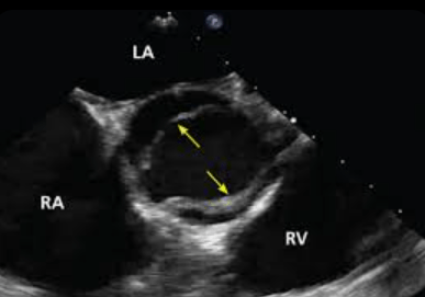
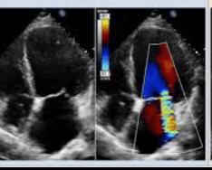
 That mitral severity and what we call the VHD.
MS / Severe
300
Patient presents with fever and the sonographer finds this on Echo. This is the pathology and the disease process.
Vegetation/Endocarditis
400
The PISA baseline values to see the hemispheric shape of the convergence of flow.
20 - 40 cm/s
400
The pressure half time of MS is 2. What is the MVA?
110 cm(squared)
220/p1/2t
400
Chaos ensues following stenosis and this happens to pressure.
Decreased Pressure
400
Valvular patho seen with connective tissue disorders like Marfan, Lupus, and Ehlers-Danlos syndrome.
MVP
400
That bright stuff seen on the mitral annulus.
MAC - mitral annular calcification
500
The flutter of the mitral valve is seen with this other pathology.
Severe AI
500
The peak gradient for AS totals 64. This is the velocity.
4
500
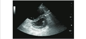
 Prime example of this class of MR following atrial dilation.
Secondary MR
500
PVA =
0.785 x RVOT(squared) x RVOT VTI / PV VTI
500
Name that congenital pathology and what it's caused by. 
Displaced tricuspid valve - Epstein's Anomaly