Embryology
Ungulates
Joints
K9 Pelvic Boney Landmarks
Pelvic Nerves/Arteries
Integument
100
The three layers of embryonic development
Mesoderm, Ectoderm, Endoderm
100
Animals that walk one all their toe bones vs an animal that walks on only the most distal toe bone.
Digitigrade and Unguligrade
100
Two types of ossification.
Intramembranous ossification (flat bones)
Endochondral ossification (long bones)
100
"A"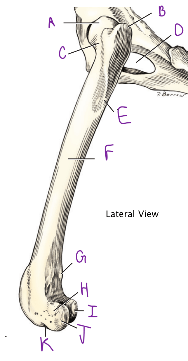
Greater trochanter
100
The spinal segments of the pelvic intumescence.
L4 to S3
100
The three pads of domesticated carnivores.
1) Carpal/Trasal pad
2) Metacarpal/trasal pad
3) Digital pads
200
The embryological cell layer making up the neural tube.
Ectoderm.
200
Evolution left horses with only one primary metatarsal(carpal) and digit bones. (Hint: it's a number)
Third/Three
200
Joint classification based on function. (3)
1) Diarthroses (aka synovial) - freely moveable
2) Synarthrosis - immoveable
3) Amphiarthroses - slightly moveable
"-arthroses" = greek, Joint
"di-" = greek, through
"syn-" = greek, together
"amphi-" = greek, both sides
200
The purple promience.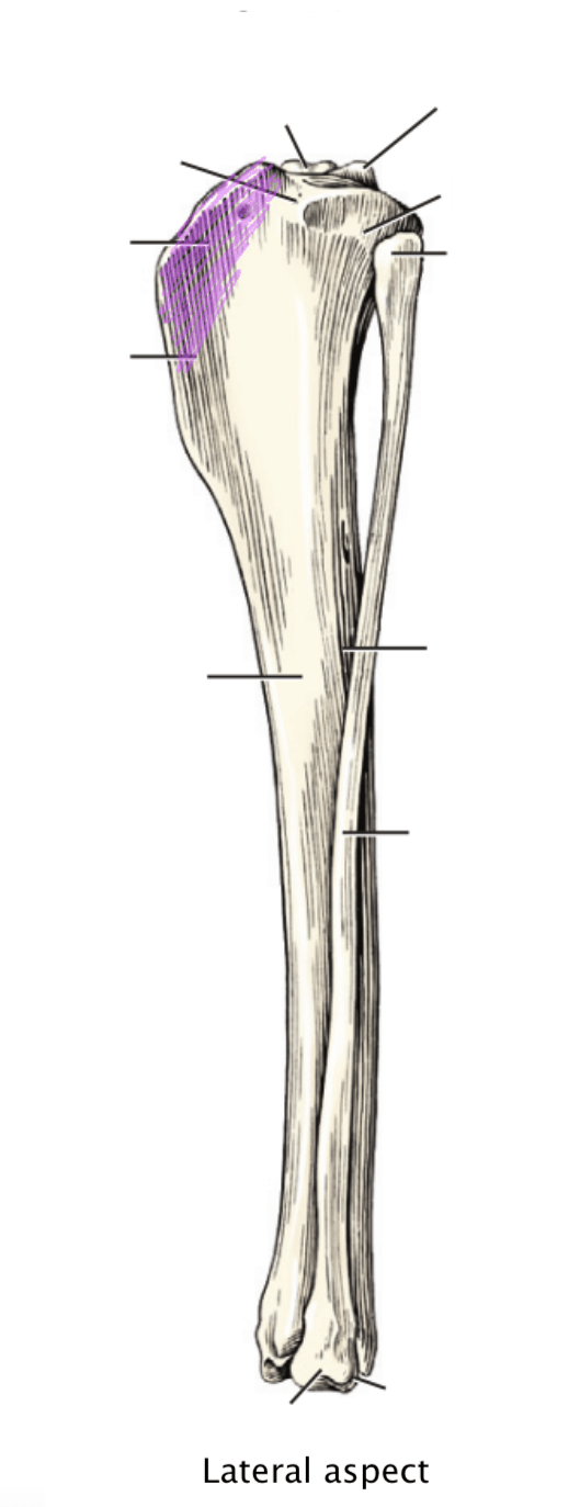
"B"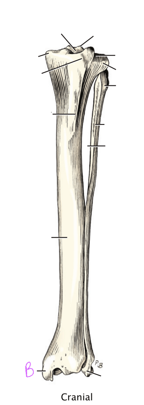
Purple: Tibial tuberosity
B: Medial malleolus
200
The nerve that runs on the dorsal aspect of the equine hindlimb along the cannon bone (M3).
Dorsal metatarsal n. (medial/lateral)
200
Two types of glands found in the skin.
Sweat glands
Sebaceous glands
300
The neural groove deepens as the neural folds grow up and towards each other. At the apex of each fold is a differentiating group of cells.
Neural crest (cells)
300
Name the red and blue colored parts.
Red = Peroneus tertius m. (Fibularis tertius m.)
Blue = Superficial digital flexor m.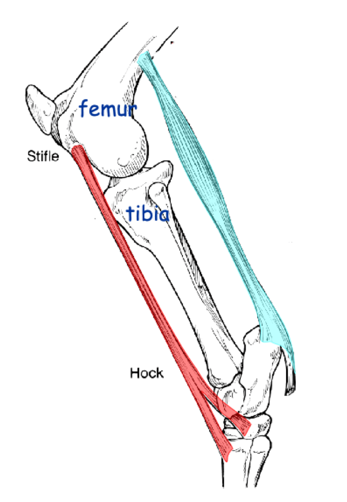
300
Ligament(s) within the joint space that assists with control of the joint movement.
Intracapsular ligaments
300
Three pelvic limb palpable landmarks proximal to the shaft of the femur.
1) Crest of the ilium
2) Greater trochanter
3) Ischial tuberosity
300
Iliopsaoas m. and Sartorius m. are innervated by this nerve.
Femoral n.
*also innervates the Quadriceps mm.
300
Gland located along the medial pelvic fold, most commonly found in sheep and rabbits.
Inguinal glands.
400
Three functions of the mammalian yolk sac.
1) Nutrient and gas transfer prior to complete placental development
2) where blood elements are formed prior to live development
3) generation of primordial germ cells for future fetal gonads
400
Function of the reciprocal apparatus.
Prevents collapse of the hindlimb while supporting the weight of the animal via a locking mechanism; also guarantees that when the stifle is in extension the hock will also be in extension.
400
A fibrocartilage or dense fibrous tissue that increases congruency between articular cartilages. Also helps absorb concussion loads.
Articular discs and/or menisci
400
Three aspects of the femur where the patella sits.
1) Medial trochlea ridge
2) Trochlea
3) Lateral trochlea ridge
400
The Sciatic n. innervates these muscles.
Biceps femoris m.
Semimembranosus m.
Semitendinosus m.
400
Connective tissue that is primarily collagen with some elastic fibers, and a similar connective tissue that is continuous with the periosteum.
Superficial fascia
Deep fascia
500
The column of paraxial mesoderm divides itself into small, paired blocks along the cranial-caudal axis. These small aggregates are visible on the dorsum of the embyo and are made of a specific embryological cell layer.
Somites and mesoderm
500
The three primary parts of the suspensory apparatus.
1) suspensory ligament
2) proximal sesamoid bones
3) distal sesamoidean ligaments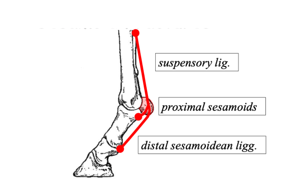
500
Thickened facia that anchors and/or stabilizes tendons, but still allows them to move.
Retinaculum
500
The origin and insertion of the gastrocnemius m.
Origin: Lateral and Medial supracondylar tuberosities (femur)
Insertion: Calcaneal tuber
500
The Cranial gluteal n. spans these spinal segments and innervates at least one muscle.
L6-S1
Middle gluteal m.
Deep gluteal m.
Tensor fascia lata m.
500
The layers of epidermis.
Stratum basal
Stratum spinosum
Stratum granulosum
Stratum corneum
600
The development of the extra-embryonic coelom divides the mesoderm into two separate layers: the Somatic Mesoderm and the Splanchnic Mesoderm, each of these two layers join with another tissue/layer creating...
(Troph)Ectoderm + Mesoderm = Somatopleure
Endoderm + Mesoderm = Splanchnopleure
600
The proximal anatomical parts that allow a horse to lock it's stifle in extension. (3)
Proximad draw of the patella so that the parapatellar cartilage and medial patellar tendon can be hooked over the femur's medial trochlear ridge.
600
Synarthroses joints, aka Fibrous joints + cartilaginous joints. (6)
Fibrous Joints:
1) syndesmosis: united by white fibrous or elastic connective tissue (metacarpal bones, costal cartilage)
2) sutures: narrow fibrous uniting of bone, skull plates
3) gomphosis: collagen fibers to bone, periodontal ligaments
4) synostosis: fusion of bone, sacrum*
Cartilaginous joints
4) synchondrosis: hyaline cartilage joint, epiphyseal junction
5) symphysis: fibrocartilage joint, pubic and mandibular symphysis
600
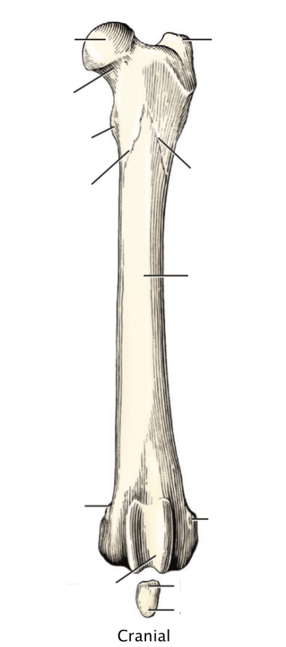
Name A through G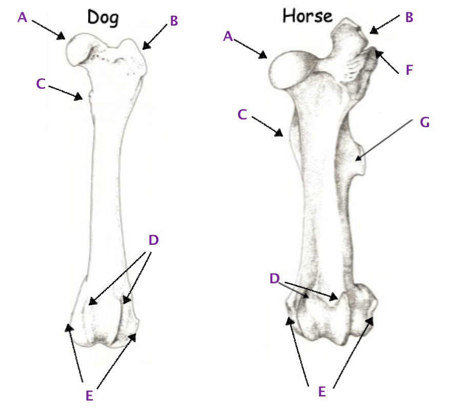
A: Head of the Femur
B: Greater trochanter
C: Lesser trochanter
D: Medial and Lateral trochlear ridges
E: Medial and Lateral epicondyles
F: Greater trochanter notch
G: 3rd trochanter
600
Name: A, B, C, D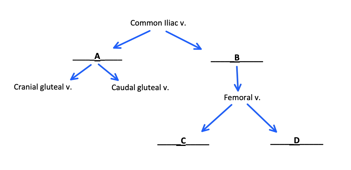
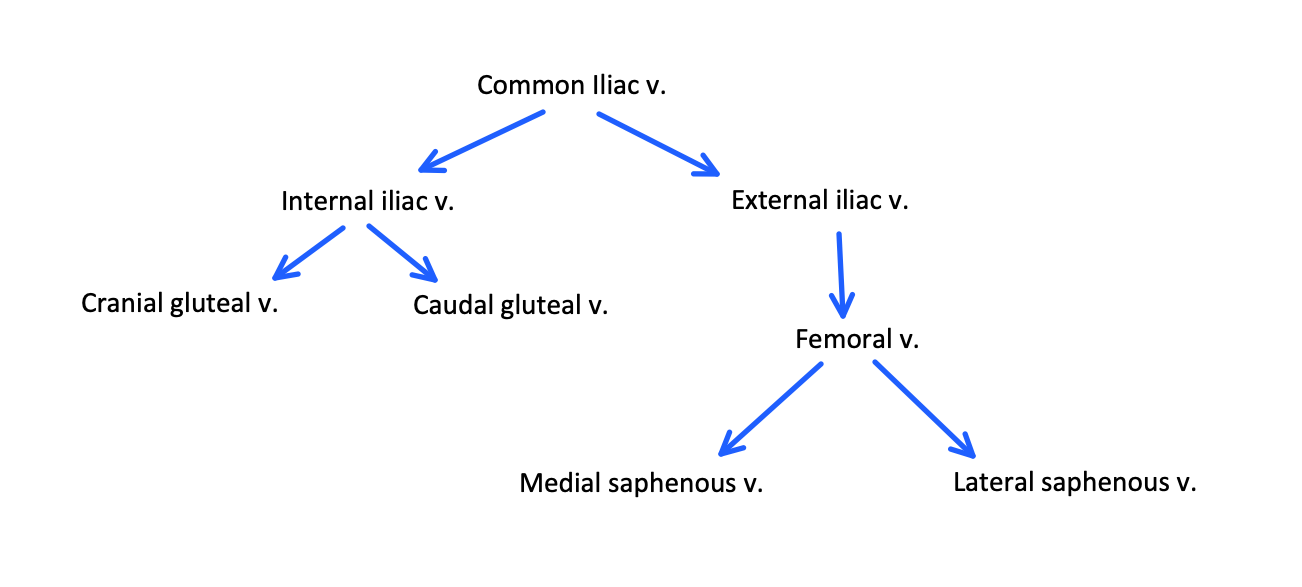
600
The two interdigitating, connective parts of a hoof wall and the dermis/periosteum.
Epidermal lamellae (insensitive)
Dermal lamellae (sensitive)