RENAL DOPPLER
AORTA
MEASUREMENTS
CAROTID/VERTEBRAL
BRAIN TEASER
100
This renal artery is longer than the other
what's the right renal artery?
100
The first branch of the Aorta
What's Celiac Trunk
100
The Aortic aneurysm is measured this way
Outer to outer
100
The right Carotid branches off of the Brachio-Cephalic
True or False?
False
100
Low resistance flow in the ECA suggestive of...
ICA severe stenosis or Occlusion
200
the R and L renal arteries originate below this vessel
SMA
200
the rupture threat for the AO aneurysm is
5 CM
200
Acceleration time
resistive Index
Hypertension
Intrarenal Doppler
200
the vertebral arteries bilaterally join to form this vessel
Basilar
200
Obesity
Smoking
Diabetes
hypertension
Modifiable risk factors for arterial disease
300
Common anatomic variant of renal vasculature
Duplicated renal artery
300
Patient signs and symptoms for an aneurysm of about 3.8 cm
Asymptomatic
300
Obesity
Smoking
Diabetes
hypertension
modifiable risk factors for arterial disease
300
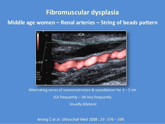
FMD
300
Bilateral Carotid exam showed:
Damped and low velocity waveforms bilaterally in all the carotid system
Where is the problem?
Cardiac. Probable aortic valve disease. Know the patient history.
400
RI=.8
Abnormal RI in the intrarenal vessel
400
The most reliable plane for the Aorta measurement
Transverse, AP and width
400
RI < 0.75
Normal acceleration time
Normal findings in the Intrarenal vessels
400
Thyroid is supplied by these arteries
Superior Thyroid Arteries
400
Severe stenosis to near occlusion in the left ICA.
Minimal plaque in the right ICA
Increased velocities in the right ICA
Compensatory flow
500
Equation for RI
PSV-EDV/PSV
500
Surveillance of the Endograft done post surgically for this reason
Endoleak
500
PSV
EDV
Ratio
Neck artery
ICA 1-3 cm
500
Flow is reversed in the left vertebral artery
Remember, blood flows from high to low resistance
Subclavian artery stenosis
500
Tardus - Parvus waveform in the right CCA, Vertebral and the right subclavian. The stenosis is here
Brachiocephalic