Determines course of the frontotemporal branch of facial nerve
A line that runs from 0.5 cm below the tragus to 1.5 cm above the lateral eyebrow
accessory nerve
lateral neck
this junction flattens with aging
dermal-epidermal junction
most common early complication of facelift
hematoma
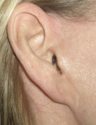
name the deformity. How does it form?

Pixie ear deformity
caused by extrinsic pull on the earlobe due to tension of the incision
Name four structures that are contiguous with the SMAS
galea aponeurosis
temporoparietal fascia
partoid masseteric fascia
platysma
superficial cervical fascia
source: G+S
what/where is McKinney's point?
what: reference point for the great auricular nerve
where: where the great auricular nerve crosses the SCM approx 6.5cm below the caudal edge of the bony external auditory canal
With aging, Type ____ collagen increases and type ____ decreases (causes fine wrinkles)
Type III collagen increases and type I decreases (causes fine wrinkles)
Name the most common nerve that is injured in facelift and how it will manifest
Great auricular nerve -> numbness to the earlobe
source: G+S
this is (in theory) the best technique for patients with potential wound healing (i.e. smokers)
deep plane/composite
the SMAS and skin are suspended as a composite flap which is thick and has robust blood supply
this is the dominant artery that supplies a facelift flap (and name its origin)
The transverse facial perforating artery (TFA) is a branch of the superficial temporal artery that perfuses the lateral face.
how can you determine an injury to the cervical branch facial nerve, rather than the marginal mandibular nerve?
In a cervical branch facial nerve injury, lip depression can be weak, but the mentalis and orbicularis oris innervation remain intact, so that the patient would be able to purse her lips. Also, patient will be able to evert the lower lip because of a functioning mentalis muscle.
In general, cervical branch weakness typically resolves within 4 to 12 weeks.
Injury to the marginal mandibular nerve can occur in either subcutaneous or superficial musculoaponeurotic system dissection. Although this injury can be permanent, as with other facial nerve injury, spontaneous recovery within 6 months is the expected outcome in most (80%) patients.
what is nanofat? what is it used for?
extraction of autologous fat from a patient, which is then transformed into “nanofat”, consisting of small fat particles that are emulsified with a diameter of less than 0.1 mm and containing high concentrations of stem cells and growth factors, it has no viable fat cells
not for volume enhancement, but for skin rejuvenation
a patient comes in to clinic 2 weeks following a facelift with a 3x3cm area of eschar. Describe the next step
Wound-healing issues and skin necrosis should initially be managed with local wound care. In many cases, the wounds will go on to heal without negative sequelae. In the remainder of the cases, a corticosteroid injection or scar revision may be all that is necessary.
Debridement of the region is not recommended because the eschar acts as a biologic dressing. Skin grafting would be indicated for a very large area of full-thickness necrosis. Re-advancement of the flap would not be indicated at this time as conservative management works well.
Furthermore, re-advancement of the flap at this time would likely place too much tension on the closure with its resulting stigmata. However, re-advancement may be indicated at the time of scar revision once the wound has healed and the skin laxity has returned.
in a dual plane facelift, the SMAS flap is elevated
______ (direction) and the skin flap ________
SMAS -> superolateral
Skin -> posterolateral
name the 3 muscles that are innervated by the facial nerve on their superficial surface
mentalis, buccinator, depressor labii
source: G+S
1) tragal pointer
2) posterior belly of the digastric muscle
3) stylomastoid foramen/mastoid process
4) tympanomastoid suture
5) styloid process
6) peripheral branches -> retrograde dissection
Describe SMAS stacking
Stacking bridges the contouring effect between the deep medial and lateral superficial malar compartments. Stacking is more powerful as an augmentative maneuver than plication because an island of SMAS is pre- served centrally and a bilaminar construct is created. DM, deep malar fat; DN-L, deep nasolabial fold.
source: Rohrich et al, Lift-and-Fill Face Lift: Integrating the Fat Compartments
what is frey's syndrome?
describes gustatory sweating after aberrant reinnervation of cutaneous sweat glands after disruption of auriculotemporal nerve branches (more likely after parotidectomy)
The first facelift was performed in (year?) in (City?)
The first facelift was performed in 1901 by Eugen Hollander in Berlin.
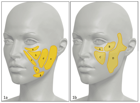
Name the fat pads (need 6 to get correct)

Superficial cheek fat compartments:
a) Infraorbital fat. b) Medial cheek fat. c) Nasolabial fat. d) Middle cheek fat. e) Lateral cheek fat. f) Superior jowl fat. g) Inferior jowl fat.
Deep cheek fat compartments:
A) Medial sub-orbicularis oculi fat. B) Lateral sub-orbicularis oculi fat. C) Deep medial cheek fat. D) Buccal fat.
This embryologic structure becomes the facial nerve at third week gestation
the facioacoustic primordium
Fill in the blanks. An aged face, when compared with a youthful face, has the following:
_____ of the malar region
______ upper orbital sulcus
______ of the lower eyelid-malar junction
______ submental angle
Concavity of the malar region
Deep-set upper orbital sulcus
Long position of the lower eyelid-malar junction
Obtuse submental angle
A 54-year-old woman comes to the office because she is unhappy with the appearance of her forehead 1 year after undergoing endoscopic brow lift surgery and upper and lower blepharoplasty. She says there is an indentation between her eyebrows when she frowns. Physical examination shows irregular dimpling in the glabellar area upon frowning. Which of the following is the most likely cause of this patient's postoperative outcome?
Inadequate removal of muscle
An observation following endoscopic forehead rejuvenation is inadequate removal of the glabellar muscles, resulting in early recurrence of glabellar lines and frowning action. This can be avoided by removal of all of the muscle fibers between the frontal bone and the subcutaneous plane and replacement with fat grafts. Application of the fat graft in this area will not only improve the contour but also reduce the potential for the full gain of muscle function, even if some fibers are left intact. The residual or regenerated muscle fibers will not be as effective or as powerful without bone insertion. Furthermore, the fat graft will eliminate the flatness of the glabella as a consequence of aging. This flaw could also be the result of contraction of retained muscle fibers in those patients with very thin glabellar skin. These irregularities may only become noticeable on animation. Complete removal of the glabellar muscles and replacement with fat grafts will prevent this undesirable outcome.
Name the Beale five R's for performing successful secondary rhytidectomy
1) resect (skin)
2) release (abnormal vectors)
3) refill (fat graft)
4) reshape
5) redrape