"Gouty" Tophi
Histo Staining Process
MSK Histo Images
100
Considered the classical hallmark of gout, Tophi present as these (size AND type) crystal aggregates
What are large, "chalk-like" monosodium urate crystal aggregates
100
In standard slide preparation for staining, specimen is fixed with this, an aqueous solution; after, the specimen is stained with H&E.
What is formalin (or formaldehyde)?
100
The left-most arrow in the below image indicates the "amorphous-appearing pale to pink" deposition of these within the joint
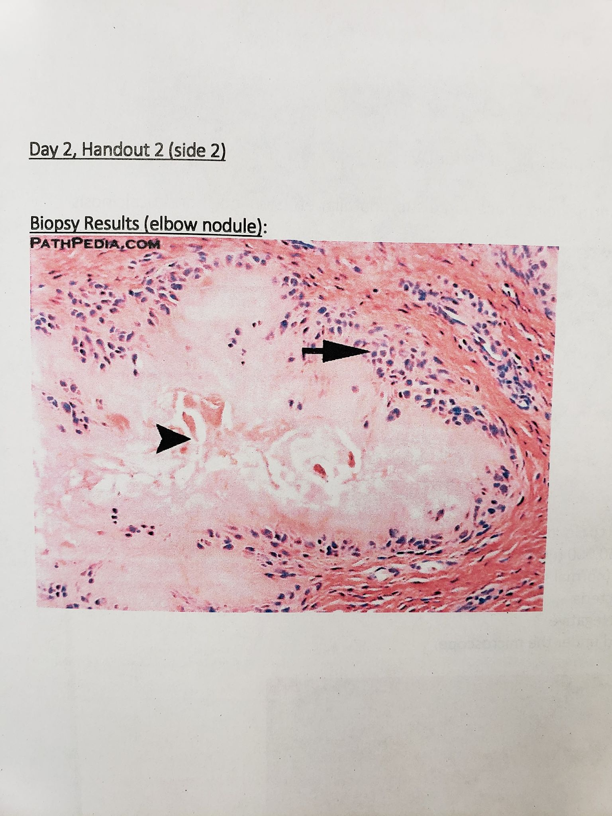
What are MSU crystals (tophi deposition)
200
Tophi often manifest in areas with periarticular tissue, such as the proximal end of the ulna, known as this
What is the olecranon
200
When preparing a gouty tophi specimen for staining, it is preferred that the tophi is fixed with this kind of solution instead of an aqueous solution, to prevent crystals from being dissolved. One such solution is called this - a mix of absolute alcohol, chloroform, and glacial acetic acid.
What is alcohol; Carnoy's solution?
200
Similar to the right-most arrowhead (in image 1), the top-most arrowhead (in image 2) indicates the presence of these inflammatory cells
Image 1:
Image 2:

What are histiocytes (arranged in a "scalloped palisade array)
300
Tophi deposits cause inflammatory responses. As monocyte-derived cells, histiocytes are unique for undergoing these 2 processes
What are "phagocytosis" and "antigen-presentation"
300
When prepared with standard fixatives, tophi are seen as pale basophilic granules with feathery needle-like “spaces” (where crystals used to be); the granule is swarmed by a ring of several macrophages and giant cells, resulting in this pathological feature.
What is a granuloma?
300
The central portion of the deposits (in image 1) may (mistakenly) resemble this important central deposit feature that is actually unique in the diagnosis of Rheumatoid Arthritis (in image 2)
Image 1:

Image 2:
What is fibrinoid necrosis
400
Sufficient MSU crystal formation can ultimately destroy articular cartilage. While high plasma urate levels have this impact on synovial joints, they also have this impact on solubility
What is supersaturation of synovial fluid and decreased solubility
400
The nature of the inflammatory cells, seen in the H&E stain above as the eosinophilic borders of the tophi, can be described as this (i.e. nuclei are still present) and this.
What are non-keratinized and stratified squamous?
400
The central portion of the deposits (in image 1) may (mistakenly) resemble this important central deposit feature that is actually unique in the diagnosis of Rheumatoid Arthritis (in image 2)
Image 1:
Image 2:
What is fibrinoid necrosis
500
Staining of tophi with eosin in absolute alcohol preserves the needle-shaped urate crystals, which appear yellow-brown; a similar stain is called this, which also prevents the crystals from being dissolved and causes them to appear brown-black (as seen in the picture).

What is the de Galantha stain?