Chest
shoulder
elbow
pelvis
100
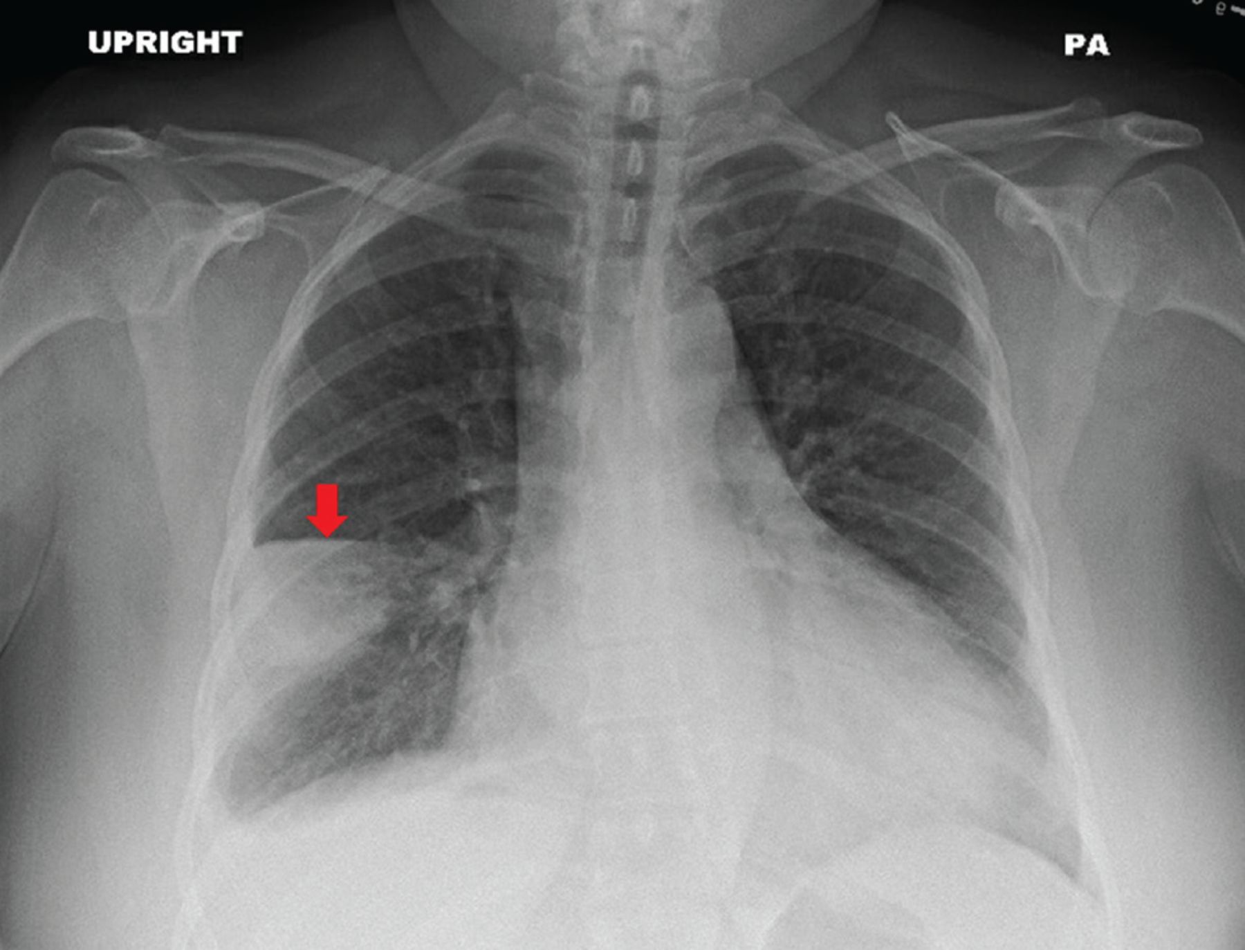
A sharp line with no lung markings beyond it—what does this represent?
Pneumothorax
100
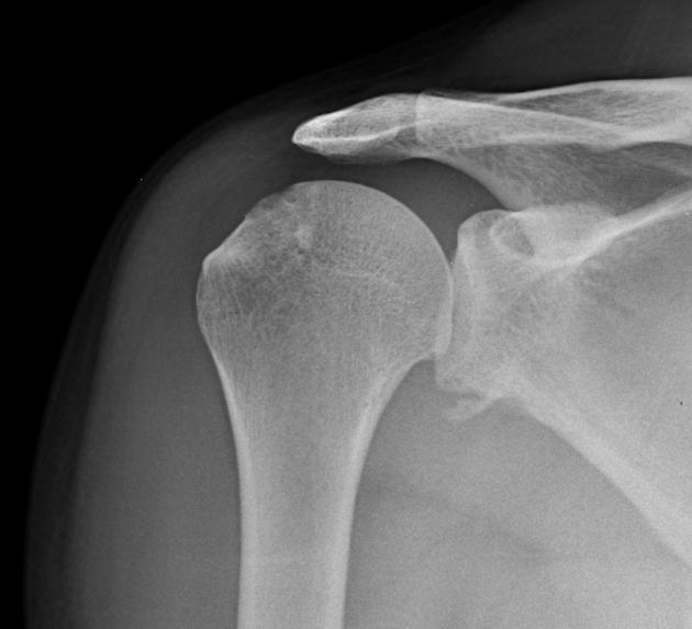
The most common direction of shoulder dislocation.
anterior shoulder dislocation
100
The most common direction of elbow dislocation.
Posterior
100
This smooth curve from the femoral neck to the superior pubic ramus should be continuous on AP pelvis; disruption suggests fracture or dislocation.
Sheltons line
200
A widened mediastinum on CXR can be a sign of this life-threatening vascular emergency.
aortic dissection- 
200
This nerve is most commonly injured in anterior shoulder dislocations.
Axillary nerve
200
 40 year old presented with history of elbow pain - sign and common fracture ?
sail sign
posterior fat pad sign
radial head fracture
200
what is this fracture called - 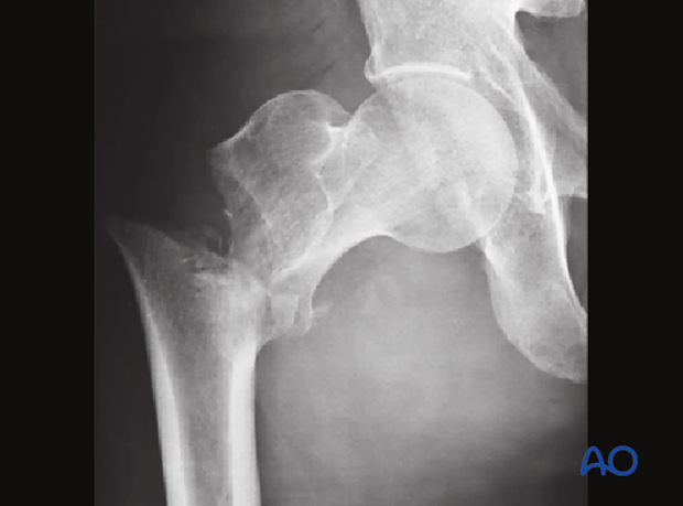
intertrochanteric fracture
300
In a trauma patient, air under the diaphragm on CXR suggests this surgical emergency.
Perforation
300
The deformity of the humeral head after repeated anterior dislocations, seen on imaging.
Hill Sach lesion- 
300
fracture of the coronoid process, radial head, and dislocation is called this triad.
Terrible triad
300
A fracture of the femoral head with associated posterior hip dislocation is named after French orthopedic surgeon.
Pipkin fracture
400
A wedge-shaped peripheral opacity with the base toward the pleura, often with a small pleural effusion, suggests this diagnosis.
 hamptons hump - PE
400

Posterior shoulder dislocation
400
This nerve is most at risk in a posterior elbow dislocation.
Ulnar nerve
400
This artery is at risk in femoral neck fractures, potentially leading to avascular necrosis.
medial femoral circumflex artery
500
This sign — air-fluid level with a thick-walled cavity
Lung abscess
500
bankarts lesion-injury of the antero inferior aspect of the glenoid labral complex .
500
A dislocation without fracture in a child often involves the radial head subluxation, also called this.
Nursemaid's elbow
500
smooth curve from the femoral neck to the superior pubic ramus should be continuous on AP pelvis; disruption suggests fracture or dislocation.
Shenton’s line