ACTIVITY 11: UPPER BODY
ACTIVITY 11: LOWER BODY
ACTIVITY 12: UPPER BODY
ACTIVITY 12: LOWER BODY
ACTIVITY 13
100
Identify the bone and whether the head of the bone is toward the top or the bottom of the bone. 
Ulna. The head of the ulna is toward the bottom.
100
Identify the bone and the specific structure "E".
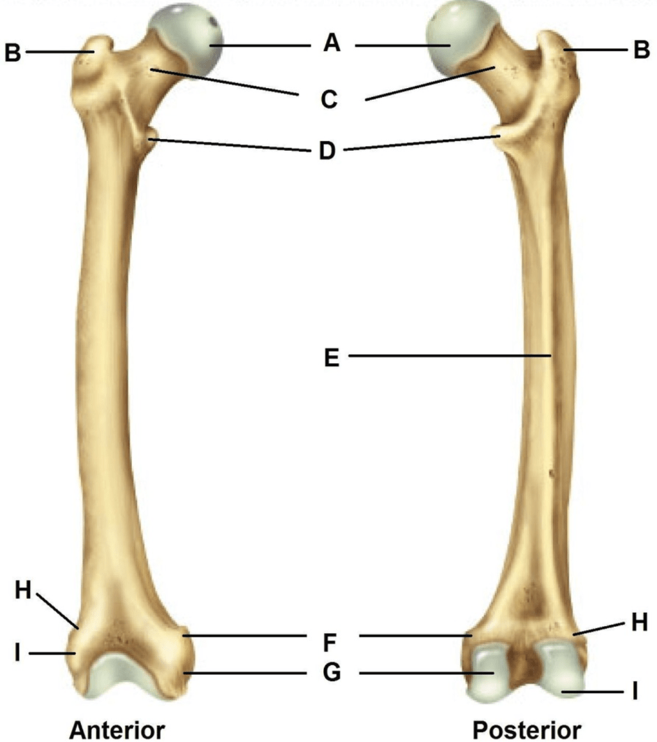
The linea aspera of the femur.
100
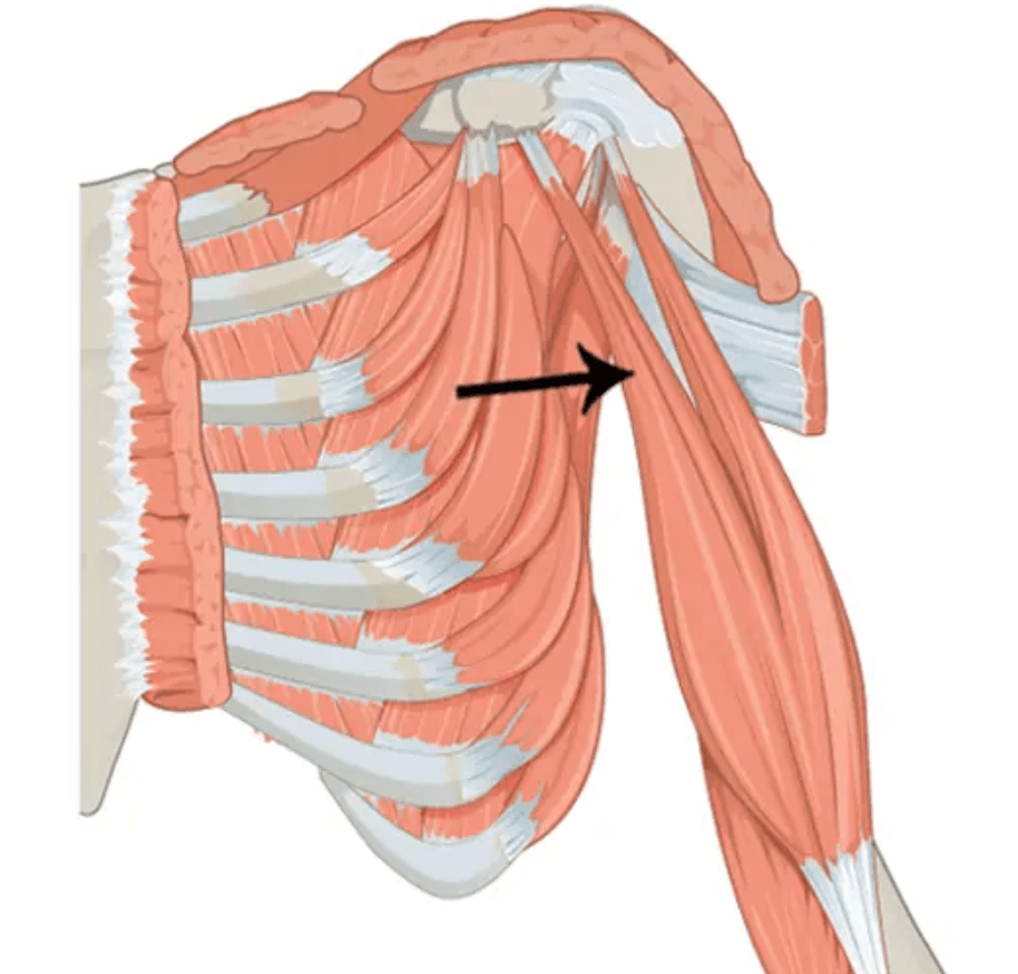
Identify the muscle and ONE action.
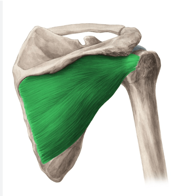
The infraspinatus and it stabilizes the glenohumeral joint as part of the rotator cuff.
100
Identify the muscle and its origin. 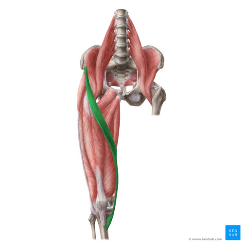
The sartorius muscle and it originates on the anterior superior iliac spine.
100
Identify the structure labeled "10".

Basilar artery
200
Identify the specific structure and which bone it is a part of. 
Surgical neck of the humerus
200
Identify the bone and the specific structure for the label "12".

The lateral malleolus of the fibula.
200
 Identify the muscle and its insertion.
The pectoralis major and it inserts on the greater tubercle of the humerus.
200
Identify the muscle and one action.
The piriformis and it abducts the thigh.
(Adbuctor muscle memory trick: George Goes To Play Golf Safely
-Gluteus maximus
-Gluteus medius
-Tensor fasciae latae
-Piriformis
-Gluteus minimus
-Sartorius
200
Identify the structure (not the valve).

Chordae tendineae.
300
Identify the SPECIFIC structure of "J".  .
.
Digit 5, proximal phalanx.
300
Identify the specific ligament.

The calcaneofibular ligament.
300
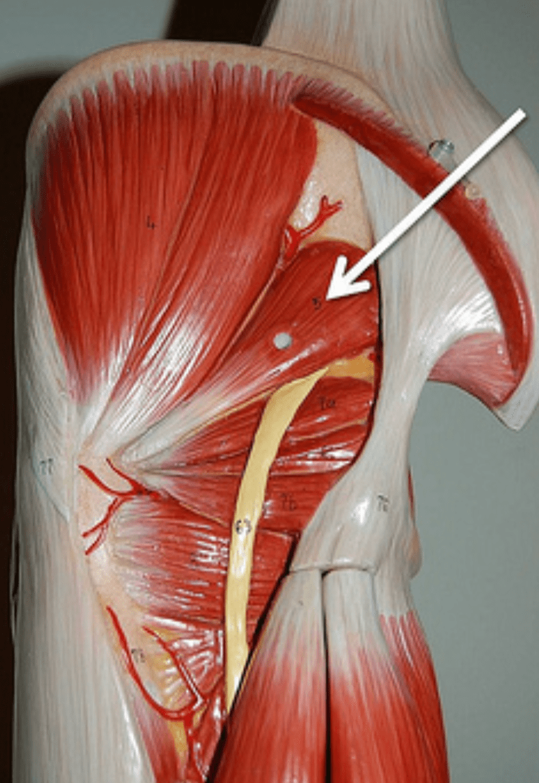
Identify the specific muscle and its origin.
The biceps brachii (short head) and it originates on the coracoid process of the scapula.
300
Identify the muscle and the origin.

The vastus medialis and it originates at the linea aspera of the femur.
300
Identify the layer of the dissected sheep heart.

The myocardium.
400
Identify the ligament "2".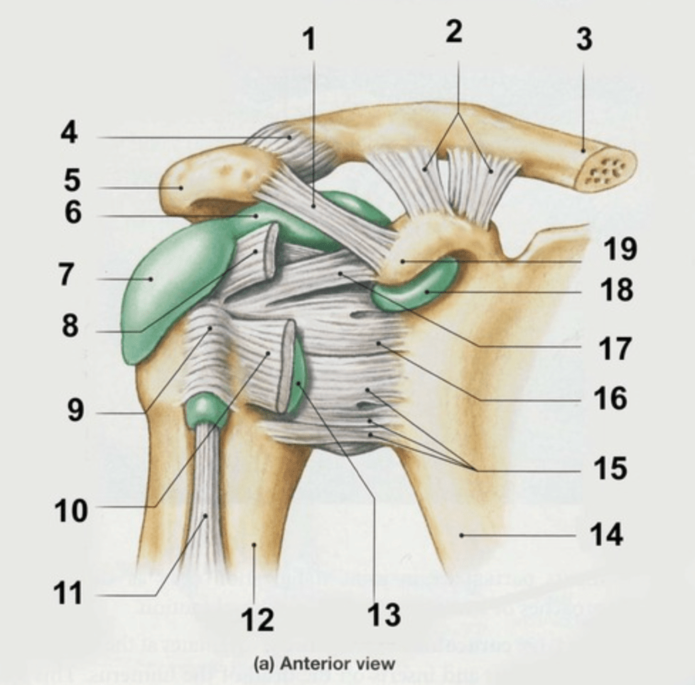
Coracoclavicular ligaments.
*The ligaments attach to the coracoid process of the scaupla and the clavicle*
400
Identify the specific ligament (this is a posterior view).

The ischiofemoral ligament.
400
Identify the specific muscle and its insertion AND its action.
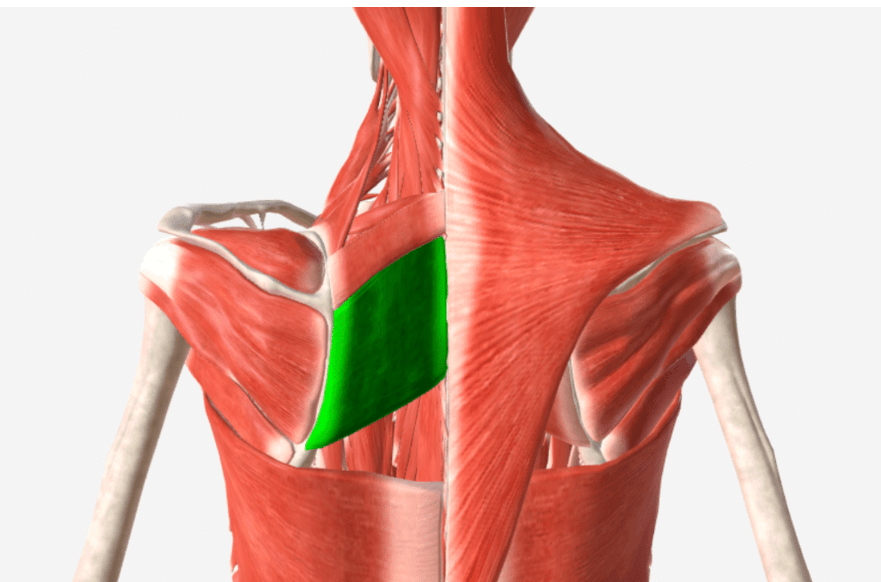
The rhomboid major and it inserts on the vertebral border of the scapula and its action is to rotate and retract the scapula.
400
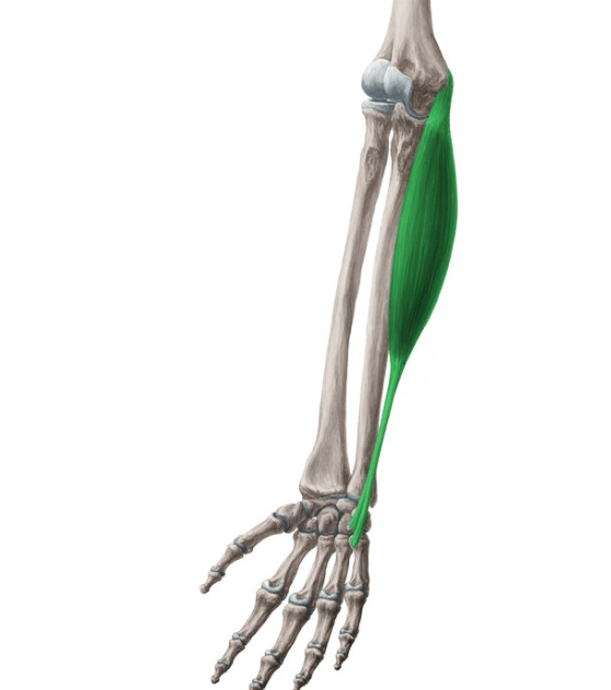
Identify the muscle and what is the fibrous band of connective tissue that holds the surroundings tendons in place.

The flexor digitorum longus and the flexor retinaculum.
400
Identify the specific wave labeled in "red" and the atrial activity at this point.

The T wave and the atria is filling (diastole).
500
MULTIPLE STEPS: MUST GET ALL CORRECT FOR THE POINTS
1. Identify the red structure
2. Identify which bone it is a part of
3. Identify which joint it is a part of and that joint's structure and function. 
Glenoid fossa of the scapula and part of the glenohumeral joint (the head of the humerus attaches)
The glenohumeral joint's structure is synovial ball and socket and function is triaxial.
500
MULTIPLE STEPS: MUST GET ALL CORRECT FOR THE POINTS
1. Identify the labeled structure
2. Identify which three structures are affected by a terrible triad injury.

1. Medial collateral ligament
2. The anterior cruciate ligament (ACL), medial collateral ligament (MCL), and the medial meniscus.
500
Identify the muscle and its action.
The flexor carpi ulnaris and it flexes and adducts the wrist.
500
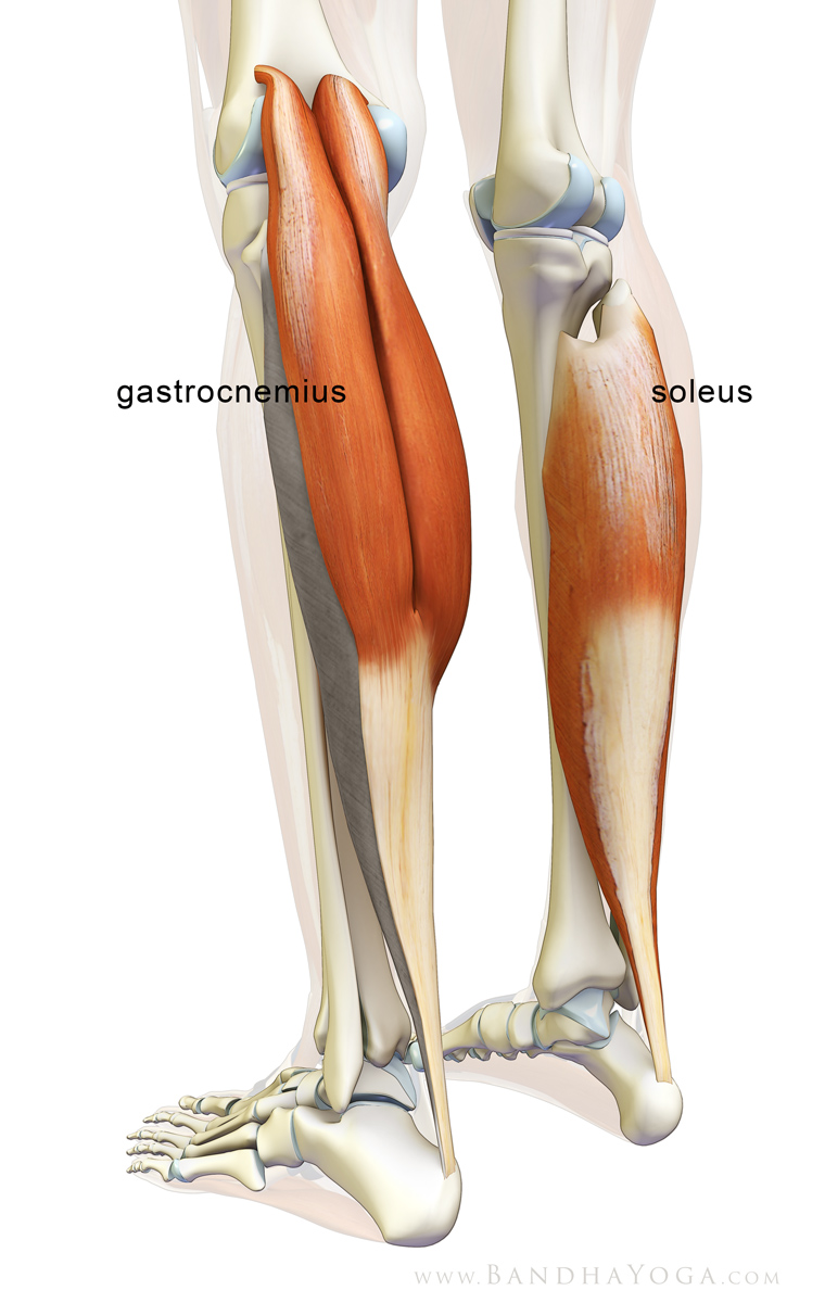
Identify the muscle, its insertion, and the muscle that is immediately DEEP to it.
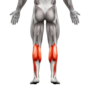
The gastrocnemius inserts at the calcaneus and the soleus is immediately deep to it (they both insert at the calcaneus).
500
Trace a drop of blood from the left femoral vein to the right subclavian artery.
1. Left femoral vein
2. Left external iliac vein
3. Left common iliac vein
4. Inferior vena cava
5. Right atrium
6. Right AV valve
7. Right ventricle
8. Pulmonary semilunar valve
9. Pulmonary arteries
10. Lungs
11. Pulmonary veins
12. Left atrium
13. Left AV valve
14. Left ventricle
15. Aortic semilunar valve
16. Aorta
17. Brachicephalic artery
18. Right subclavian artery