Tibia or Not Tibia, That is The Question
Bone to Be Wild
Osteo-Introductions (Benign Bone Tumors Describe Themselves)
Radiology Isn't Black or White (Imaging)
Bad to the Bone
100
A 24-year-old male presents to the clinic complaining of right leg pain. He states the pain is worse at night, but does improve after taking Advil. On X-ray, you see a radiolucent nidus with a ring of surrounding sclerotic woven bone in the cortex of the femur. The lesion measures < 2 cm. This most likely represents an...
a. Osteoid osteoma
b. Osteoblastoma
a. Osteoid osteoma
Explanation
Osteoid osteomas:
< 2 cm (Mnemonic: "Big Blast," Osteoid is a shorter word than osteoblastoma)
Improve w/ NSAID use
Often occur in surface of cortex/diaphysis of long bones
Thick sclerotic rim (Imagine concentric Os; outer sclerotic ring is capital O, nidus ring is lowercase o)
Affects < 25 years old
100
A 35-year-old male presents to the clinic with pain in the third metacarpal bone of his right hand. X-ray reveals a radiolucent lesion in the medullary cavity of the third metacarpal bone. The lesion appears to have a "rings and arcs" appearance on X-ray. This most likely represents which benign bone tumor?
a. Enchondroma
b. Osteochondroma
a. Enchondroma
Explanation:
Typically affect those in the first through fourth decades of life
"Rings and arcs"/endosteal scalloping on imaging
Typically present in medulla of hands/feet but can occur in long bones
100
My stromal tumor cells overexpress RANK-L; this causes the creation of large osteoclastic cells with multiple nuclei. Some people call me an osteoclastoma. What am I?
Giant cell tumors
Explanation:
Osteoclasts express RANK and are activated by the overexpression of RANKL by stromal cells. Osteoclastic activity causes destruction of surrounding bone.
100
This imaging finding is suggestive of a benign bone tumor called a...
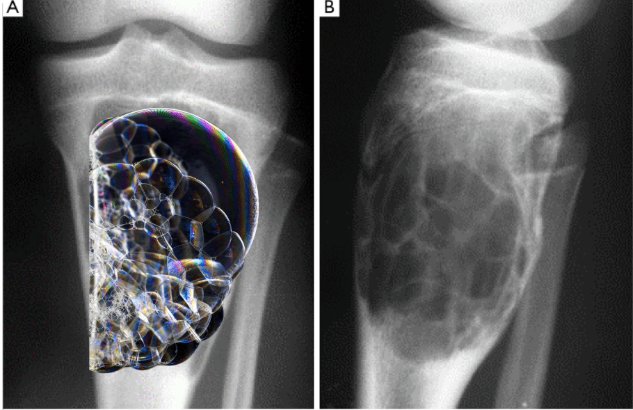
Giant cell tumor
Explanation
Endosteal scalloping results in ridges on the endosteum; resulting in the "soap bubble appearance"
100
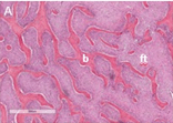
You're a pathologist examining a sample of a benign bone tumor under the microscope. This sample came from the medulla of the femur of a 14-year-old boy. On examination, you see intermittent trabeculae surrounded by fibrous tissue. An image is shown below (b = bone and ft = fibrous tissue). This most likely represents:
a. Simple bone cyst
b. Endochondroma
c. Fibrous dysplasia
d. Osteoid osteoma
c. Fibrous dysplasia
Explanation
Fibrous dysplasia:
Woven bone surrounded by fibroblasts
Often occurs in early adolescence; stops after growth plate closes
Often occurs in medulla diaphysis of long bones
200
A 14-year-old male presents to the clinic with back pain. He states his back pain does not improve even after taking Ibuprofen. X-ray of his lumbar spine reveals a 3.5 cm lesion w/ cortical expansion and a thin rim of surrounding sclerosis. This most likely represents a(n)...
a. Osteoid osteoma
b. Osteoblastoma
c. Osteochondroma
d. Giant cell tumor
b. Osteoblasteoma
Explanation:
Osteoblastomas are:
> 2 cm (Mnemonic "Big Blast" or Osteoblastoma is longer word than osteoid)
Cortical expansion and surrounding rim of sclerosis sometimes seen; however, osteoid osteoma will have a thicker sclerotic rim
Unresponsive to NSAIDs (Mnemonic, unlikely to treat pain from blast/explosion with Advil)
Most cases occur in those 10-30 years old; peak incidence in 20s
Typically occur in axial spine (Mnemonic: osteoblastoma has a lowercase l, the axial spine is linear(ish)
200
An 11-year-old boy is brought to the office by his parents with a 2-month history of progressive swelling of his right upper arm. He has otherwise been healthy. Medical history is unremarkable and he takes no medications. Immunizations are up-to-date. He appears well-nourished and well-developed. He is at the 60th percentile for height and weight. Vital signs are within normal limits. Physical examination reveals an immobile mass on the right proximal humerus that is nontender to palpation. Laboratory studies reveal an erythrocyte sedimentation rate of 10 mm/h (reference range: 0–15 mm/h) and an alkaline phosphatase of 35 U/L (reference range: 25–100 U/L). A radiograph of the right humerus reveals a 3-cm sessile bony protrusion with a cartilage cap on the proximal metaphysis. Which of the following is the most likely diagnosis?
a. Osteoid osteoma
b. Osteochondroma
c. Osteosarcoma
d. Chondroblastoma
e. Ewing Sarcoma
f. Chondrosarcoma
b. Osteochondroma
Explanation:
< 25
Cartilage cap
Grows on long bones out of growth plate, found typically foud on metaphysis
Can be sesslie (flatter) or pedunculated (mushroom-like)
200
Hi! I typically grow on the surface of facial bones. I'm associated with Gardner Syndrome (which occurs due to a mutation in the APC gene). What am I?
a. Osteochondroma
b. Endochondroma
c. Osteoma
d. Osteoid osteoma
e. Osteoblastoma
c. Osteoma
Explanation:
Osteomas are a symptom of Gardner Syndrome, a familial adenomatous polyposis disease
APC (adenomatous polyposis coli) is a tumor suppressor gene
200
This benign bone tumor that typically grows from the metaphysis of bones and has a distinctive appearance (shown below). It's is called a(n)...
a. Osteosarcoma
b. Endochondroma
c. Chondrosarcoma
d. Osteochondroma
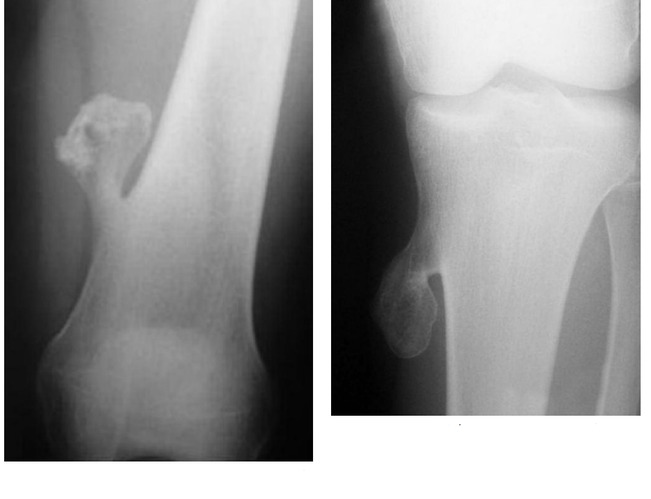
d. Osteochondroma
Explanation
Osteochondromas:
Typically appear in adolescence
Grow from the metaphysis of long bones (near the growth plate)
Have a “pedunculated” (mushroom-like) appearance on imaging
Have a cartilage cap
200
A 12-year-old patient presents to the clinic with his mother complaining of pain in his left arm after stumbling into a tree. Surprisingly, X-ray reveals a fracture to the metaphysis of his proximal humerus at well as fibrous-lined fluid filled spaces at the site of the fracture. This most likely represents:
a. Osteoblastoma
b. Endochondroma
c. Unicameral bone cyst
d. Chondroma
c. Unicameral bone cyst
Explanation
Unicameral bone cysts:
Often occur in metaphysis
Fluid-filled space; fibrous lining
Can lead to pathologic fractures
Typically occur in those <20
300
Based on appearance on X-ray, this benign bone lesions is likely an...?
a. Osteoblastoma
b. Osteoid osteoma

b. Osteoid osteoma
Explanation:
Osteoid osteomas typically present in the cortex/diaphysis of long bones
Thick sclerotic rim (Mnemonic: imagine concentric Os; outer sclerotic ring is capital O, nidus ring is lowercase o)
300
This imaging finding is suggestive of...
a. Osteochondroma
b. Enchondroma
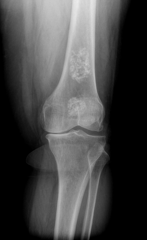
b. Enchondroma
Explanation:
"Rings and arcs" appearance on X-ray
300
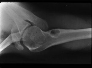
I'm > 2 cm. I usually present in the spine, but sometimes I present in long bones. Here's a selfie. What am I?
a. Giant cell tumor
b. Enchondroma
c. Osteoblastoma
d. Osteoid osteoma
c. Osteoblastoma
Explanation
Osteoblastomas are:
> 2 cm (Mnemonic "Big Blast" or Osteoblastoma is longer word than osteoid)
Typically occur in axial spine (Mnemonic: osteoblastoma has a lowercase l, the axial spine is linear(ish)
300
This benign tumor can occur after trauma. The imaging below is most suggestive of:
a. Myositis ossificans
b. Enchondroma
c. Osteoid osteoma
d. Osteoma
e. Osteoblastoma
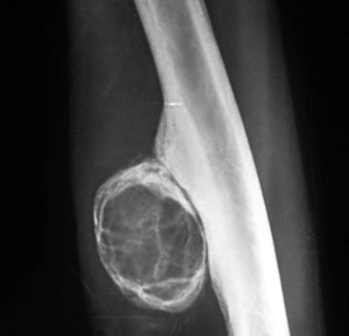
a. Myositis ossificans
Explanation
Myositis ossificans:
Bone growth in muscle
Example of "hetertropic ossification"
Can occur several months after blunt trauma to muscle
Common in larger muscles (e.g. quadriceps, brachialis, etc)
"Eggshell calcification" on imaging
Painful mass of soft tissue
300
A 20-year-old female semi-professional softball player presents to her PCP after noticing a painful lump on her quadriceps 1 week prior. When asked what she thinks caused her injury, she states she is unsure; however, she notes that she was struck in the thigh by a very fast softball pitch approximately 3 months prior. On X-ray, you note a soft tissue mass with eggshell calcification; on histology, you see zonal architecture and fibroblastic proliferation surrounded by metaplastic bone formation. Her diagnosis most likely represents:
a. Fibrous dysplasia
b. Chondroma
c. Endochondroma
d. Myositis ossificans
d. Myositis ossificans
Explanation
Myositis ossificans:
Bone growth in muscle
Manifests as painful mass of soft tissue
Common in larger muscles (e.g. quadriceps, brachialis, etc)
Example of "heterotropic ossification"
Can occur several months after blunt trauma to muscle
"Eggshell calcification" on imaging
Fibroblasts seen on histology slides because bone is formed from cartilage
"Zonal architecture" refers to the concentric circles seen on imaging/histo; these distinct zones correspond to the different stages of bone growth
400
A 22-year-old male presents to his PCP complaining of right arm pain. He states he doesn't believe in western medicine, so he's unsure whether NSAIDs improve his pain. On X-ray, you see a lesion in his radius, but the computer you're using to view the X-ray shuts down for a required update before you get a proper look at it. Based on the location of the lesion, this is most likely an...
a. Osteoblastoma
b. Osteoid osteoma
b. Osteoid osteoma
Explanation
Osteoid osteomas:
Occur more frequently in the appendicular spine; osteoblastomas favor the axial spine