Landmarks of the chest
Lung Assessment
Lung Assessment
Heart Assessment
Vascular Assessment
100
Accurate physical assessment of the thorax and Lungs requires
What is a review of the ventilatory and respiratory functions of the lungs
100
The organization of an examination
What is always compare both sides
What is assess Body parts at most risk first
What is Give rest periods when needed
What is usee common accepted medical terms
What is always record quick notes
100
The four types of adventitious breath sounds
What are crackles, rhonchi, wheezes, and pleural friction rub?
100
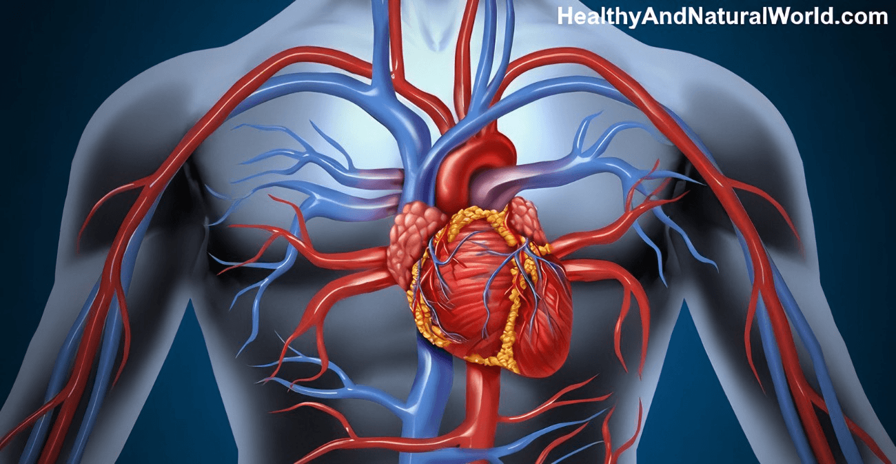
The location of the heart?
What is the center of the chest (pericordium) behind to the left of the sternum, with a small section of the right atrium extending to the right of the sternum.
100
The carotid arteries
What reflects heart function better than peripheral arteries because their pressure correlate with the aorta
200
Anatomical chest wall landmarks
What are the posterior, Lateral; and anterior chest landmarks?
200
The order assessment of the thorax
What is Inspection, Palpation, Chest excursion, Tactile Fremitus, Auscultation of the lungs
200
Coarse, low pitch may clear with cough
what is Ronchi?
200
The Apex of the heart between the 4th and 5th intercostal space
 What is the point of maximal impulse?
200
Syncopy could develop from doing this to your patient?
What is palpating or massaging the carotid arteries vigorously
300
Where are the Lung borders located in the chest?
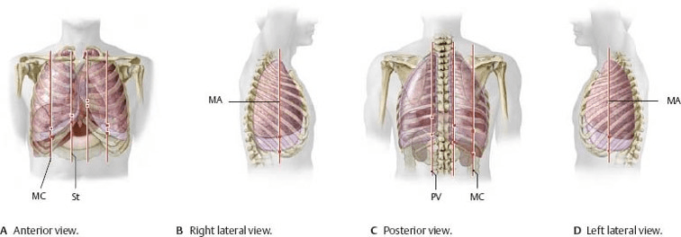
Anteriorly -Apex 3-4 cm above the inner 1/3 of the clavicles. Base - rest on the diaphragm, 6th rib MCL
Laterally- Extends from the axilla apex to 7th-8th rib
Posteriorly -Apex of the Lung is at C7- base T10 ( on deep inspiration to T2)
300
Heard over the trachea and Larynx, high pitch, loud, harsh.
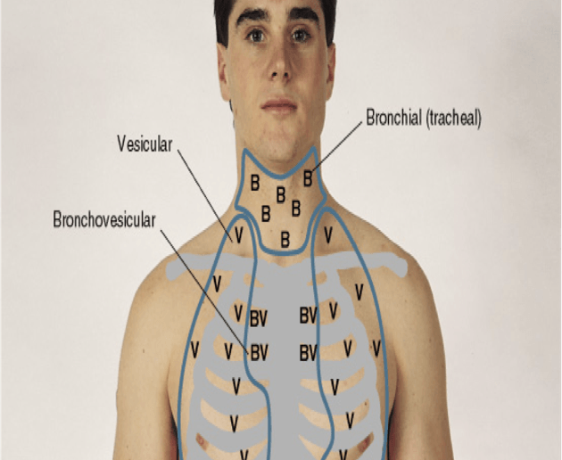 What are Bronchial Breath sounds?
300
Whistling high pitched bronchus
What is a wheeze?
300
Phases in the cardiac cycle
What is systole and diastole?
300
The most accessible veins for examination
What is the internal and external Jugular vein in the neck?
400
Before beginning a physical assessment what is some information you may want to obtain?
What is your smoking history
what is your work environment? Do you have a history of coughing? infections, weight loss, bleeding, night sweats, fever, TB, Pneumonia, Covid, HIV, homeless, recently incarcerated?
All risk factors related to lungs and heart issues.
400
Heard over major bronchi. Moderate pitch and loudness.
 What are Brochiovesicular Breath sounds?
400
low-pitched, grating, or creaking sounds that occur when there is an inflamed pleural surface?
What is a Pleural rub?
400
S1 "Lub"
S2 "Dub"
S1 Lub- what are the first heart sound, caused by the closure of AV valves (mitral & tricuspid) beginning of systole ( ventricle contraction)
S2 Dub- what are the second heart sound closed by the closure of the aortic and pulmonic beginning of diastole (filling)
Abnormal heart sounds extra sounds (S#) and murmurs come between S1 and S2
400
The peripheral arteries?
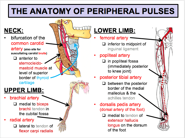 What is Radial pulse: Thumb side of the wrist
Ulnar Pulse: Little finger side of the wrist
Brachial Pulse: Inside of the elbow
Femoral Pulse: runs below the inguinal ligament between the symphysis pubis and the anterosuperior iliac spine.
Popliteal Pulse: behind the knee
Dorsalis pedis pulse: top of the foot line
Posterior tibial pulse: inner side of each ankle behind and below the medial malleolus (ankle bone)
500
What impacts an assessment?
What is age, gender? Pregnancy, medications, lifestyle choices. past surgeries, Family history, culture and ethnicity, The environment the patient lives in.
500
Heard over lung fields Low pitch, soft sound
What are Vesicular breath sounds.
500
Fine crackling high pitched breath sounds
What are crackles?
500
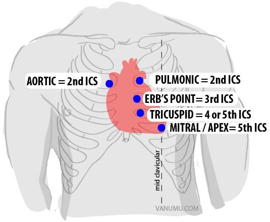
The steps and heart landmarks of a heart assessment
What is to make sure the patient is relaxed and explain what you are doing?
Patient in a supine or 45-degree angle position
no talking
start assessment at the base and move towards the apex
1. 2nd intercostal space on the right (aortic area
2. 2nd intercostal space on the left is the (pulmonic area)
3. 4th intercostal space on the right is the (Tricuspid area)
4. 5th intercostal on left is the (mitral area)
500
The grading is +1 to +4 depending on the degree of pitting
+1 2mm
+ 2 4mm
+3 6mm
+4 8mm
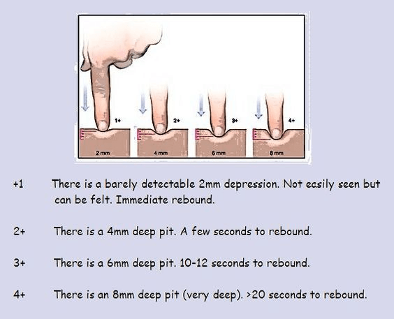 What is edema grading?