What is AIDP??
Lumbar Puncture/CSF Analysis
MRI and Other Test
100
Is AIDP characterized by ascending or descending weakness and hypo/areflexia?
Ascending.
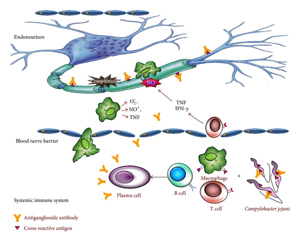
Autoantibodies (most commonly IgG) and complement attack Schwann cell and the surfaces of the myelin sheath of peripheral nerves. This initial damage recruits macrophages to the area which exacerbates the problem.
This results in blockages/reduced conduction velocity in nerves (specifically large myelinated motor and sensory fibers).
Attack is concentrated at spinal nerve roots and proximal nerve trunks/terminals, where the blood–nerve barrier is least effective and circulating antibody gains access. The longest fibers are affected earliest giving rise to the ascending pattern of the illness.
100
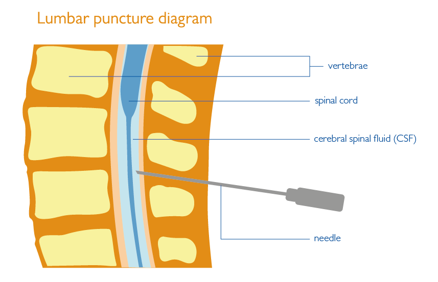
At what spinal level is it safe to perform a lumbar punture?
L3-L4 or L4-L5.
The spinal cord of an adult ends near the L1-L2.
A commonly used landmark is Tuffier's Line.
Tuffier's Line = Line from highest points of the iliac crests, most commonly intersects the L4 spinous process.
100
Is MRI considered a routine or necessary test in the diagnosis of AIDP?
No, AIDP is most commonly diagnosed through a combination of CSF analysis and and electrodiagnostic studies.
MRI can be helpful to support the diagnosis of AIDP but is non-specific on its own.
200
What is the proposed mechanism by which Camplobacter jejuni precipitates AIDP?
Molecular mimicry. Antibodies against bacterial antigens react with peripheral nerve myelin and Schwann cells.
200
What is the most common appearance of the CSF in a person with a bacterial infection of the CSF?
61185-4/asset/c80846aa-ebdc-4dd4-94ea-aee3693b9dfe/main.assets/gr2_lrg.jpg) It will be cloudy and turbid (panel B).
It will be cloudy and turbid (panel B).
Viral infections typically result in clear CSF.
200
What is the difference between a Nerve Conduction Study (NCS) and an Electromyograph (EMG)
NCS measures how fast an electrical impulse travels through a nerve.
EMG specifically looks at the electrical activity inside of a muscle.
An electrodiagnostic study (EDX) isthe combination of the results from a NCS and an EMG
EDX = NCS + EMG

300
A patient presents with ascending paralysis over the past week. Lumbar puncture and CSF analysis shows elevated CSF protein but no elevated WBCs. What is the next best test to support a diagnosis of AIDP?
Nerve Conduction Studies and Electromyography (EMG).
300
What are the two solutes that are measured in a normal CSF analysis?
Glucose: Normal = 2/3 the serum glucose value
Protein = Normal Range = 15 - 45 mg/dL
300
Nerve conduction studies are most informative at what point after weakness onset? How many nerves should be tested?
Abnormalities are most readily detectible around 2 weeks after symptom onset.
Tests should include:
- 4 motor nerves
- 3 sensory nerves
Additionally, F-waves and H-reflexes should be tested.
400
A patient presents within 24 hours of symptom onset. CSF protein and cell counts are normal. Why specifically can you not exclude AIDP based on these findings?
Because it takes at least 3 days for the albuminocytologic dissociation to develop.
50% of patients by 3 days.
80% of patients by day 7.
90%+ by two weeks.
Thus if a lumbar puncture is performed too close to symptom onset it can yield a false negative.
400
What is a normal lumbar puncture opening pressure in a healthy adult? What does it represent?
Normal opening pressure = 10 - 20 cmH20
Represents the pressure measurement of the CSF at the moment the needle enters the subarachnoid space.
400
During nerve conduction studies, a patient exhibits slowed conduction velocity, prolonged distal latencies, and conduction block. What pathophysiologic process do these findings suggest?
Peripheral nerve demyelination.
500
What NCS finding paradoxically remains normal in AIDP?
The sural SNAP (sensory nerve action potential).
Measured via NCS it shows how well the sural nerve is working (tests amplitude, latency, and conduction velocity).
In AIDP SNAPs of effected regions are typically reduced or completely absent. Most polyneuropathies are length-dependent, meaning longer nerves are affected earlier and more severely. In AIDP the issue is primarily at the nerve roots however meaning that we can observe a paradoxical preservation of the sural SNAP even when shorter nerves are affected.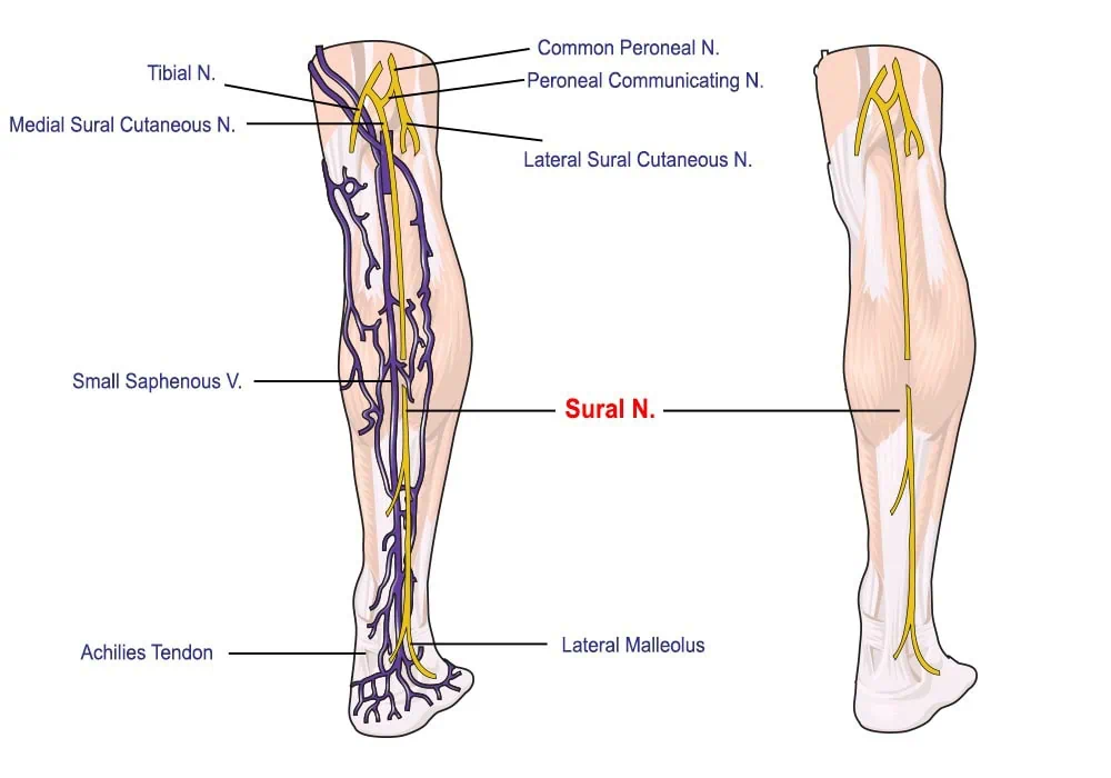
500
Analysis of a sample of a patients CSF yields elevated protein and >75 WBC/uL. Are these values suggestive of a diagnosis of AIDP? Explain your answer.
No, a dx of AIDP would be unlikely in this patient.
In AIDP the elevated CSF protein level is due to a disruption in the blood-nerve barrier due to autoantibody production.
Pleocytosis (elevated CSF WBCs) with elevated CSF protein typically suggests that there is an infection.
500
When a MRI is performed on a patient with AIDP where is nerve root enhancement most commonly seen?
Intrathecal spinal nerve roots.
These include the cauda equina/lumbar nerve roots.
Requires the use of a Gadolinium contrast agent in order to visualize.
