Ischemia & Angina
Congenital Diseases
Mitral & Aortic Valvulopathies
Cardiomyopathy
Heart Failure
100
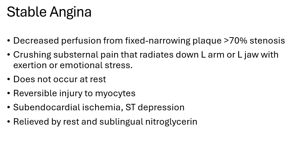
This presents as crushing substernal pain that radiates down the left arm, but is alleviated by rest or nitroglycerin.
stable angina
100
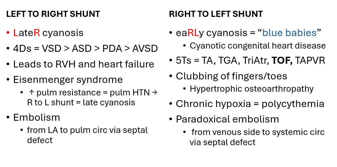
A 22-year-old woman is found to have a wide, fixed split S2 during a routine physical. She is asymptomatic.
Echocardiography demonstrates this type of shunt in the atria.
left-to-right shunt
100
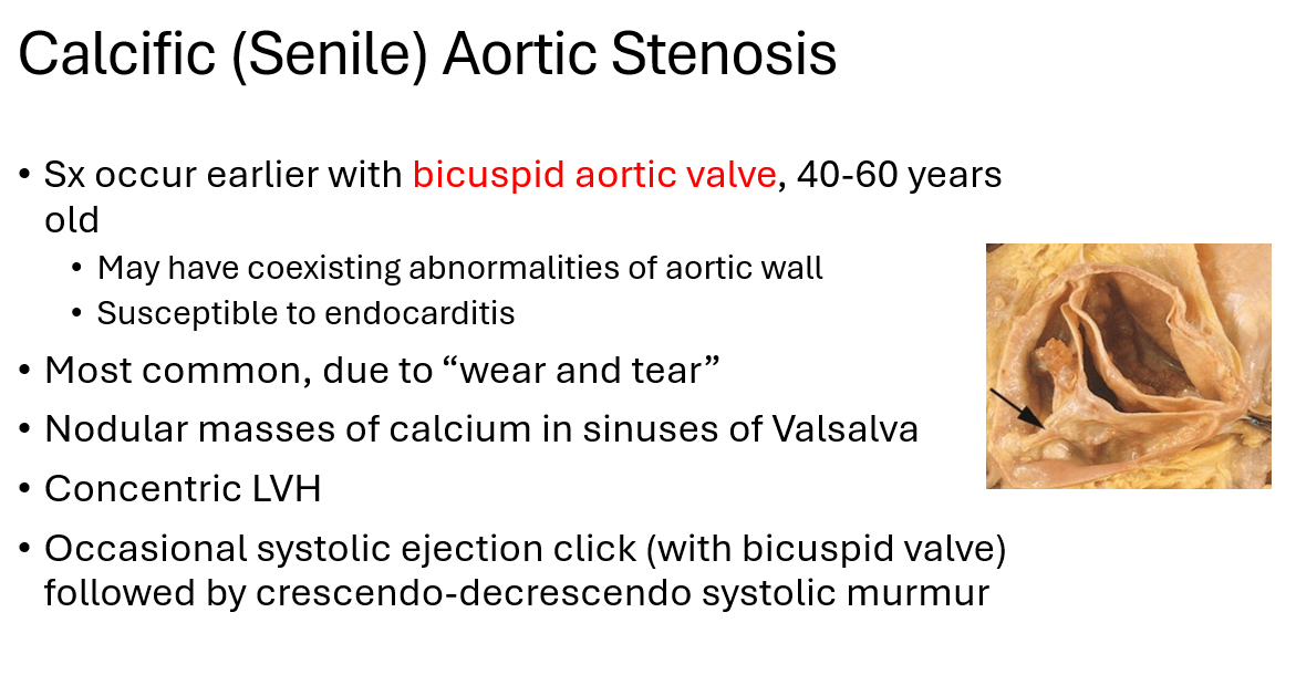
A 55-year-old man presents with progressive exertional dyspnea, chest pain, and several episodes of syncope while walking. Physical examination reveals a systolic ejection click and crescendo-decrescendo systolic murmur.
Echocardiography reveals aortic stenosis and this other valvular abnormality.
congenital bicuspid aortic valve
100
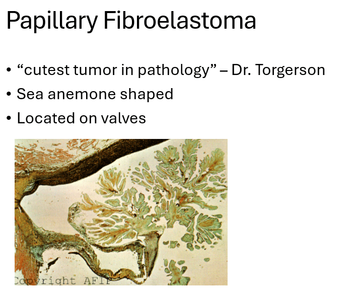
A 67-year-old woman undergoes echocardiography that reveals a mass on the aortic valve. Surgical excision reveals this sea-anemone–shaped endocardial tumor.
papillary fibroelastoma
100
A 68-year-old man presents with progressive dyspnea, orthopnea, and bilateral pulmonary crackles. Laboratory testing demonstrates a markedly elevated level of this hormone released from ventricular myocytes.
B-type (or Brain) natriuretic peptide (BNP)
200
A 41-year-old woman who smokes cigarettes experiences recurrent episodes of substernal chest pain while resting at night. Coronary angiography reveals no significant fixed atherosclerotic obstruction. Her symptoms improve after administration of nitroglycerin.
She is most likely experiencing this type of angina.
Prinzmetal (variant) angina
200
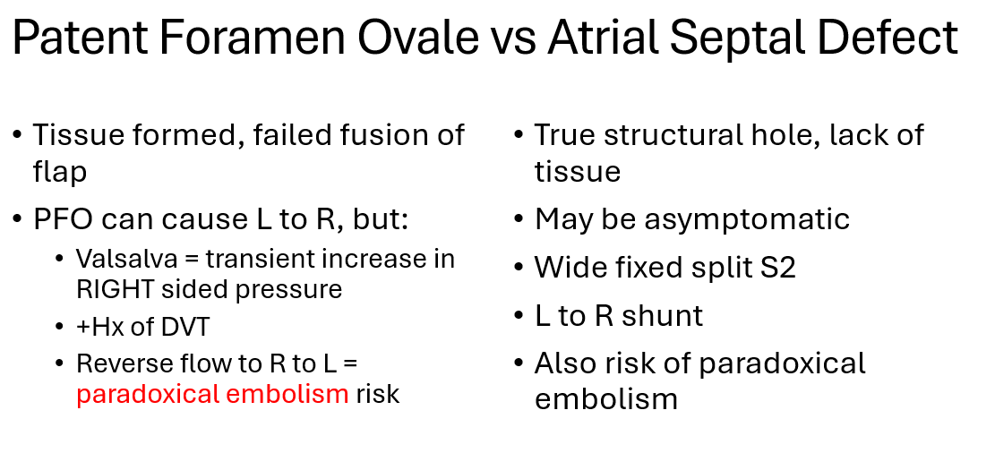
A 38-year-old man with a history of DVT suddenly develops right-sided weakness shortly after performing a Valsalva maneuver. Echocardiography demonstrates incomplete fusion between the septum primum and septum secundum.
His right-sided weakness is caused by this complication.
paradoxical embolism due to PFO
200
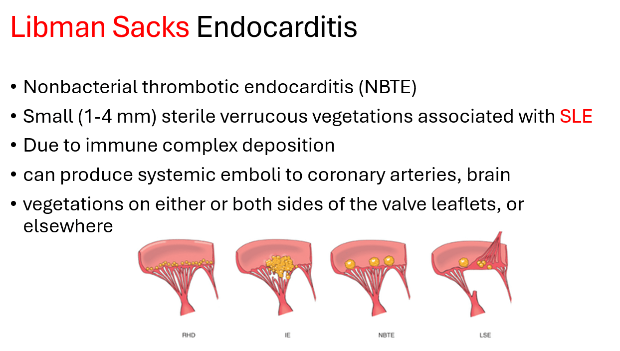
A 36-year-old woman with systemic lupus erythematosus develops sudden-onset right-sided weakness. Blood cultures are negative. Echocardiography demonstrates small vegetations on the mitral valve.
She is diagnosed with this type of nonbacterial thrombotic endocarditis.
Libman Sacks endocarditis
200
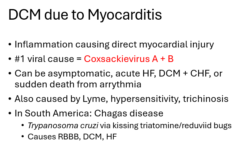
A 32-year-old man develops a viral illness with fever, fatigue, and myalgias. Several weeks later, he develops progressive dyspnea, exercise intolerance, and signs of congestive heart failure. Echocardiography demonstrates dilation of all four cardiac chambers and a markedly reduced left ventricular ejection fraction.
This virus is the number one cause of his condition.
Coxsackievirus A + B
200
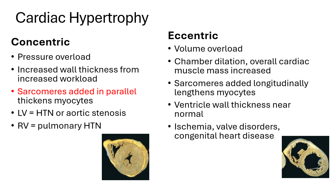
A 62-year-old man has a 15-year history of poorly controlled systemic hypertension. Echocardiography demonstrates the cardiomyopathy shown below. Thickened myocytes result from this cellular adaptation.
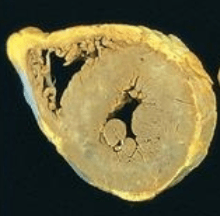
sarcomeres added in parallel
300
A 50-year-old man develops sudden severe substernal chest pain radiating to his neck. He is tachycardic and hypotensive. Coronary angiography demonstrates an acute thrombotic coronary occlusion.
This complication presents the highest risk of unexpected death within the first hour.
ventricular fibrillation
300
A newborn has cyanosis, hypocalcemia, and recurrent infections. Examination reveals abnormal facies and a cleft palate. Imaging reveals the following.
Abnormal development of these embryonic cells caused the cardiac defect.
neural crest-derived cells
300
A 12-year-old boy presents with fever, migratory pain and swelling of his knees and ankles, and sydenham chorea following a sore throat. Cardiac examination reveals a new murmur and a pericardial rub.
You expect this histological finding.
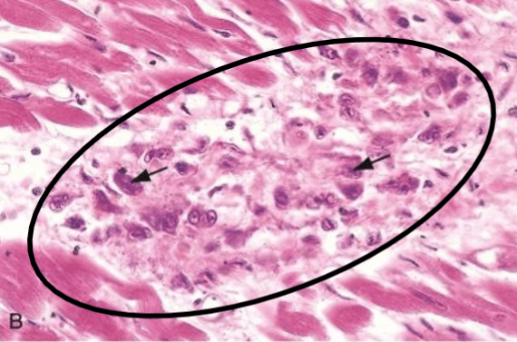
Aschoff body
300
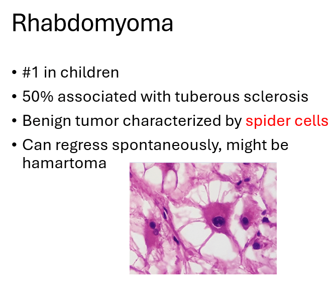
A 6-month-old infant with tuberous sclerosis presents with fatigue and poor feeding. Echocardiography demonstrates a well-circumscribed mass arising within the ventricular myocardium.
Histologic examination of the mass reveals this characteristic finding.
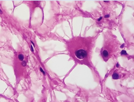
spider cell
300
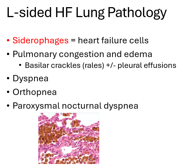
A 71-year-old woman with long-standing ischemic heart disease presents with worsening dyspnea, orthopnea, and episodes of severe shortness of breath that awaken her at night. Examination reveals bilateral basilar crackles.
A lung biopsy finds these characteristic cells.
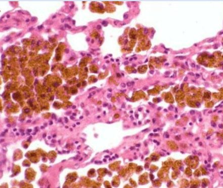
siderophages
400
A 64-year-old man is hospitalized following an ST-elevation myocardial infarction. 5 days later, he suddenly develops severe dyspnea and hypotension. Echocardiography demonstrates acute severe mitral regurgitation.
Occlusion of the right coronary artery most likely led to this complication.
papillary muscle rupture
400
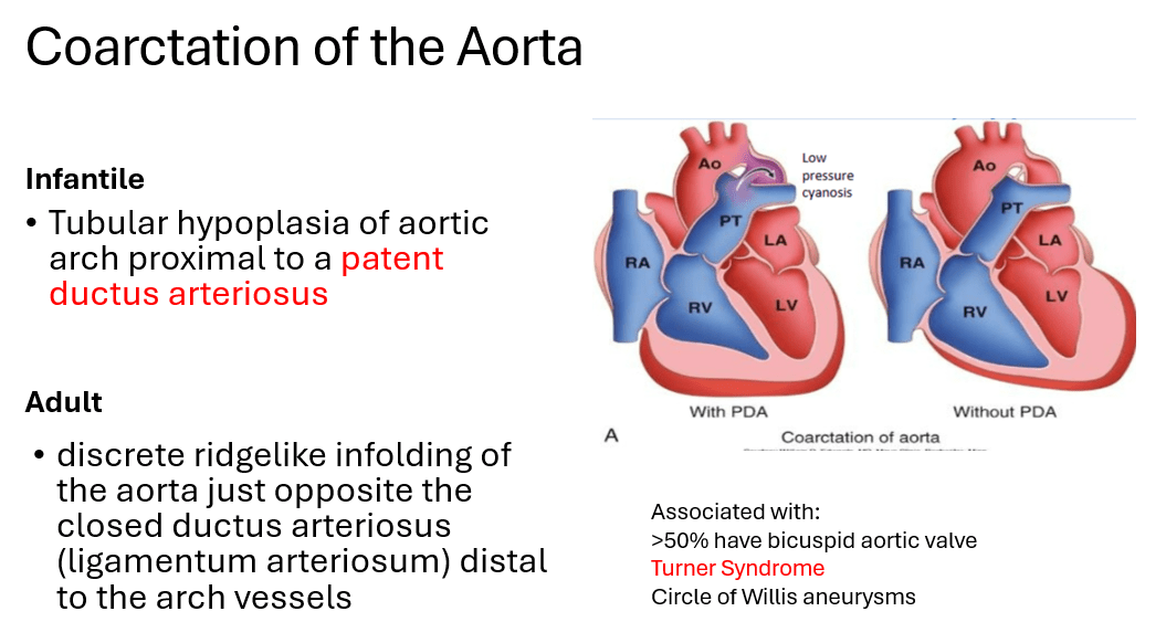
A newborn girl has a webbed neck, broad chest, and congenital lymphedema. Examination reveals strong upper-extremity pulses but weak femoral pulses and cyanosis of the lower extremities.
Echocardiography demonstrates narrowing of the aorta proximal to this malformation.
patent ductus arteriosus
400
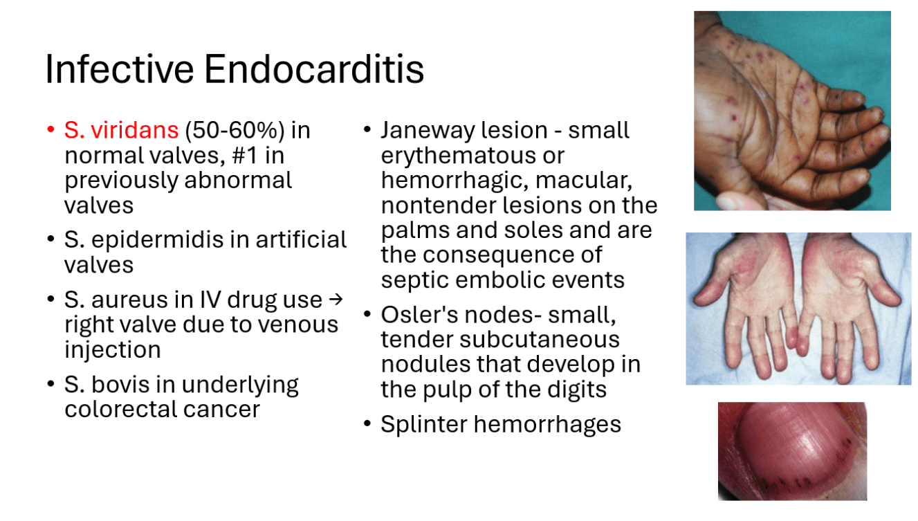
A 34-year-old man presents with Janeway lesions, splinter hemorrhages, and Osler's nodes. Echocardiography reveals a large vegetation on a previously normal cardiac valve.
Blood culture results most likely identify this organism.
S. viridans
400
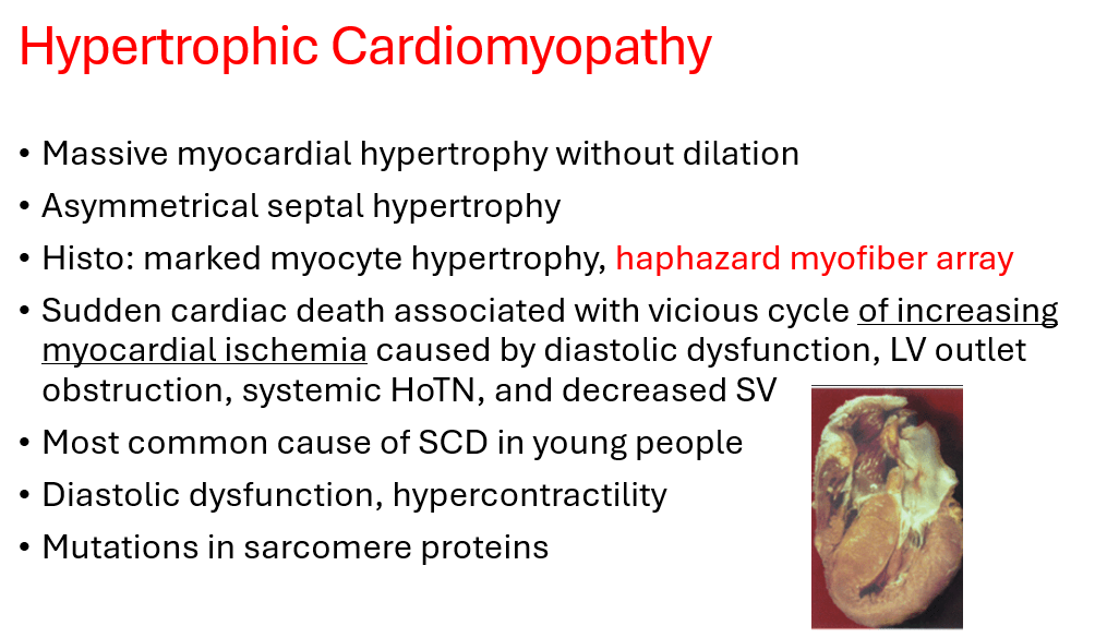
A 19-year-old male collegiate basketball player suddenly collapses during a game and cannot be resuscitated. He had previously experienced several episodes of exertional dyspnea and lightheadedness but had no history of hypertension.
Autopsy demonstrates the following histological findings most consistent with this cardiomyopathy.
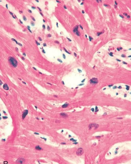
Hypertrophic cardiomyopathy
400
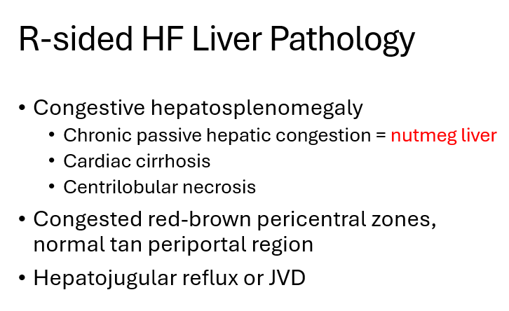
A 66-year-old man with severe COPD presents with progressive lower-extremity edema and abdominal distention. Examination demonstrates jugular venous distention, pitting edema, and hepatomegaly.
Gross examination of the liver would reveal this finding.
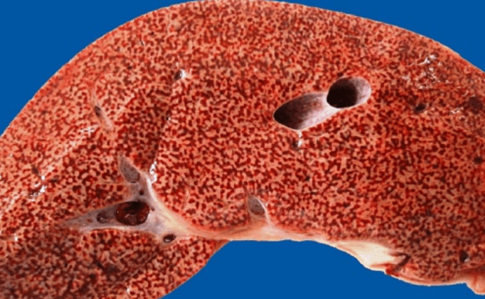
Nutmeg liver
500
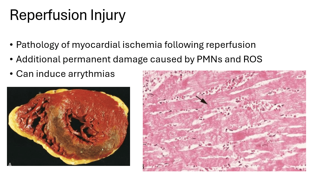
A patient presents with an acute transmural myocardial infarction caused by complete coronary thrombosis. He receives thrombolytic therapy shortly after presentation and coronary blood flow is successfully restored. The patient subsequently dies.
Histologic examination of the infarcted myocardium demonstrates hemorrhage and prominent contraction bands within necrotic myocardial fibers, consistent with this complication of MI treatment.
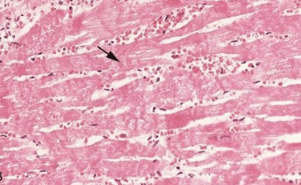
Reperfusion injury
500
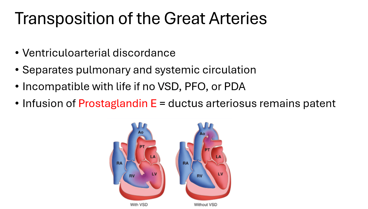
A full-term newborn develops profound cyanosis shortly after birth. Despite supplemental oxygen, his oxygen saturation remains critically low. The infant becomes progressively cyanotic and unresponsive and ultimately dies. Autopsy of the heart reveals the abnormality shown below. No ventricular septal defect is present.
This infusion could have prolonged the infant’s survival.
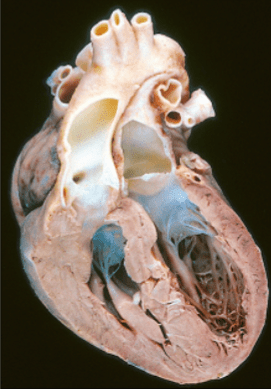
prostaglandin E
500
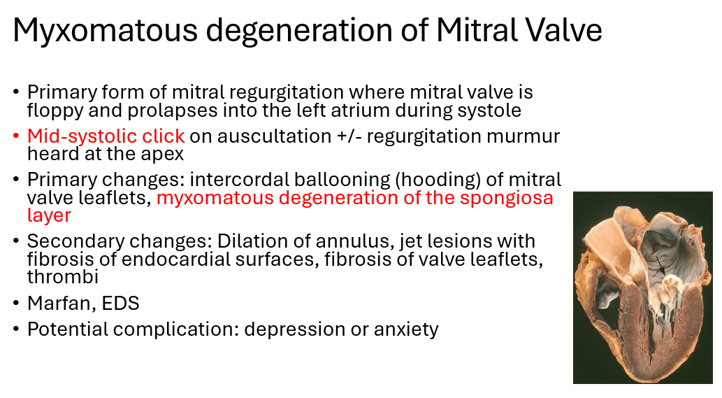
A pathologist examines a mitral valve removed from a young woman during valve replacement surgery. Histologic examination demonstrates the following.
The patient most likely exhibited this abnormal heart sound.
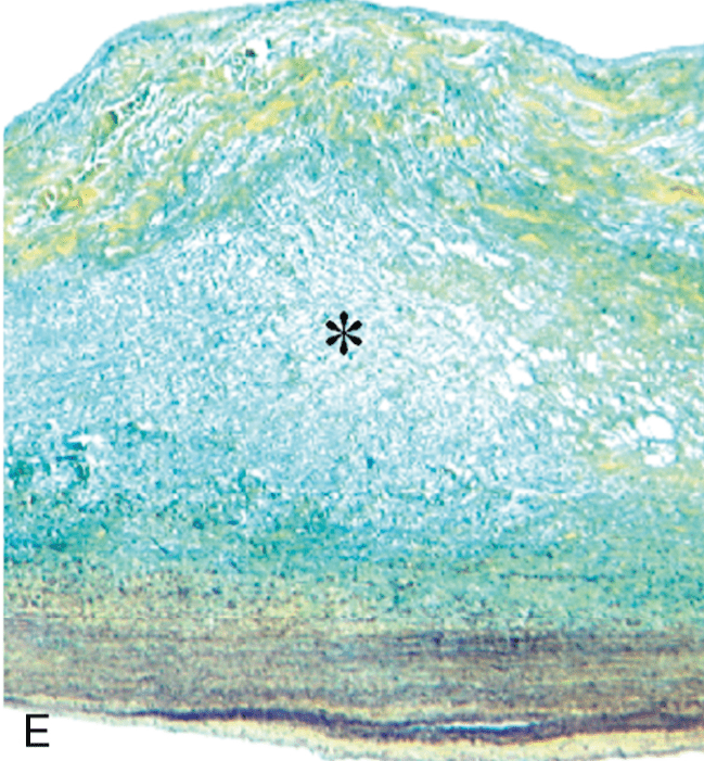
mid-systolic click
500
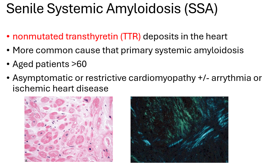
A 74-year-old man presents with progressive exertional dyspnea, peripheral edema, and fatigue. ECG demonstrates low-voltage QRS complexes. Echocardiography shows impaired ventricular filling, and restrictive cardiomyopathy is suspected. Endomyocardial biopsy shows the histological findings below. The tissue demonstrates amorphous extracellular material that exhibits apple-green birefringence with Congo red staining. Further evaluation reveals no evidence of multiple myeloma or plasma cell dyscrasia.
This protein is found in the cardiac deposits.
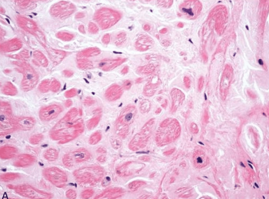
Nonmutated transthyretin (TTR)
500
A 67-year-old man with chronic heart failure has decreased cardiac output. As a compensatory response, activation of the renin-angiotensin-aldosterone system causes sodium and water retention, increasing ventricular filling volume.
Initially, this increase in ventricular filling improves cardiac output through this mechanism.
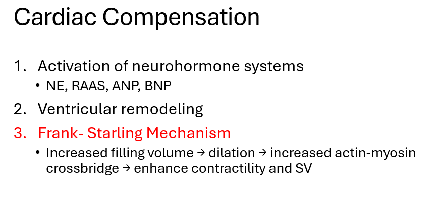
Frank-Starling Mechanism