Gross Anatomy
Gross Anatomy
Micro Anatomy
Describe the position & orientation of the heart in the thoracic cavity.
Then, describe how it is isolated from its more delicate neighbors.
Heart is located in the mediastinum, central portion of the thoracic cavity.
Heart occupies space where 3rd lobe of Left Lung would be situated
Apex oriented inferiorly & to the left.
Great vessels exit the base of the heart & proceed superiorly.
There are 3 layers of pericardial serous membranes which serve to minimize friction & limit the impact of continuous heart contractions on the lung tissues.
Identify the 3 layers of the heart wall.
Endocardium
Myocardium
Epicardium
Bonus 100
Which of these is also a pericardial serous membrane?
T/F
Each side of the heart will contract & relax independently of the opposite side.
False
Both sides of the heart will contract & relax at the same time.
Which region of the respiratory tract regularly comes into contact with solid, liquid, & gaseous materials?
[Food, water, air]
Oropharynx
Bonus 100
Which structure ensure that only air is able to enter the larynx?
T/F
Respiration occurs throughout the entire respiratory tract.
False
True respiration (gas exchange) only occurs once air is conducted to microscopic structures called alveoli.
100 each
Identify one chamber of the heart, then specify which valve will prevent blood from reentering that chamber.
Left atrium >>> bicuspid [L AV] valve
Right atrium >>> tricuspid [R AV] valve
Left ventricle >>> aortic semilunar valve
Right ventricle >>> pulmonary semilunar valve
Bonus 100
Which 2 valves would be responsible for making the second heart sound?
Which structure unique to cardiac muscle cells allows them to coordinate & synchronize their contractions with adjacent cardiac muscle cells?
Intercalated discs
Give the clinical terms for "contraction" & "relaxation" when referring to heart activity.
"Contraction" = systole
"Relaxation" = diastole
Inferior to the laryngeal cartilage, this region of the respiratory tract proceeds inferiorly to the carina, where the primary bronchi originate & proceed toward each lung.
Trachea
Bonus 100
Describe the structure of the trachea.
SPELLING BEE
Identify the basic functional cell of the respiratory system.
Then spell it out loud!
A-L-V-E-O-L-I
Bonus 100
Identify the function of a type II pneumocyte.
Identify the blood vessels which enter each atrium.
Left atrium
pulmonary veins (usually 4)
Right atrium
superior vena cava
inferior vena cava
coronary sinus
Describe the pathway of the electrical conducting system of the heart.
Sinoatrial node
Atrioventricular node
AV bundle (bundle of His)
L/R bundle branches
Purkinje fibers
Bonus 100
What makes the cardiac muscle cells present in the SA & AV nodes unique amongst all other cardiac muscle cells?
During ventricular diastole, which describe what is happening in the heart.
Where is blood moving?
Which valves are open/closed?
Ventricular relaxation creates negative pressure, which helps facilitate passive ventricular filling.
AV valves open
Semilunar valves closed
Secondary & tertiary bronchi supply which regions of the lung?
Secondary
directs air toward each lobe of the lung
Tertiary
directs air toward each segment in a lobe
Bonus 100
Which of these bronchi allows for gas exchange to occur?
Describe the respiratory membrane.
-systems interacting
-epithelial types
-process for gas exchange
Respiratory & cardiovascular interaction
2 layers of simple squamous epithelium
> 1 = alveolar cells
> 2 = capillary walls
O2 / CO2 move across via basic diffusion
Identify both great vessels.
Pulmonary trunk
Aorta
Bonus 100
Identify the first branches off each great vessel.
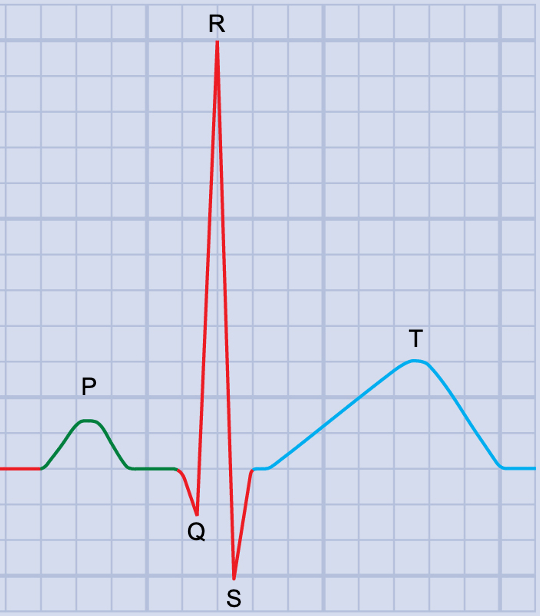
Identify which heart actions are "correlated" with the characteristic EKG.

P wave - atrial contraction
QRS - ventricular contraction
T wave - ventricular relaxation
Bonus 100
Which action is missing, & when does it occur?
During ventricular systole, which describe what is happening in the heart.
Where is blood moving?
Which valves are open/closed?
Ventricular contents are being squeezed into their respective great vessels.
AV valves closed
Semilunar valves open
Describe the muscles which facilitate each of the following actions:
-Passive breathing
-Forceful inhalation
-Forceful exhalation
Passive breathing
diaphragm
Forceful inhalation
diaphragm
external intercostals
scalenes
Forceful exhalation
internal intercostals
abdominal muscles
Bonus 100
Identify where these muscles attach to the lungs to allow for inhalation & exhalation.
Describe how air enters & exits the lungs.
Be sure to compare barometric pressure to the pressures inside the lungs.
How are these changed?
Barometric pressure is usually a constant variable (1atm).
Air will usually move from areas of higher pressure to areas of lower pressure.
To pull air into the lungs, internal pressure needs to drop below 1atm.
To force air out of the lungs, internal pressure needs to rise above 1atm.
Describe the pathway of blood through the heart.
Begin as an RBC entering the right atrium.
R atrium
R AV valve
R ventricle
R semilunar valve
Pulmonary trunk
Pulmonary artery
Lung
Pulmonary vein
L atrium
L AV valve
L ventricle
L semilunar valve
Aorta
Rest of the body!
Identify the key difference between how skeletal muscle cells & cardiac muscle cells conduct action potentials.
Skeletal muscle rapidly depolarizes & repolarizes, with almost no observable refractory period.
~500x per second
Cardiac muscle has a significant delay during repolarization.
~2x per second
Bonus 100
Which ion is responsible for this delay, & what is the delay called?
At brief moments at the beginning & end of ventricular systole, what happens inside of the heart?
How can we examine this using a stethoscope?
Periods of isovolumetric systole & diastole
>>> All 4 valves close / No blood movement
The characteristic "heart sounds" occur during these moments.
Describe the entire respiratory pathway.
Nostril
Nasal cavity (turbination & humidification of air)
Pharyngeal regions
Larynx
Trachea
Main bronchi
Secondary bronchi
Tertiary bronchi
Terminal bronchioles
Respiratory bronchioles
Alveoli
The medulla oblongata houses the cardioregulatory center, the vasomotor center, as well as the dorsal & ventral respiratory groups.
Chemoreceptors here are highly sensitive to which molecule?
CO2
Bonus 100
Describe the function of each respiratory group.
[Note the pontine respiratory group as well.]