diameter of coronary artery
1/8 inch
what part is #14

Fourth intercostal space at the right sternal border
V1
Left midaxillary line on the same horizontal plane as V4 and V5
V6
this interval measures the time from the initial depolarization of the atria to the initial depolarization of the ventricles and reflects a physiological delay in AV conduction imposed by the AV node
PRI
this encloses the heart
pericardial sac
what part is #2

Fourth intercostal space at the left sternal border
V2
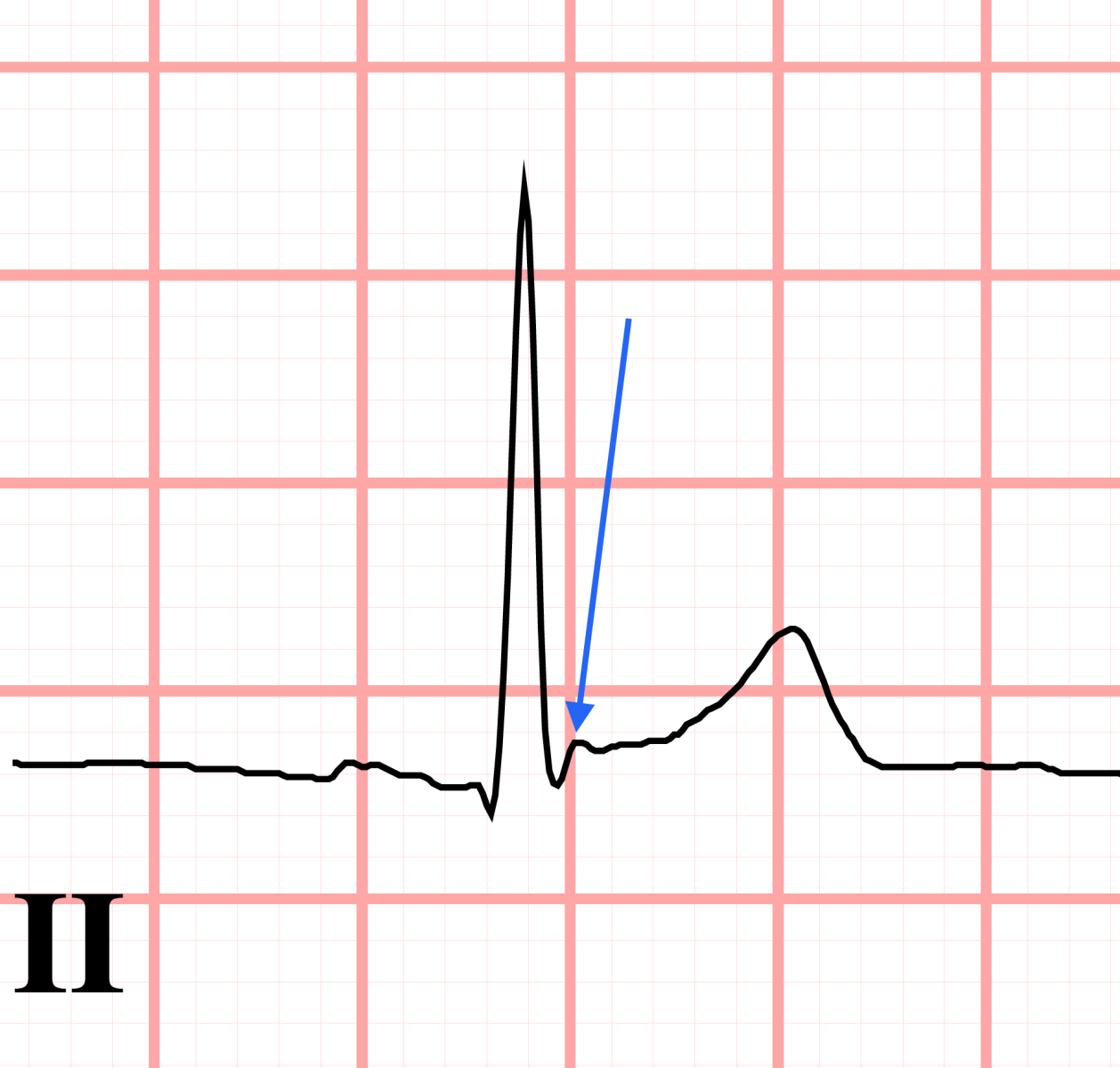
represents the interval between ventricular depolarization and repolarization.
ST segment
what is this on the EKG

what part of the heart is #3

what part is #7

Halfway between leads V2 and V4
V3
represents atrial depolarization
P wave
what is this on the EKG
what part of the heart is #1

Flow of blood from body tissue to the heart and then from the heart back to body tissues
SYSTEMIC CIRCULATION
Fifth intercostal space in the midclavicular line
V4
represents ventricular depolarization
QRS
Leads and electrodes are different:
electrodes are the "stickers" and leads are the
"pictures" of the heart from various directions
what structure is #11

Once the oxygen diffuses across the alveoli, it enters the bloodstream and is transported to the tissues where it is unloaded, and carbon dioxide diffuses out of the blood and into the alveoli to be expelled from the body
TRANSPORTATION
Left anterior axillary line on the same horizontal plane as V4
V5
represents repolarization of the ventricles
T
Standard calibration or gain is set at
10 mm/mV