Anatomy
Lab Values
Vascular
Pathology
Case Studies
100
The caudate lies on the ___/____ surface of the liver.
a. anterior/superior
b. posterior/superior
c. posterior/inferior
b. posterior/superior
100
More specific to the liver
a. ALP
b. AFP
c. ALT
d. LDH
c. Alanine Aminotransferase (ALT)
100
The veins that run between the segments and lobes and have non-echogenic walls
Hepatic Veins
100

Which pathology presents as a "Starry Night" on ultrasound?
Acute Hepatitis
100
A 59 y/o male with a BMI of 48 presents with elevated LFTs seen on recent blood work. His provider orders an ultrasound of his liver. When collecting his history, he has no complaints, essentially asymptomatic. What pathology are you most likely to find?
Hepatic Steatosis/Fatty Liver
200
The layer of connective tissue that surrounds the liver
Glisson's Capsule
200
An increase in GGT with ALP indicates ...
Biliary Obstruction
200
The caudate lobe is drained by ...
a. the right renal vein
b. emissary veins
c. IVC
d. hepatic veins
b. emissary veins
200

Is this liver in the acute or chronic stage of cirrhosis?
Chronic
200
A 35 y/o male is getting a scan of his liver after being treated at the ER for jaundice and elevated LFTs. He stated he is active recovery for alcoholism. The sonographic findings include an echogenic, heterogeneous liver with surface nodularity, mild ascites, and decreased visualization of vasculature.
Likely diagnosis?
Cirrhosis
300
 What is the purple arrow pointing to?
Ligamentum Venosum
300
If AFP is elevated, what liver pathology would be first suspected?
Hepatocellular Carcinoma (HCC)
300
The Hepatic Arteries arise from the ...
a. Celiac Trunk
b. SMA
c. Aorta
a. Celiac Trunk
300
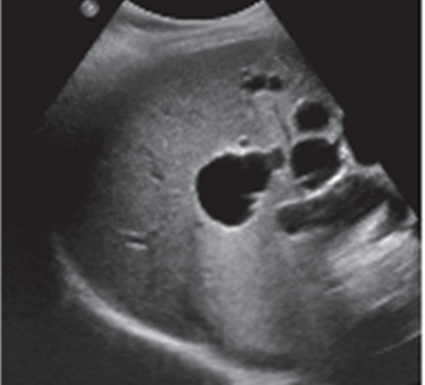
Most likely diagnosis?
Adult Polycystic Kidney Disease
300
A 28 y/o female comes in for her ultrasound of the liver, gallbladder, and pancreas. Her chart states that she has been complaining of intermittent right upper quadrant pain that has worsened in the last week. After further review of her chart, you note that she has been taking oral birth control for the past 3 years. Her pancreas and gallbladder were within normal limits; however, there was a solid, hypoechoic mass, with mixed echoegnicity in the right lobe of her liver.
Likely diagnosis?
Liver Cell Adenoma with possible hemorrhage
400
Hepatomegaly is assumed when the liver measures greater than ...
15.5 cm
400
What are the two most important lab values to check when performing procedures?
Prothrombin Time (PT) and INR (Internal Nationalized Ratio)
400
Which word best describes the flow of the Main Portal Vein?
a. Biphasic
b. Continuous
c. Pulsatile
d. Hepatofugal
b. Continuous
400

Most likely diagnosis?
Echinococcal/Hydatid Cyst
400
A 2.5 year old male comes into the ER with abdominal pain, jaundice, and nausea and vomiting. His mom states he has lost 15 pounds in a month. His lab work shows elevated LFTs and AFP. While performing his ultrasound, you find a solid, heterogeneous mass with internal calcifications in his right lobe.
Possible diagnosis?
Hepatoblastoma
500
Normal comparison of anatomy and their echogenicity (Least echogenic to most echogenic)
a. spleen/liver, renal sinus, pancreas, renal cortex
b. renal cortex, pancreas, spleen/liver, renal sinus
c. pancreas, spleen/liver, renal sinus, renal cortex
d. renal cortex, spleen/liver, pancreas, renal sinus
d. renal cortex, spleen/liver, pancreas, renal sinus
500
An increase in AST without ALT could indicate...
a. myocardial infarction
b. biliary obstruction
c. acute pancreatitis
d. celiac artery occlusion
a. myocardial infarction
500
What part of the portal triad/Mickey Mouse sign forms Mickey's left ear?
a. Common Bile Duct
b. Portal Vein
c. Hepatic Artery
Hepatic Artery
500

Most common benign tumor of the liver
Cavernous Hemangioma
500
A 32 y/o female comes through the ER with complaints of right upper quadrant pain, diarrhea, and a 101.6 fever. Her lab work comes back with elevated LFTs and WBCs. The patient states that she has just come back from a month long trip to South America. While you are scanning, you see the liver is mildly enlarged and there is a round hypoechoic structure in the right lobe.
Possible diagnosis?
Amebic (Parasitic) Abscess