What is the pathophys of Intracerebral Hemorrhage?
HTN → Charcot - Bouchard microaneurysms / rupture of small vessels of brain
*Most commonly the putamen is affected
Ex: Basal Ganglia Hemorrhage

What are some common symptoms seen with increased intracranial pressure?
- General symptoms: Headache, lethargy, vomiting
- Papilledema (optic disk swelling)
- Cushing's Triad: hypertension, bradycardia, and irregular respirations
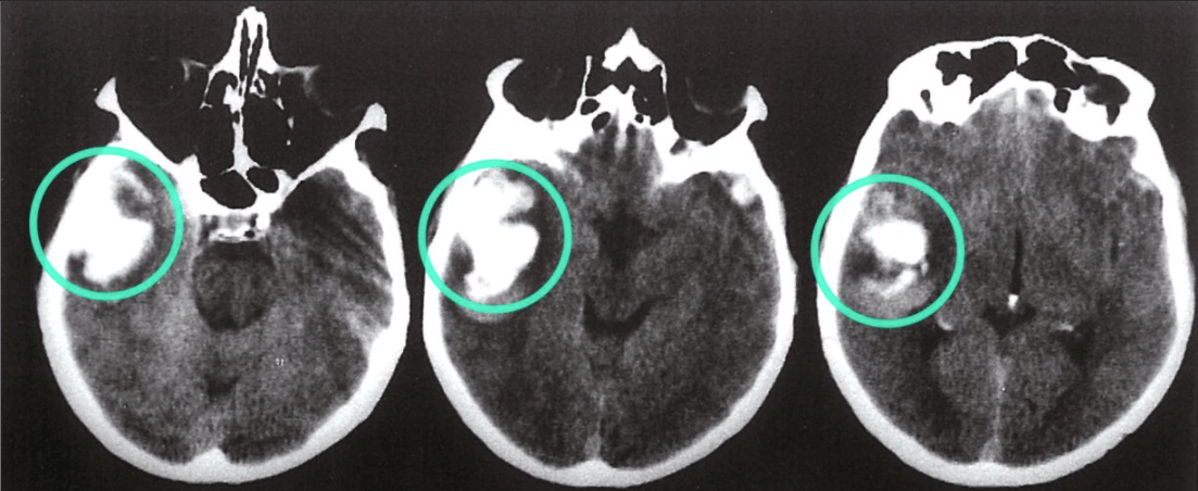
What is the diagnosis?
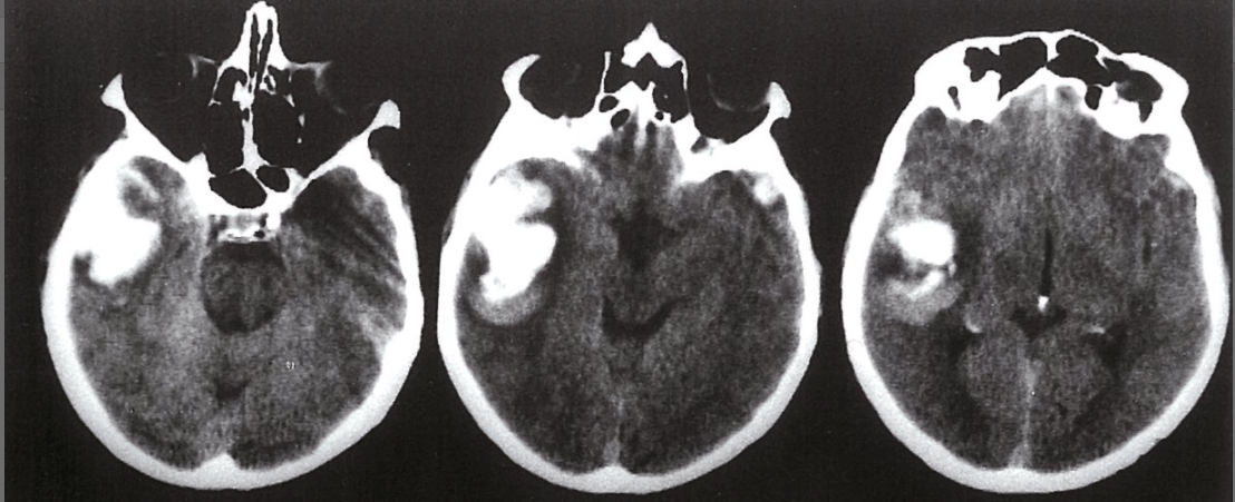
Intracerebral Hemorrhage of right temporal lobe
What is the treatment for intracranial hemorrhages?
Craniotomy to drain hematoma
- Mannitol promotes osmotic diuresis + decreases cerebral edema
- IV fluids if hypotensive
Which lobe of the cortex processes visual information?
Occipital lobe
What are some common etiologies of Intracerebral Hemorrhage (aka Intraparenchymal bleed)?
- HTN
- Anticoagulation
- Malignancy
- Trauma
What are some clinical features a patient with an intracerebral hemorrhage may present with?
- General symptoms: headache, drowsiness, loss of consciousness
- Focal Neurologic signs
- Stroke symptoms
Which intracranial hemorrhage presents with xanthochromia upon lumbar puncture?
Subarachnoid hemorrhage
Xanthochromia -- Yellowish discoloration of CSF due to pigments released from dead RBCs.
*Remember that LP results can be falsely negative in the first few hours when xanthochromia has not yet developed.
What type of posturing is this?
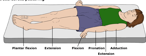
Decerebrate Posture — indicates damage to the brainstem
The following is an example of which aphasia?
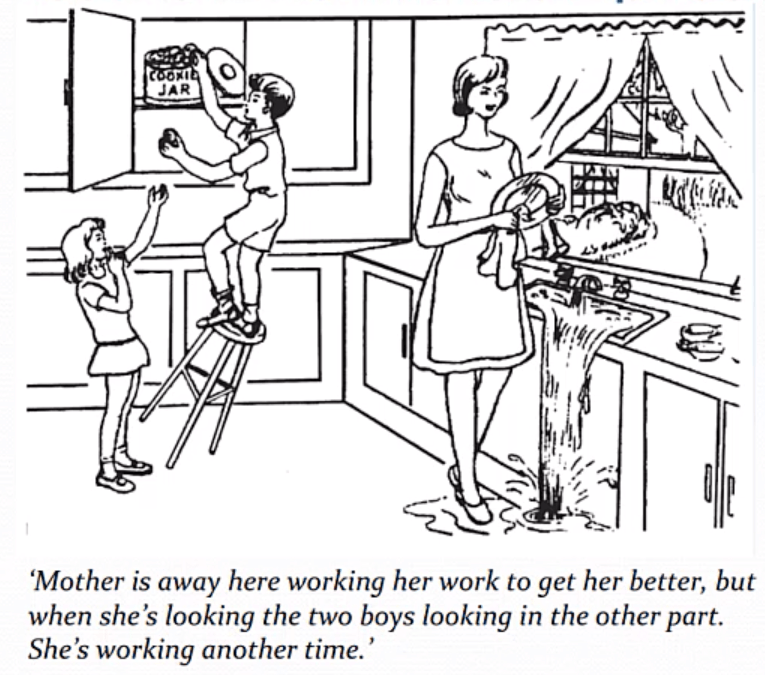
Wernicke's Aphasia
Wernicke's Area
Functions in language comprehension
Usually located on dominant (left) temporal lobe
Lesion → “fluent aphasia” / meaningless speech
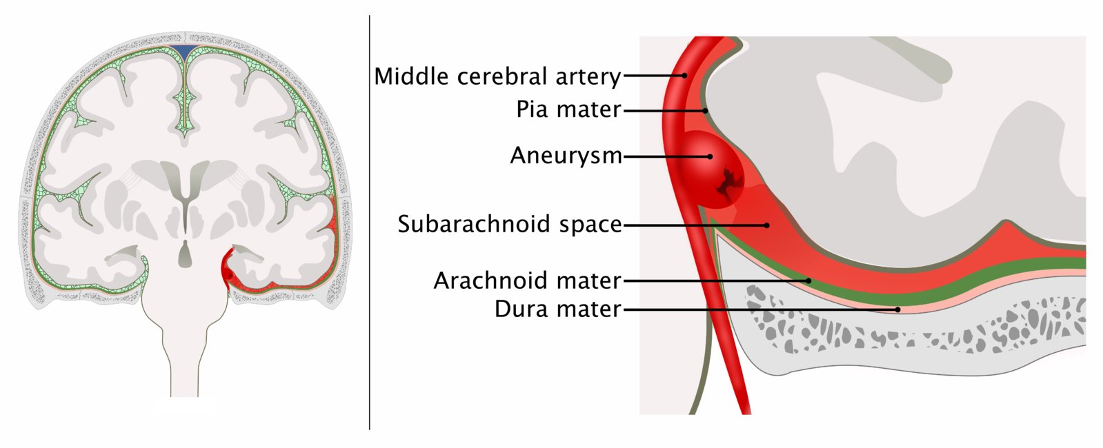
Explain the etiologies and pathophys of a subarachnoid hemorrhage.
Etiologies: Berry aneurysm in circle of willis (80%) or AV malformation
Risk Factors: Smoking & Hypertension
What are some clinical features a patient with a subarachnoid hemorrhage may present with?
- Sudden, severe headache
- Fever, nuchal rigidity
- No focal deficits
- General symptoms: drowsiness, loss of consciousness
What type of intracranial hemorrhage is this?

Left Subdural Hematoma
Hyperdense, crescent shaped lesion over lateral aspect of the left hemisphere. Also diffuse hypodense areas in left hemisphere due to white matter edema, and midline shift to green line

What type of posturing is this?
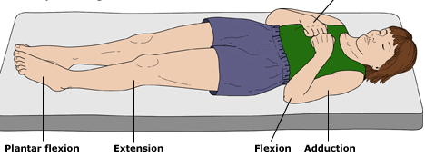
Decorticate Posturing — indicates damage to cerebral hemispheres / cortical spinal tracts.
The primary somatosensory cortex is a part of which lobe?
Parietal lobe
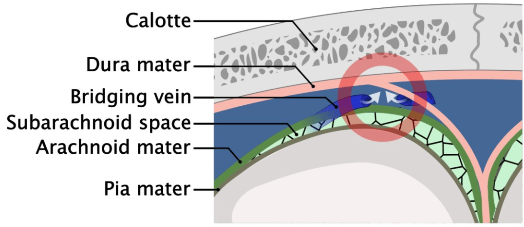
Explain the etiologies & pathophys of a subdural hematoma.
Etiologies include: blunt head trauma, frequent falls (elderly, alcohol abuse, epilepsy), and shaken baby syndrome.
Pathophys: Rupture of bridging veins → low-pressure venous bleeding.
*Quick changes in velocity can cause accumulation of venous blood in bridging veins
What are some clinical features a patient with a subdural hematoma may present with?
- General symptoms: headache, drowsiness, loss of consciousness
- Changes in mental status
- Memory impairment
*Subdural hematoma & epidural hematoma can NOT be differentiated based on clinical appearance alone
What is the diagnosis?

Subarachnoid Hemorrhage
Hyperdensities around circle of willis

If the patient is stable, what first line diagnostic tool is used to diagnose intracranial hemorrhages?
Cranial CT
Visualize:
- Skull fractures
- Dura mater rupture
- Midline shift of the brain
- Hemorrhage
Lesions to the non-dominant side of this lobe may present with hemineglect
Parietal lobe (non-dominant side functions in spatial awareness)
Hemineglect — patients fail to be aware of items to one side of space
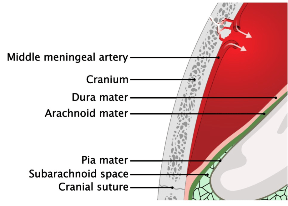
Explain the most common etiology and pathophys of an epidural hematoma.
Traumatic rupture of middle meningeal artery
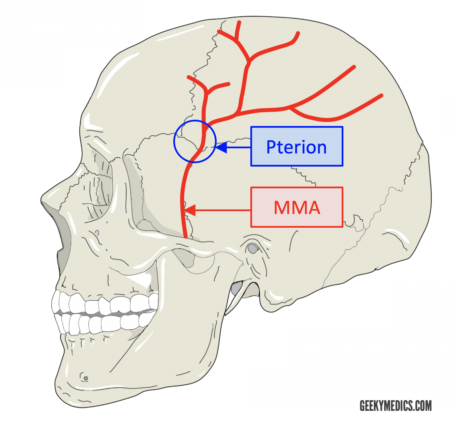
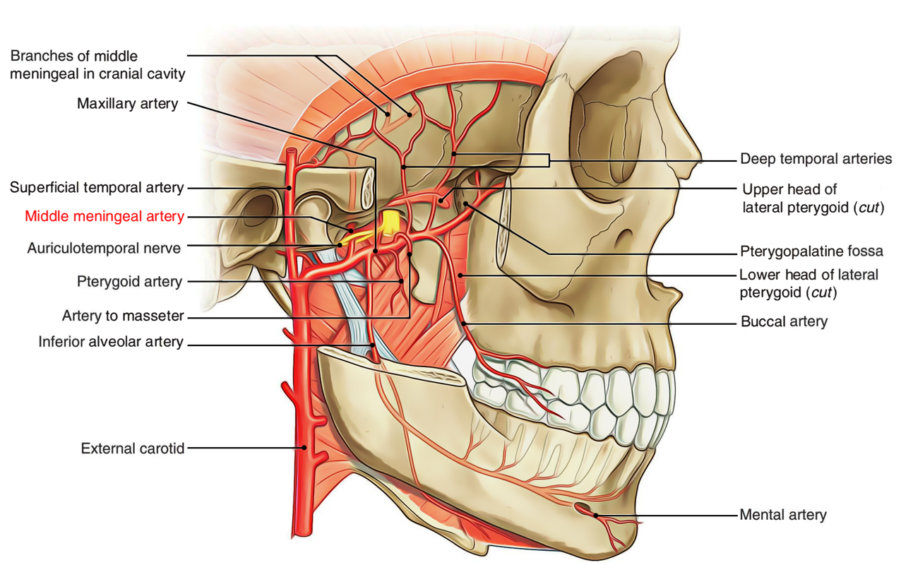
What are some clinical features a patient with an epidural hematoma may present with?
- General symptoms: headache, drowsiness, loss of consciousness
- Lucid Interval
- Hemiplegia
How does an epidural hematoma present on cranial CT?
Biconvex hyperdense lesion between the brain and skull that is limited by the suture lines (dura mater is very tightly adherent at the suture lines)

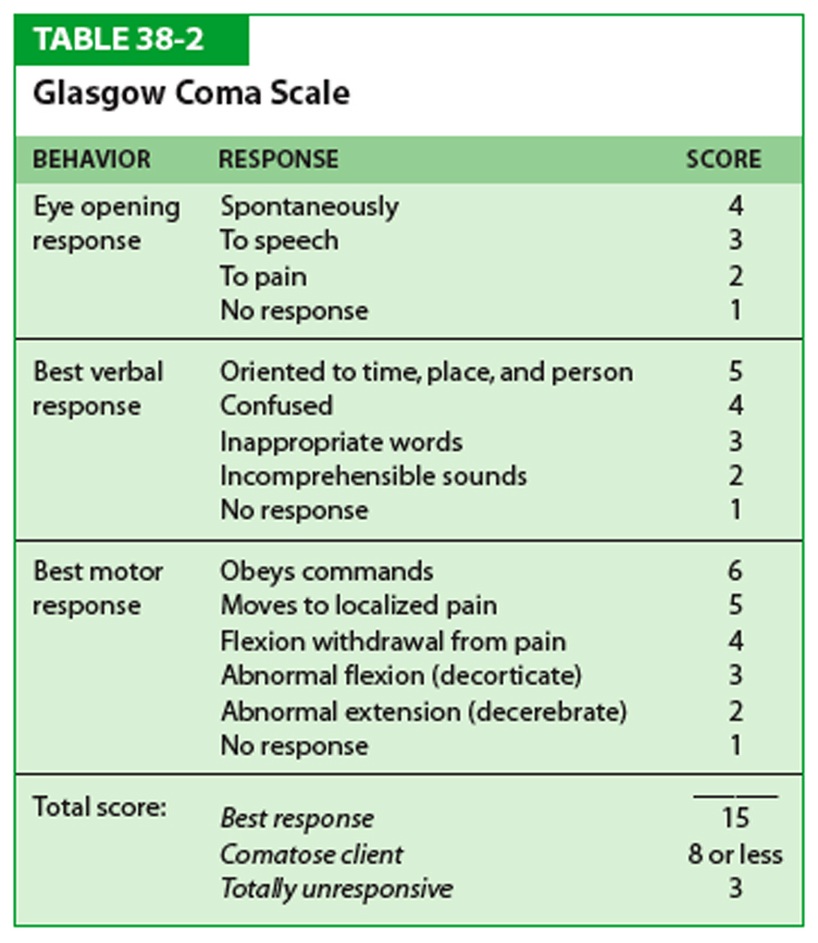
What diagnostic tool is used to evaluate consciousness in patients with traumatic brain injury (TBI)?
Glasgow Coma Scale
Mild head injury: GCS score 13–15
Moderate head injury: GCS score 9–12
Severe head injury: GCS score ≤ 8 (Indication for endotracheal intubation)
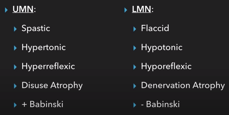
Does an UMN lesion present with hyperreflexia or hyoreflexia.
Hyperreflexia due to loss of inhibitory signals.
