External Anatomy (Anterior)
External Anatomy (Posterior)
Vascular Supply and Relationships to Ventricular System
100
What brainstem structures do labels 1, 2, and 3 correspond to respectively?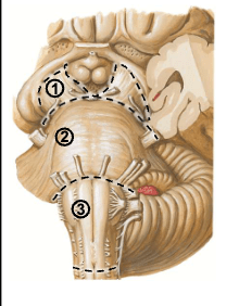
1. Midbrain
2. Pons
3. Medulla
100
What brainstem structures do labels 1, 2, and 3 correspond to respectively?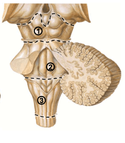
1. Midbrain
2. Pons
3. Medulla
100
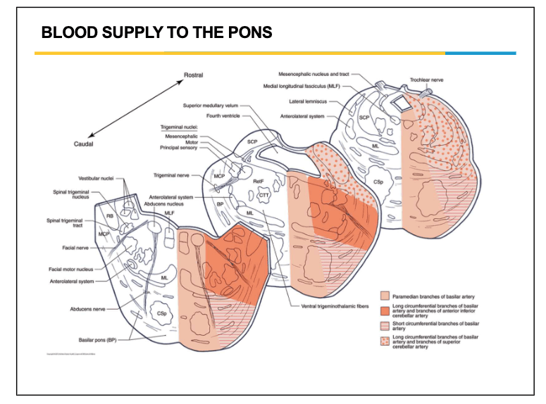
The primary source of arterial blood for the Pons is supplied by this vessel.
Pontine Branches of the Basilar Artery
Paramedial Branch = medial pons -> corticospinal tracts, medial lemniscus, abducens nucleus
Short Circumferential Branch = ventrolateral pons
Long Circumferential Branch = lateral pons and parts of the cerebellum
200
CN III (Oculomotor) emerges from this depression located between the cerebral peduncles
Interpeduncular fossa
200
What structure of the midbrain is indicated by the red star?
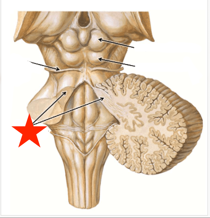
Superior Cerebellar Peduncles
Massive bundles of both afferent and efferent white matter tracts. Connect midbrain to cerebellum. Notably contain the spinocerebellar tracts.
200
What artery branches from the vertebral artery to supply the cerebellum?
What is Posterior Inferior Cerebellar Artery (PICA)
The Anterior Inferior Cerebellar Artery (AICA) branches from the Basilar Artery.
300
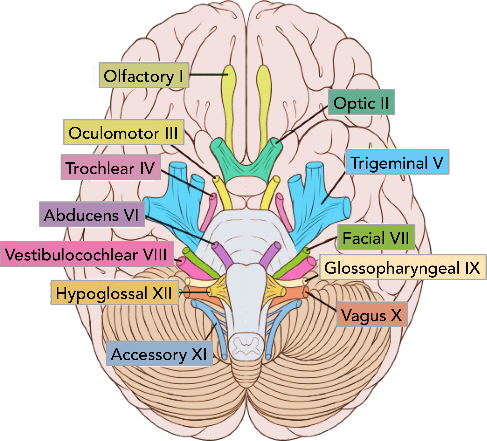
What four cranial nerves are visible in this image? Which three exit at the pontomedullary junction?
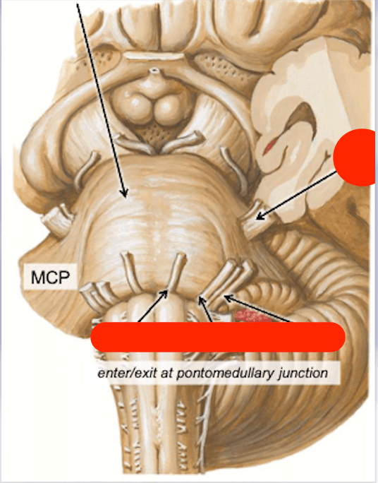
CN V: Trigeminal (branches not really shown)
CN VI: Abducens
CN VII: Facial
CN VIII: Vestibulocochlear
300
The arrows point to four structures in the medulla. Correctly identify all four structures.
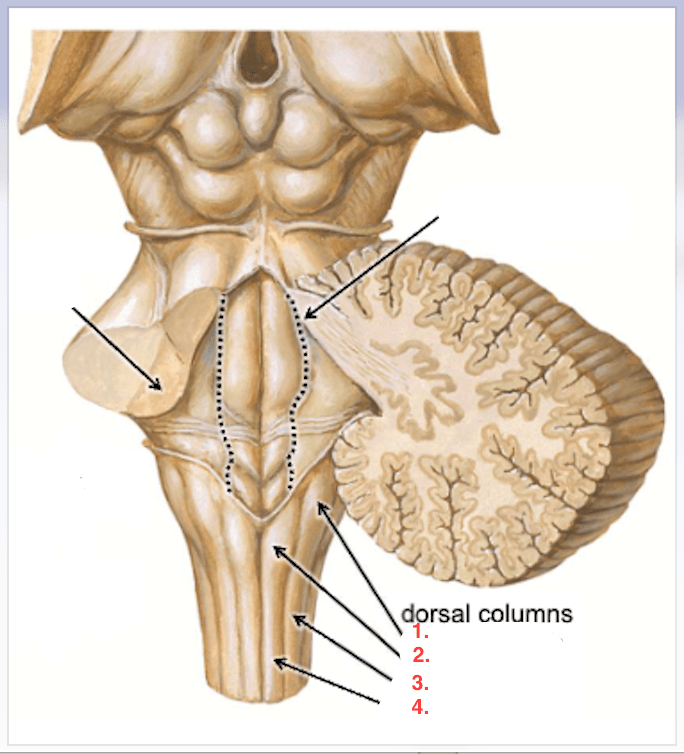
1. Cuneate tubercle
2. Gracile tubercle
3. Cuneate fasciculus
4. Gracile fasciculus
Tubercles are elevations formed due to the cuneate and gracile nuclei of the dorsal column/medial lemniscus
300
Congenital defects in the structure labeled with the red star is one of the most common causes of non-communicating hydrocephalus in children.
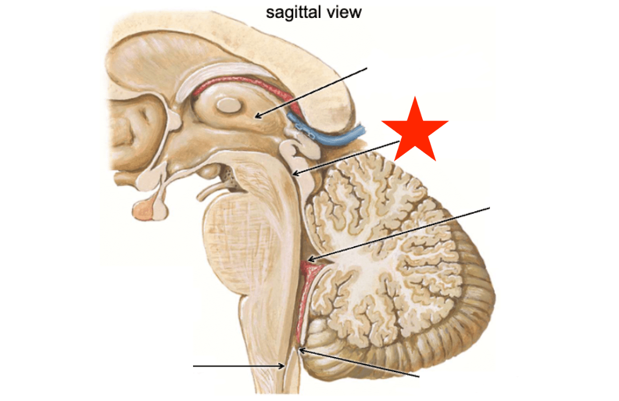
Cerebral Aqueduct (Aqueduct of Sylvius)
400
What is the importance of the structure indicated by the arrow labeled with the Green Star? What spinal tract is it associated with?
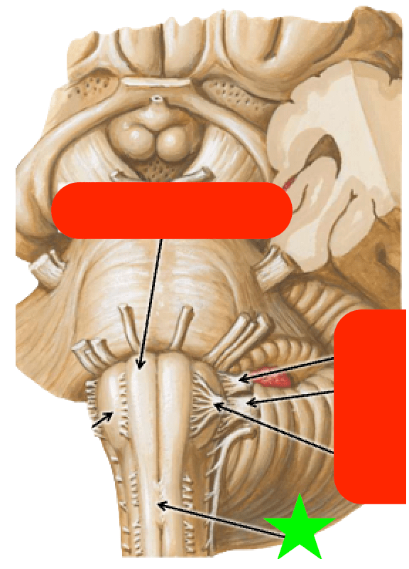
Green star labels the pyramidal decussation. This is where 85% of corticospinal fibers cross to the contralateral side.
400
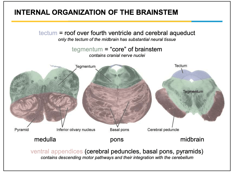
The superior and inferior colliculus are important formations for our special senses. What internal region of the brainstem do they belong to? (Not midbrain, pons, or medulla)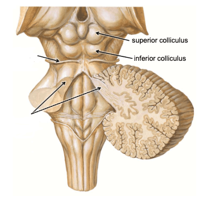
Tectum
Only has significant tissue in the midbrain (very sparse in pons and medulla).
400
A patient has contralateral hemiparesis, contralateral loss of fine touch/proprioception, and ipsilateral tongue weakness due to a brainstem infarct. Which artery is most likely occluded?
Anterior Spinal Artery (ASA)
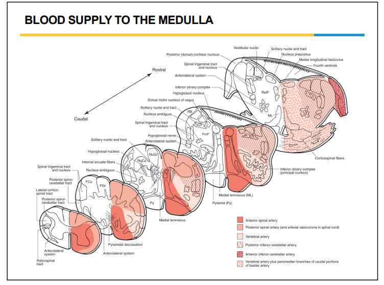 This one confused me at first because when you're looking at infarcts of the ASA in the spinal cord the dorsal columns are spared (meaning so is proprioception and fine touch) but in the brainstem because the medial lemniscus comes medially then moves laterally it is susceptible to ischemia.
500
VENTRAL VIEW!!!
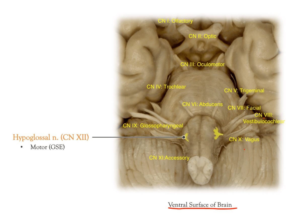
Which cranial nerve is highlighted in this image? What part of the brainstem is it emerging from?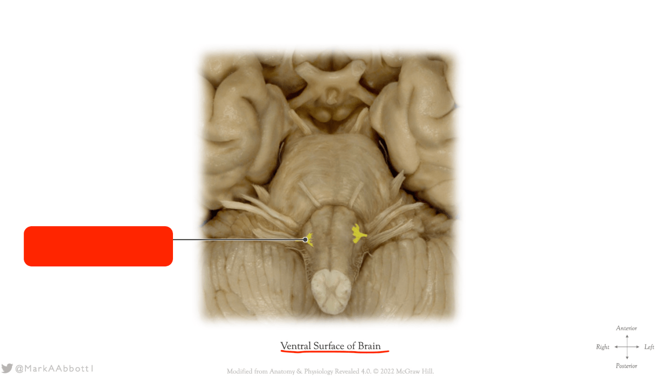
CN XII: Hypoglossal
Emerging from the anterior medulla.
500
What structure is indicated by the arrow labeled with the red star? What is its significance?
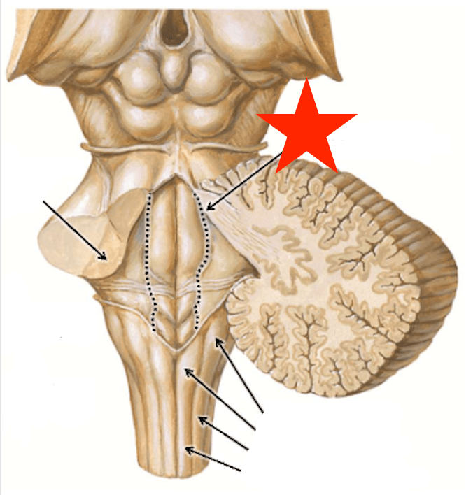
Sulcus Limitans
The developmental and functional boundary between the sensory plate and the motor plate of the neural tube and brainstem. Persists into adulthood as a landmark on the floor of the fourth ventricle.
Separates the medially located motor cranial nerve nuclei from the laterally located sensory nuclei.
500
A patient presents with ipsilateral CN III palsy and contralateral hemiparesis from a medial midbrain infarct of the paramedian branches of the basilar artery. Name the syndrome
Weber Syndrome
