Projections
Positioning Corrections
Pathology
Anatomy
Positioning
Random
Image Anatomy
100
What is the projection below?
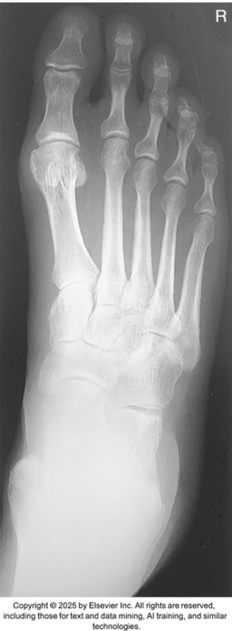
What is AP Projection: foot
(Dorsoplantar Projection)
100
A lateral projection of the patella demonstrates a closed patellofemoral joint space, with the patella superimposed over the distal femur. What positioning error most likely caused this appearance?
What is Excessive flexion of the knee
100
Weight-bearing foot series will best demonstrate what type of injury?
What is Lisfranc Injury
100
How many bones are in the foot?
What is 26 bones
100
What is the method for the AP axial projection of the intercondylar fossa of the knee?
What is the Béclere Method?
100
What modality will best evaluate injuries to the minisci and ligaments of the knee joint?
What is MRI
100
What anatomy is Letter L?
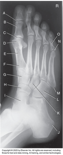
What is the cuboid
200
What projection is the image below?
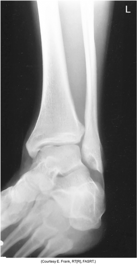
What is AP oblique projection - 45° medial rotation ankle
200
A radiograph of an axial plantodorsal projection of the calcaneus reveals foreshortening of the bone. The technolgist used 60 kVp, 6 mAs, 40 SID, and a 30 degree cephalad angel. What change(s) need to be made in order to produce a more diagnostic image of the calcaneus?
What is increase CR angle to 40° cephalad
200
What views will best demonstrate Osgood-Schlatter Disease?
What is AP and Lateral knee
200
Part on the calcaneus that provides medial support for the talus?
What is the sustentaculum tali
(sus-ten-tak-u-lum)
200
Where is the CR directed for an AP projection of the knee?
what is 1/2 inch below the patellar apex
200
What do the three bones of the distal tibia, distal fibula, and talus form?
What is the mortise
200
What anatomy is letter E?
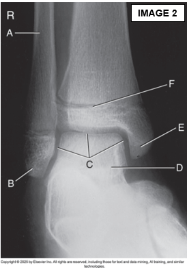
What is the medial malleolus
300
What is the projection below?
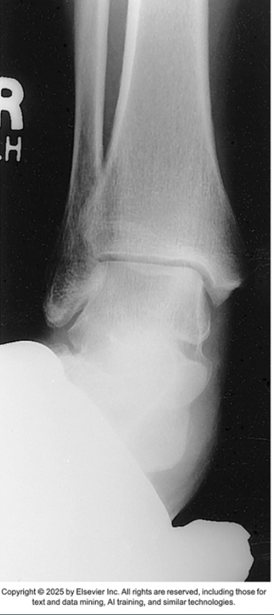
What is AP stress projection ankle - eversion
300
A radiograph of a lateral ankle shows that the distal fibula is superimposed over the anterior portion of the distal tibia. What must the technologist do to correct this positioning error on the repeat exposure?
What is Rotate lower leg/foot externally or towards the IR.
300
What pathology show bony erosions at the Achilles tendon insertion for young men?
What is Reiter Syndrome
300
The three calcaneal articular facets make up what joint.
what is the subtalar joint
aka: talocalcaneal joint
300
How should the CR be directed to properly demonstrate the joint spaces on an AP projection of the foot?
what is 10° posteriorly, perpendicular to the metatarsals
300
How much do you rotate the leg and foot for an AP oblique toe and which way for 1-3 digits and 4-5 digits?
What is 30° to 45°
1-3 toe medially
4-5 toe laterally
300
What anatomy is letter J?
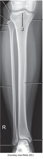
What is Intercondylar eminence
400
What projection is shown in the image below?
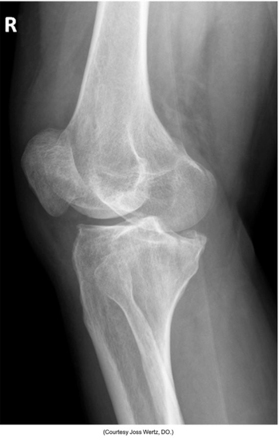
What is AP oblique - lateral (external) rotation knee
400
A radiograph of an AP projection of the third toe reveals that the interphalangeal joints are not open. What are the most likely causes for this radiographic outcome?
What is incorrect angle or CR centering. (angle should be 10° to 15°)
400
A benign, neoplastic bone lesions filled with clear fluid that appears on an x-ray like a circumscribed lucency.
What is a bone cyst
400
This arch in the foot has medial and lateral components.
What is longitudinal arch
400
How is the leg and foot rotated for an AP oblique 2nd toe projection?
What is 30° to 45° medially
400
When positioning for an AP knee, how much do you rotate the leg and in which direction?
What is 3° - 5° internally
400
What anatomy is letter B?
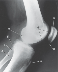
What is the Apex of the patella
500
What is the projection in the image below?
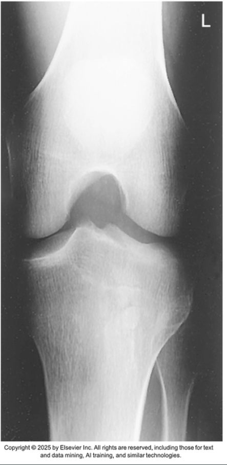
What is PA axial projection (Tunnel View): knee-intercondylar fossa
500
A radiograph of an AP oblique foot with medial rotation demonstrates considerable superimposition of the third though fifth metatarsals. What does the technologist need to do to fix this problem?
What is Decrease obliquity of the foot.
500
Malignant primary bone tumor occurring from childhood to a young adulthood that produces a sunburst pattern on an x-ray image.
What is Osteogenic Sarcoma
(Osteosarcoma)
500
What is the joint classification and mobility for the distal tibiofibular joint?
What is fibrous and amphiarthrodial (slightly moveable)
500
Which position/projection of the ankle will best demonstrate the distal tibiofibular joint?
What is AP oblique with 45° medial rotation
500
What pathology is a form of arthritis that may be hereditary, starts in the 1st MTP joint, and mostly occurs in men?
What is Gout
500
What anatomy is letter A?
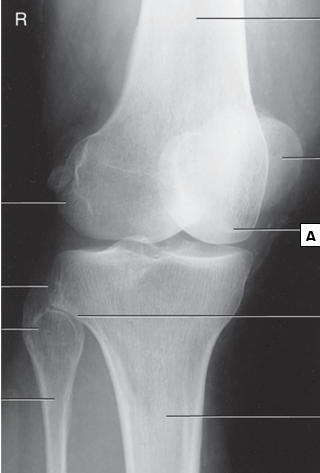
What is the medial condyle
600
What projection is in the image below?
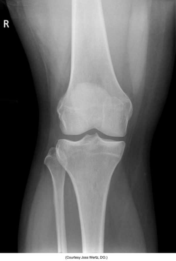
What is AP knee
600
A patient has osteoarthritis of both knees, what projection will evaluate the damage to the articular facets?
What is PA axial weight-bearing bilateral knee projection: knee
Rosenberg Method
600
what is another name for exostosis?
What is osteochondroma
600
What is the name of the deep depression that separates the femoral condyles posteriorly?
What is intercondylar fossa
600
What is the recommended SID for the superioinferior sitting tangential projection of the patellofemoral joint?
What is 48-50 inches (120-130cm)
This is to reduce magnification because of the increased OID.
600
What projection/method will best demonstrate loose bodies within the patellofemoral joint space?
What is Tangential axial or sunrise/skyline projection: patella
Bilateral Merchant Method
600
What anatomy is letter A?
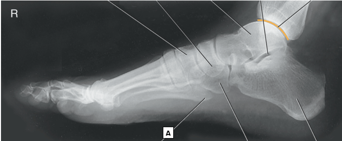
What is the tuberosity of the base of 5th MT