Perdiéndola
"Estoy frontal"
"y pico"
Wheeliando por el circuito de Papez
Ponme a prueba
100
A patient with unilateral dyspraxia, alien limb phenomenon, cortical sensory loss (graphesthesia, stereognosis), and behavioral changes is expected to have this finding on imaging.
What is asymmetric atrophy of the premotor cortex?
100
Damage to this frontal region classically results in disinhibition, impulsivity, socially inappropriate behavior, and emotional lability.
What is the orbitofrontal cortex?
100
A patient is being evaluated due to an inability to visually fixate on an object. When asked to explain what they see in the following image, they consistently report seeing several "U" s. Bilateral damage to this cortex is the cause.
What is the parieto-occipital cortex?
100
A pregnant patient presents with confusion, ataxia, and nystagmus. An imaging study shows hyperintensity of these diencephalic brain structures.
What are the mammillary bodies?
100
This cognitive function is assessed in this exercise, in which the patient is asked to name the color of the word.

What is executive function? (Stroop test)
200
Visual hallucinations, fluctuating cognition, and neuroleptic intolerance arise in an alpha-synuclein pathology, which shows hypometabolism of these regions in the FDG-PET.
What are the posterior parietal and occipital regions? (Lewy Body Dementia)
200
A patient with diminished or absent movement and speech production may have a lesion in this region.
What is the dorsomedial prefrontal cortex (anterior cingulate)?
200
Named after an Austrian neuroscientist, the syndrome that is characterized by agraphia, acalculia, finger agnosia, and left-right disorientation localizes to this region. (Be specific).
What is the dominant supramarginal and/or angular gyrus?
200
It is a target for neuromodulation in patients with medically refractory epilepsy.
What is the anterior thalamic nucleus?
200
This test, in which a patient is asked to connect the shapes in ascending order while alternating the colors of the circles, assesses this domain.

What is attention? (Trial making test)
300
A patient presents with progressive episodic memory loss. A posthumous brain biopsy shows the below pathology. What lobes are expected to be most affected in this disease?


What are the temporal and parietal lobes? (Alzheimer's Disease)
300
A patient presents with attention and working memory issues. They also refer that, although they are motivated to do various tasks, they rarely complete them. Could a lesion in this area of the prefrontal cortex be the answer? (Be specific).
What is the right dorsolateral prefrontal cortex (posterior cingulate)?
300
A patient is asked to identify his wife from a lineup of pictures, but is unable to do so. He is, however, able to recognize her later due to her voice. He likely has a lesion in this area, as famous neurologist Oliver Sacks described in his book.

What is the fusiform gyrus?
300
This white matter tract connects the anterior thalamic nucleus with the parahippocampal region (entorhinal cortex).
What is the cingulum?
300
A patient with AD is asked to draw a clock showing 10 past 11 PM. His effort is shown below with a control result. This domain is affected in our patient.
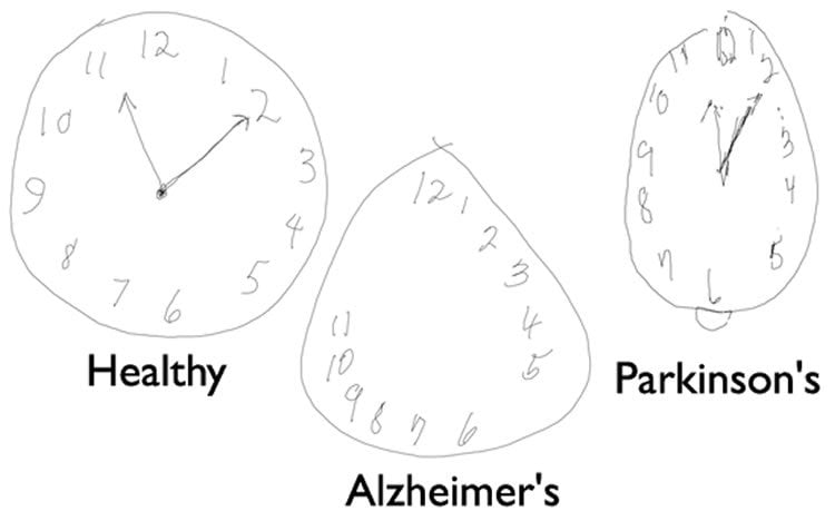
What is visuospatial? (Clock test)
400
A patient is brought by their daughter due to concerns that the patient frequently hits himself against the walls of his home. On physical examination, he is seen with left-right confusion, finger agnosia, dyscalculia, and dysgraphia. Brain MRI shows atrophy of this region.
What are the occipital and parietal cortex? (Posterior Cortical Atrophy)
400
This numeric sign, seen in seizures, correlates with this region.
What is the supplementary motor area?
400
A patient is instructed to read the title of a newspaper, but is unable to do so. He is then instructed to write a sentence, and he writes "Today is a sunny day!" He is later found with a mass involving these regions.
What are the left occipital cortex and splenium?
400
This structure, which connects the hippocampus to the mammillary bodies, is highlighted by the yellow arrow in the picture.

What is the fornix?
400
In this assessment, patients are shown several common and uncommon objects and are asked to name them, thus evaluating this cognitive domain.

What is language? (Boston naming test)
500
A patient presents with difficulty with whole word recognition and spelling, especially when the words have irregular spelling-sound (e.g., “yacht” read as “yatchet”). They are also seen with fasciculations in the limbs and diffuse hyperreflexia. A PET scan shows hypometabolism of these regions.
What is frontal and temporal regions? (Semantic Aphasia variant of FTD with ALS)
500
After being diagnosed with a left MCA stroke, a patient is instructed to elevate his left arm, but is unable to do so, in spite of having adequate strength in this arm. Infarction of this tract may explain the patient's presentation.
What is the anterior commissure?
500
A high-functioning executive presents with the concern that everyday tasks have become more difficult. For example, she recently arrived at work with her shirt backwards and has attempted to fit both legs through the hole of one pant leg. This may be explained by a lesion in this area.
What is the non-dominant parietal region?
500
DOUBLE JEOPARDY!
This artery supplies a "seahorse" shaped structure that is crucial in memory consolidation.
What is the posterior choroidal artery?
500
When a patient is asked to tap their index finger and thumb as rapidly as possible for 10–30 seconds, this is the domain under evaluation.
What is psychomotor speed? (Finger tapping test)