What is the tissue Type?
Where is it Located?
What is its function?
What is the notable structure?
MISC
10

Loose Areolar Connective Tissue
10

ligaments
10
Protects, Pads, stores fat (energy), and insulates
10
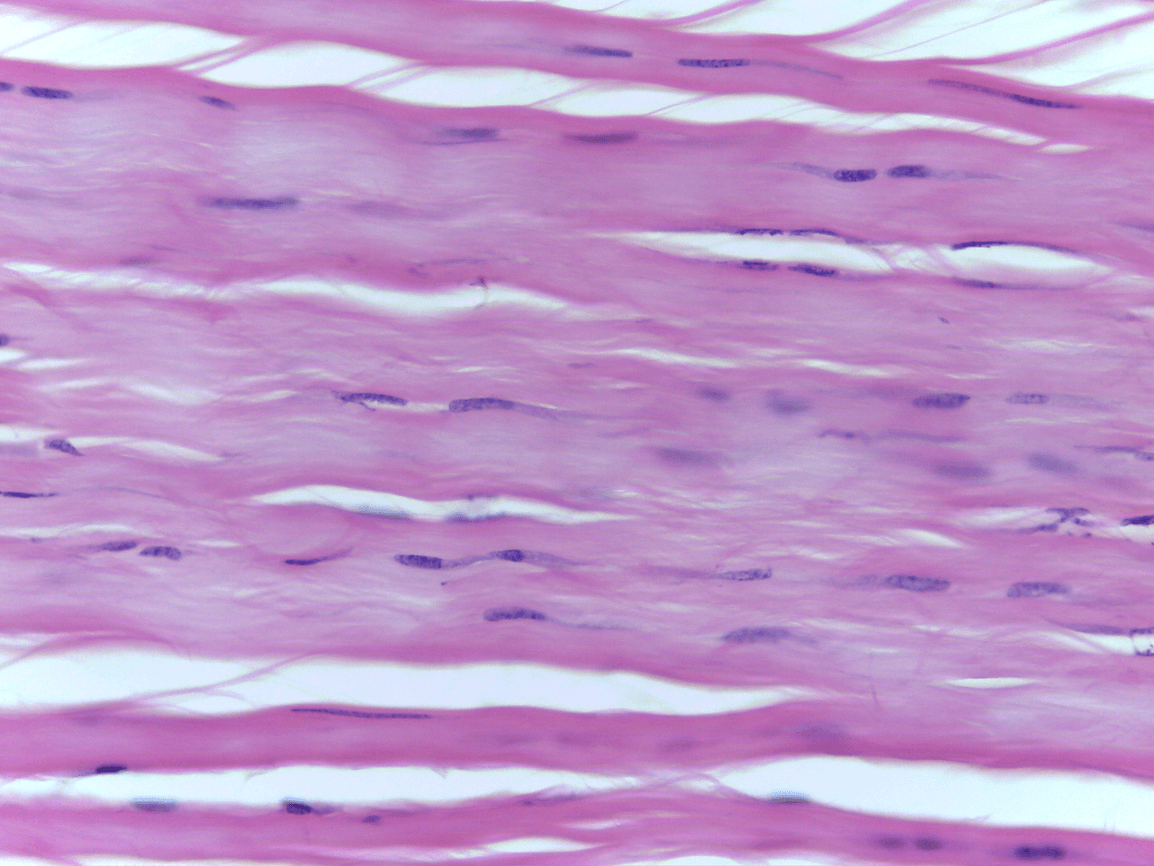
What Tissue type is this?
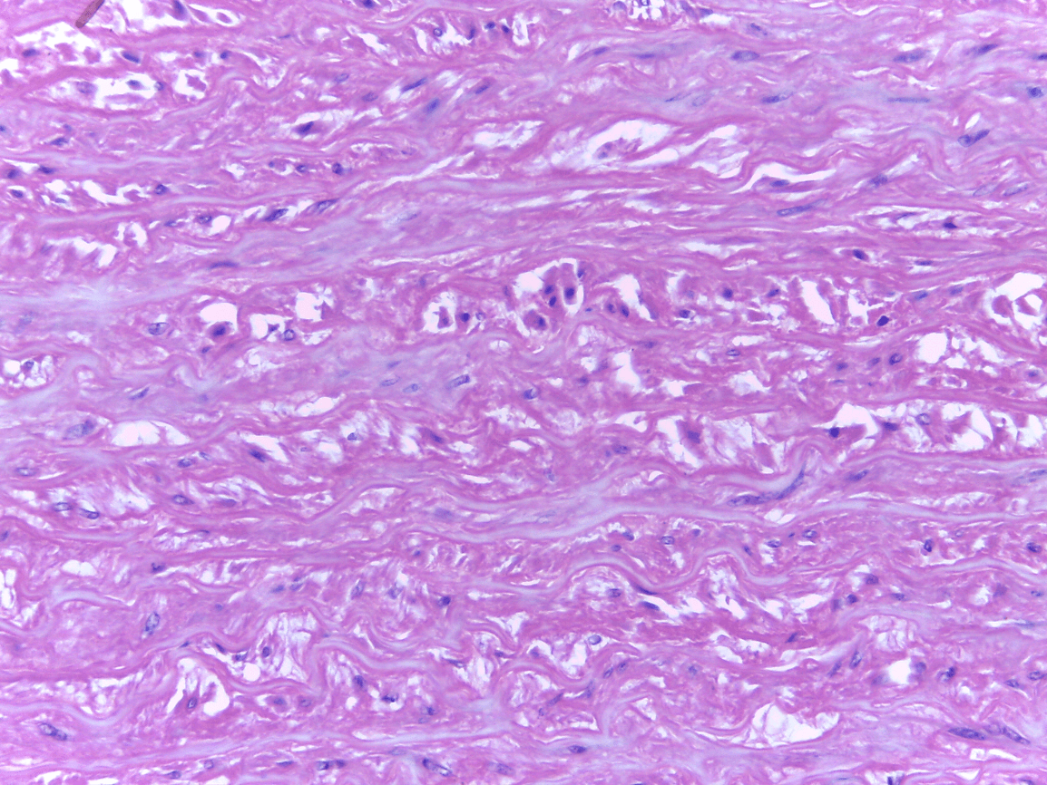
Dense Regular CT
20

Loose Adipose CT (deep)
20

Wall of Aorta
20

gives ability to stretch without tearing
20
Sinusoid- hold red blood cells
30

Mainly in hypodermis, but found throughout the body
30

easily move material up to epithelial
30
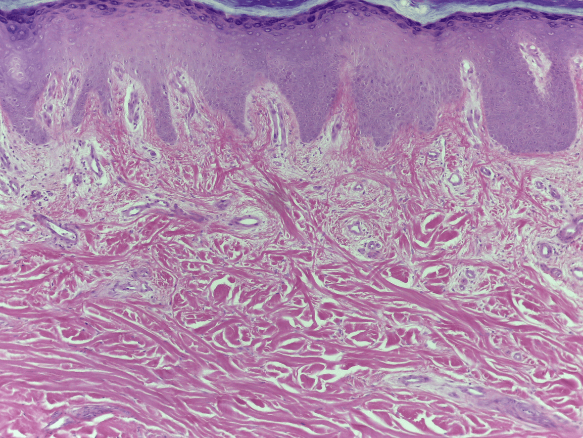
Name the three Tissues found here in order from superficial to deep:
1.) Keratinized stratified squamous ET
2.) Loose CT
3.) Dense Irregular CT
40

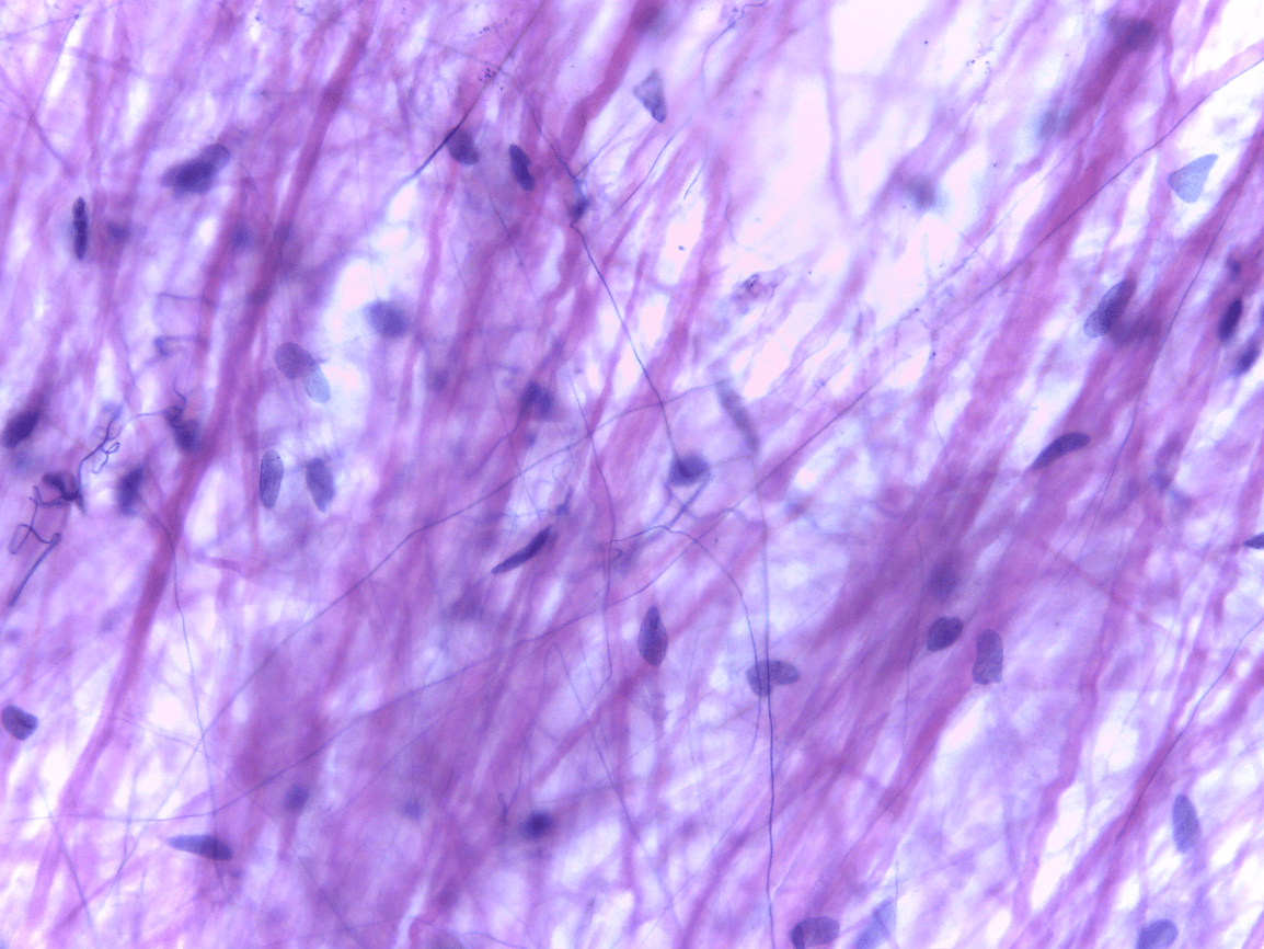
Dense irregular CT
40

Dermis of the skin
40
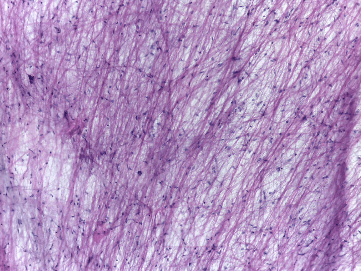
Wide collagen fibers, thinner elastic fibers, dark spots are nuclei of mast cells-immunity
40
Name the function of the three tissue types found here in order from superficial to deep: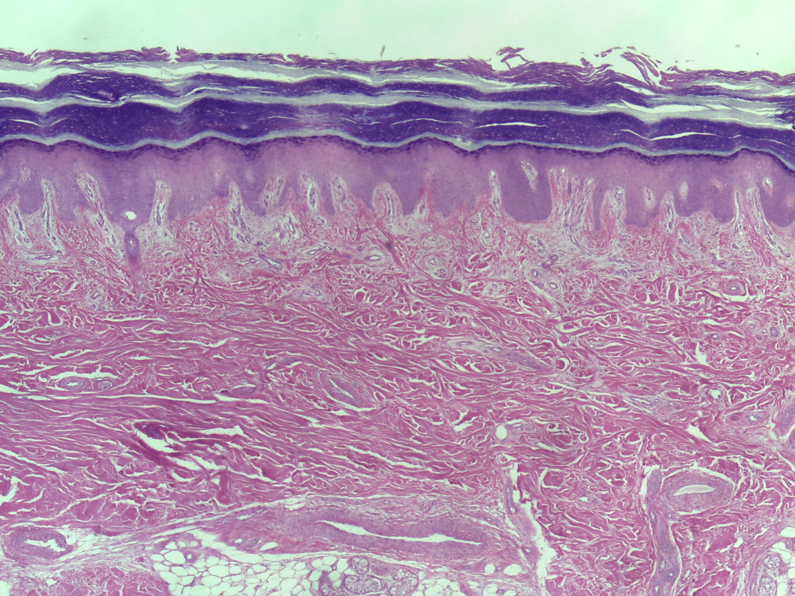
1.) strength and waterproofing
2.) Binds skin to muscles and surrounds muscles/strength/flexibility
3.) provide strength and resistance to tearing
50

Loose Areolar Connective Tissue
50

Hypodermis mainly, but also found throughout the body
50
provide strength and resistance to tearing
50
densely packed collagen fibers, parallel to the long axis of the tendon or ligament
60

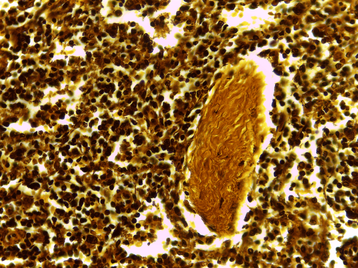
Dense Reticular CT
60
Wall of the Aorta
60

build organs-liver and spleen
60
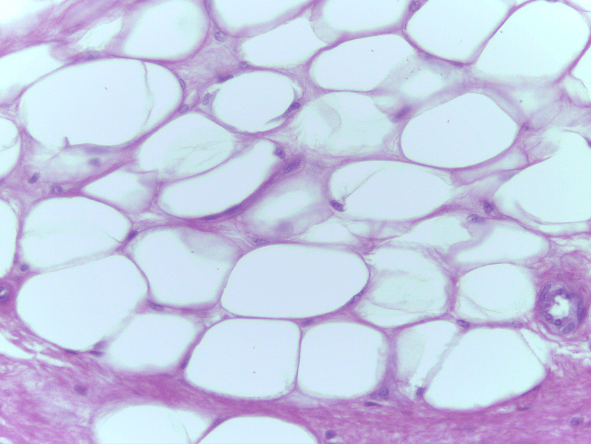
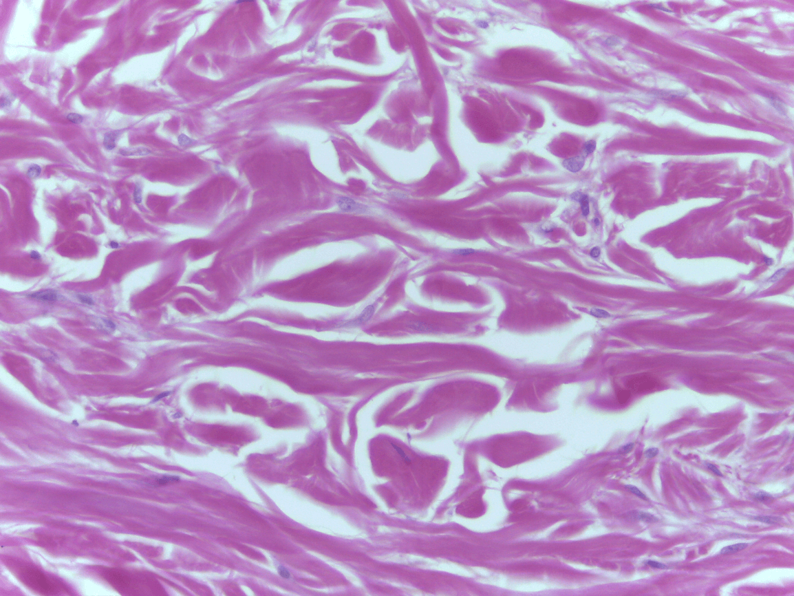
Large "bubbles"- adipocytes- which store triglycerides in the hypodermis to make up adipose CT
60
What Tissue type is this?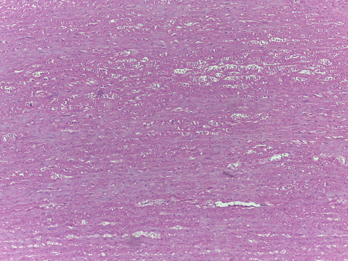
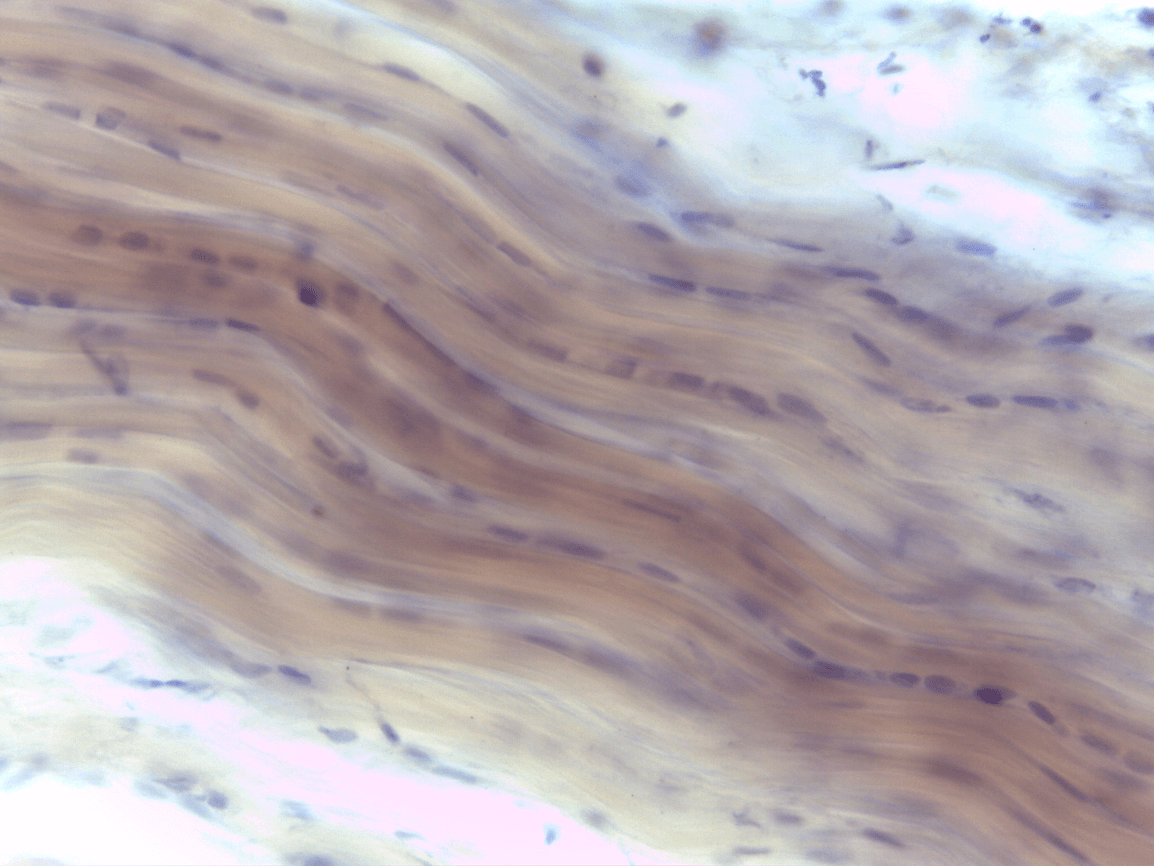
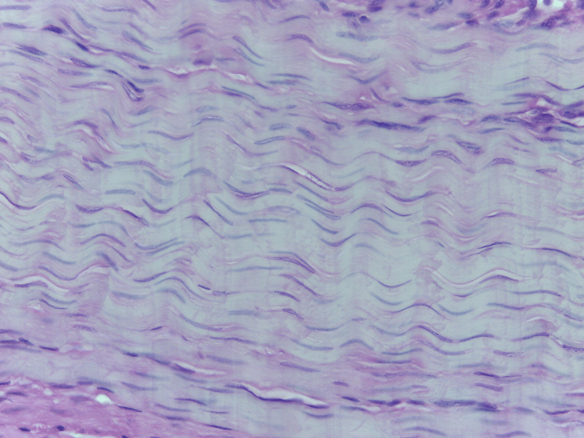
Elastic CT
70
Dense Regular CT
70
Reticular region of the dermis
70

Protects, Pads, stores fat (energy), and insulates
70
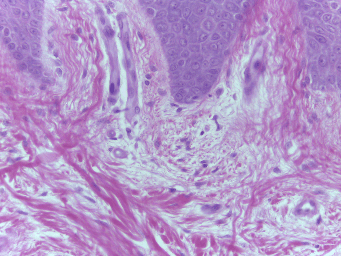
epidermal parallel pegs, dermal papillae
70
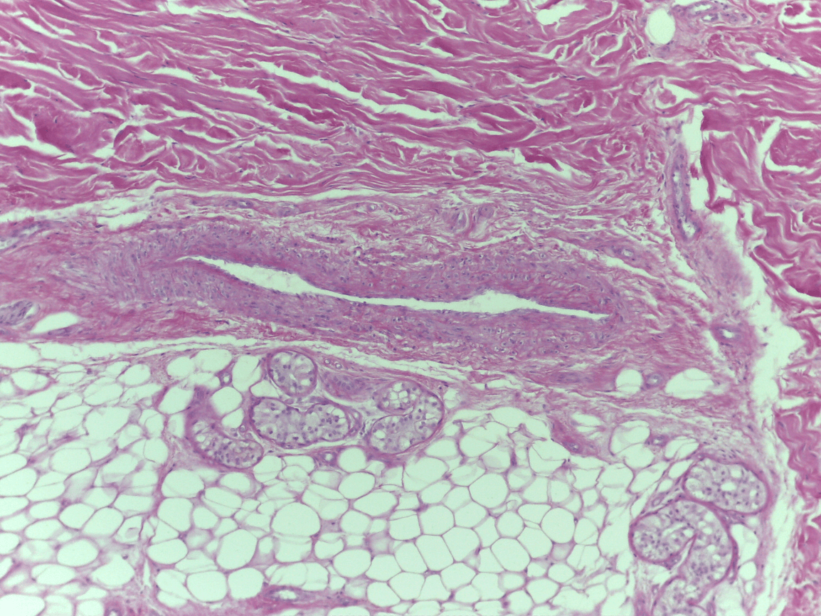
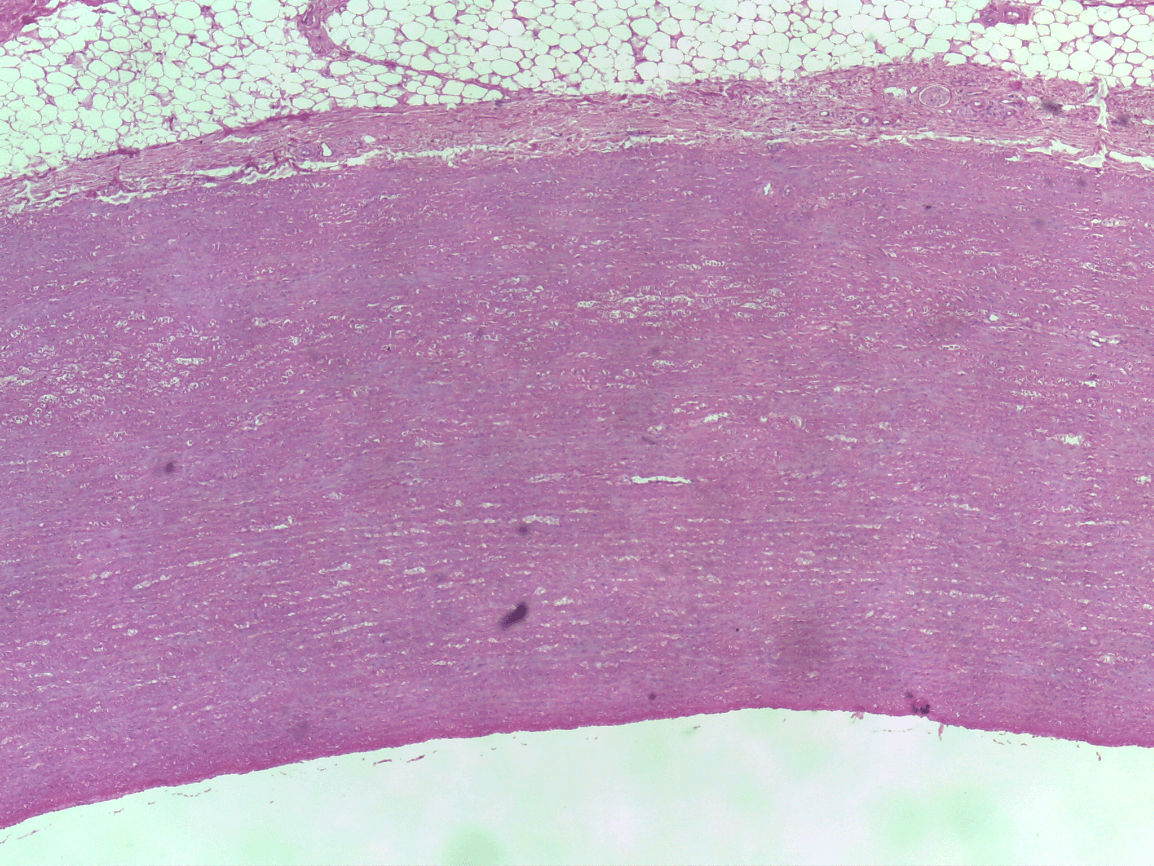
What two tissue types can you see here from superficial to deep?
1.) Adipose Tissue
2.) elastic CT
80

tendons
80

easily move material up to epithelial
80
What two Tissues can you see here from superficial to Deep?
1.) keratinized stratified squamous ET
2.) Loose CT
90

Dense Irregular CT
90

Wall of the aorta
90

return to its original length after stretching
90

the lumen of the aorta
100

Loose Adipose CT
100

Dermis of the Skin
100

gives ability to stretch without tearing
100
What Tissue type is this?
Elastic CT
110
Dense Regular CT
110

Papillary region of dermis, around blood vessels and nerves
110
return to its original length after stretching
110
What part of the dermis is adipose located in?
Hypodermis
120

Dense irregular CT
120

ligaments
120

return to its original length after stretching
120

Wide collagen fibers, thinner elastic fibers, dark spots are nuclei of mast cells-immunity
120
Which part of the dermis is the two Tissues located in?
1.) keratinized stratified squamous located in the epidermis
2.) Loose CT located in the dermis
130
Papillary region of dermis, around blood vessels and nerves
130

gives ability to stretch without tearing
140

Elastic CT
140

Liver, Spleen, Lymph nodes, Thymus, Bone Marrow
This particular slide is the spleen
140

Gives strength in multi-direction
140

densely packed collagen fibers, parallel to the long axis of the tendon or ligament
140
What two tissue types can you see here from superficial to deep?
1.) Dense irregular CT
2.) Adipose CT