Doppler & Hemodynamics
Multiple choice
MMode
EKG
Random
100
what happens to measured blood-flow velocity when blood passes through a narrowed valve opening?
Velocity increases
100
Fibrin strands seen within a pericardial effusion are frequently present with
A. long-standing effusions
B. metastatic diseases
C. A & B
D. None of the above
C. A & B
100
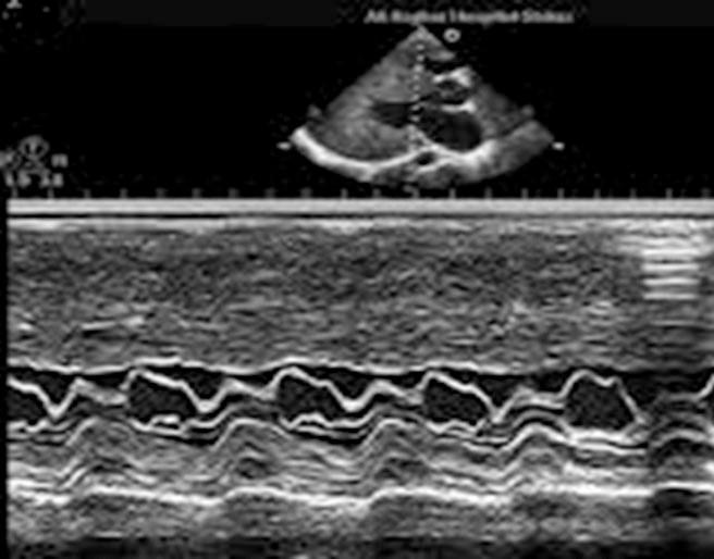
hypertrophic cardiomyopathy with SAM
100
Put the steps to EKG Rhythm Interpretation in order.
- QRS Duration & Morphology
- PR-Interval
- Rate
- Rhythm
- P-Wave Morphology
- Rhythm
- Rate
- P wave morphology
- PR interval
- QRS duration and morphology
100
What does the A wave on mitral inflow Doppler represent?
Late ventricular filling caused by atrial contraction.
200
A sonographer sees Doppler aliasing on CW. What two adjustments can help display the jet more clearly?
Increase the color scale/Nyquist limit and adjust the baseline as needed.
200
A patient develops sudden shortness of breath and hypotension several days after a myocardial infarction. Echocardiography shows a new, large amount of mitral regurgitation. Which complication could explain this finding?
A. Aortic valve sclerosis
B. Papillary muscle rupture
C. Mild left atrial enlargement
D. Physiologic tricuspid regurgitation
B. Papillary muscle rupture
200
How will the mitral valve appear on mmode with AI
diastolic fluttering of the anterior mitral valve leaflet
200
A rhythm strip shows no consistent P waves and irregularly irregular R-R intervals. What rhythm is most likely
Atrial fibrillation; it lacks consistent organized atrial activity and has irregularly irregular ventricular intervals.
200
Mitral inflow Doppler appears normal, but tissue Doppler and other findings suggest impaired relaxation and elevated filling pressures. What pattern of diastolic dysfunction can explain this?
Pseudonormal filling (typically Grade II diastolic dysfunction).
MV flow will be big e and little a. TDI will not mirror MV flow
300
Which Doppler method uses a single crystal to measure blood velocity at a specific location, but cannot reliably measure very high velocities because of aliasing?
Answer: Pulsed-wave (PW) Doppler.
300
Which statement correctly differentiates isovolumic contraction time (IVCT) from isovolumic relaxation time (IVRT)?
A. IVCT occurs after the aortic valve closes, while IVRT occurs before the mitral valve closes.
B. IVCT occurs after the mitral valve closes and before the aortic valve opens, while IVRT occurs after the aortic valve closes and before the mitral valve opens.
C. IVCT occurs during ventricular filling, while IVRT occurs during ventricular ejection.
D. IVCT and IVRT both occur while the mitral and aortic valves are open.
A. M-mode
B. Color Doppler only
C. Continuous-wave Doppler
D. Tissue Doppler imaging
B. IVCT occurs after the mitral valve closes and before the aortic valve opens, while IVRT occurs after the aortic valve closes and before the mitral valve opens.
300
a 38 year old male is sent to the echo lab for evaluation after complaining of severe dyspnea on exertion for two months. M-mode findings include a dilated left ventricle, increased EPSS, B-notch on the mitral valve and overall hypocontractile left ventricular motion. What type of cardiac abnormality do these findings suggest?
dilated cardiomyopathy
300
On an ECG rhythm strip, 6 seconds contain 9 QRS complexes. Estimate the heart rate using the 6-second method.
Approximately 90 beats/minute (9 × 10).
300
During a Doppler assessment, mitral inflow changes markedly with respiration, and the interventricular septum shows a characteristic bounce. What pericardial condition should be suspected?
Constrictive pericarditis.
400
A patient has a peak tricuspid regurgitation velocity of 3.0 m/s. The IVC is dilated but collapses. Calculate the RV systolic pressure, assuming no pulmonic stenosis
RVSP = 4(3.0²) + 8 = 44 mmHg.
400
A patient presents with progressive shortness of breath and lower-extremity edema. Echocardiography shows a thickened pericardium and exaggerated respiratory variation in ventricular filling. Which physiological process best explains these findings?
A. Increased LV contractility during exercise
B. Restricted ventricular filling due to an inelastic pericardium
C. Decreased pulmonary vascular resistance
D. Increased LV compliance during diastole
B. Restricted ventricular filling due to an inelastic pericardium
400
Absent or diminished "A" wave and mid-systolic notching represented by a "flying w" pattern are both m-mode findings associated with which cardiac abnormality?
pulmonary hypertension
400
On a standard ECG, which electrical landmark is commonly used to identify end-diastole, and which is used to approximate end-systole for routine echocardiographic measurements?
End-diastole is commonly identified at the end of the QRS complex; end-systole is commonly identified near the end of the T wave.
400
A patient has a dilated IVC with less than 50% inspiratory collapse. What does this suggest about right atrial pressure?
It suggests elevated right atrial pressure. 15 mmHg
500
What happens when a patient develops acute decompensated heart failure (ADHF)?
The heart cannot pump enough blood to adequately meet the body's needs.
500
The parasternal long-axis view allows identification of all the following wall
segments EXCEPT:
A. Basal and mid-interventricular septum
B. Basal and mid-posterior wall of the left ventricle
C. Infero-lateral wall of the left ventricle
D. Left ventricular apex.
D. Left ventricular apex.
500
True or false:
In order to accurately measure while using mmode the cursor should be aligned parallel to the structure of interest.
False
500
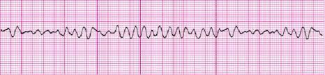
vfib
500
A Doppler tracing shows a late-peaking, dagger-shaped systolic velocity profile in a patient with a thickened basal septum. What condition is this pattern associated with?
Dynamic left ventricular outflow tract obstruction (LVOTO), commonly associated with hypertrophic obstructive cardiomyopathy (HOCM).