Ischemic Heart Disease
Measure This!
Function Function what's the function?
Picture This!
M-mode
What Time Is It?
100
These are indications of an old myocardial infarction via echocardiography and ECG.
What is a wall motion abnormality, thinning/scarring, increased echogenicity and Q waves on the ECG?
100
The type of 2D measurement used for the Left Ventricular Outflow Track
What is leading edge to leading edge?
100
Ejection fraction < 30%
What is severely reduced?
100
These two vessels (name the vessels and corresponding letter) feed.tje coronary arteries, supplying the heart with oxygenated blood.
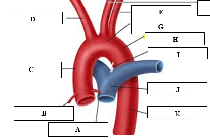
What are the coronary sinus's, letters A and B?
100
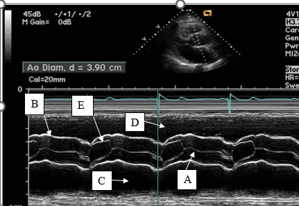
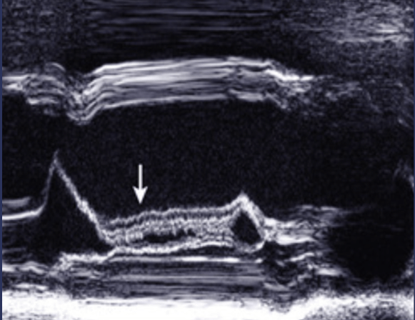
This line represents aortic valve closure on m-mode.
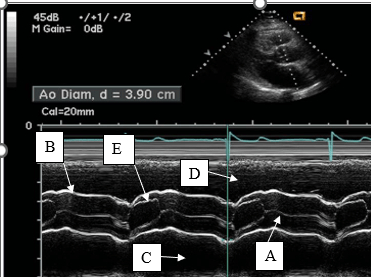
What is A?
100
This is the ECG timing of the left ventricular outflow track measurement.
What is early to mid-systole?
200
A small pericardial effusion 1-12 weeks after a myocardial infarction.
What is Dressler's syndrome?
200
The wall normal thickness of the posterior wall in diastole?
What is 0.6-1.1 cm?
200
This is one of the most important quantitative parameters of right ventricular systolic function and how it is obtained?
What is the measure of pulmonary artery pressure using the peak velocity TV regurgitant and the respiratory collapse and size of the inferior vena cava seen from the subcostal window?
200
This vessel supplies the anterior wall and the apex with blood. (Vessel name and corresponding letter)
What is the left anterior descending (LAD), letter A?
200
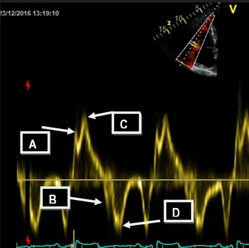
Letters A and C represent these waveforms.
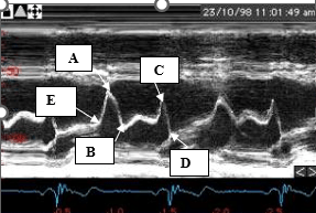
What is a- rapid or early filling and c - atrial systole or atrial kick?
200
The intraventricular septum and the posterior wall are measured at this time in the ECG.
What is end diastole?
300
The 5 steps in the sequence of events in order of myocardial ischemia and when it can be detected by echocardiography.
What is perfusion abnormalities, diastolic dysfunction, systolic dysfunction, ECG changes, angina and at the point of systolic dysfunction?
300
LVIDs is performed at this time
What is end-systole?
What is when the LV volume is the smallest?
The frame after AV closure?
300
The heart contractions in this direction at the apex and this direction at the base?
What is counterclockwise at the apex and clockwise at the base? From base to apex
300
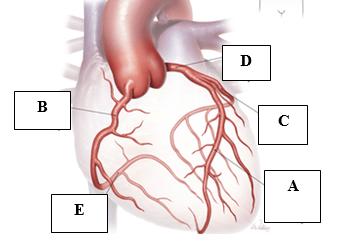
This layer of the heart muscle produces the pericardial fluid, which acts as a lubricant between the inner and outer layers.
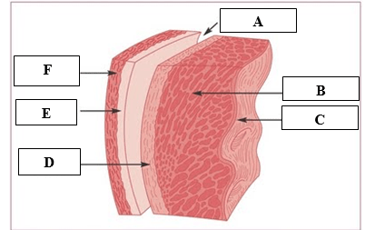
What is the visceral pericardium or the epicardium, D?
300
The name and description of how this measurement is performed.
What is Tricuspid Annulus Systolic Excursion - TAPSE, m-mode (zoomed preferred) through TV annulus in a right focused view in the apical 4 chamber, measure systolic waveform excursion in a vertical line?
Normal 17 mm or greater.
300
This is the ECG timing of the left atrial measurement.
What is end systole?
400
Occlusion of this artery will cause myocardial infarction of these walls.
What is anterolateral (lateral) and inferolateral?
400
These are normal ranges for an aortic root measurement
What is 2.1-3.5 cm?
400
These are 4 qualitative assessments of the left ventricle.
What are regional and global wall motion, shape of the left ventricle, wall thickness of the left ventricle and chamber size
400
This is the point AND the name of the measuremnt that is measured for right sided Tissue Doppler Imaging (TDI), a measurement of the maximum velocity of the tricuspid lateral annulus during systole.
What is S' (S prime) and pont C?
400
Letter E represents this aortic valve cusp and it's position during systole.
What is the right coronary cusp in the open position?
400
These valve actions begin and end isovolumetric relaxation.
What is aortic valve closure and mitral valve opens?
500
The walls represented by A and B
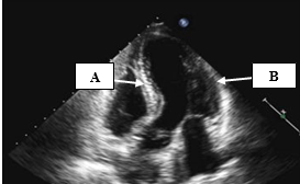
What is A - Antero septum and B- anterolateral?
500
This is from where and when you measure LA volume and it's normal value
What is AP2 and AP4, end-systole and 34 ml/m2?
500
This septal motion pattern in PSAX, a qualitative assessment of right ventricular systolic function, is indicative of a pressure volume overload pattern.
What is systolic and diastolic septal flattening?
500
The ejection period is represented by these to points on the Wigger's Diagram.
DAILY DOUBLE : What two valves are opening and closing at these same points?
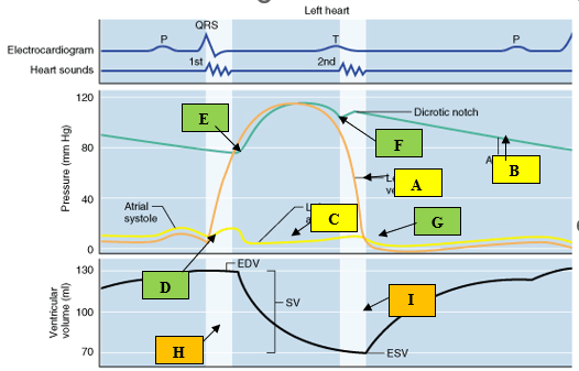
What is E - F?
DD: What are the Aortic and Pulmonic valves?
500
The name for this indirect sign of aortic valve regurgitation on M-mode.
What is anterior mitral valve leaflet diastolic fluttering?
500
The valve action that begins and ends isovolumetric contraction.
What is mitral valve closure and aortic valve opening?