Basic Anatomical Terminology
Organization of the Body Wall
Skeletal System
Cardiovascular System
Lymphatic System
100
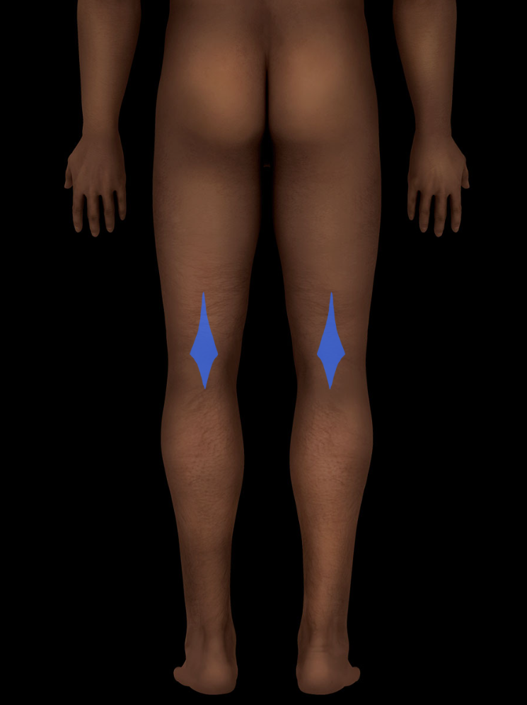
What is this region called?
Popliteal fossa
100
What is the dermis composed of?
Collagen and elastic fibers
100
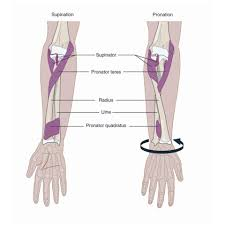
Describe the position of the ulna and radius when the antebrachium is supinated vs pronated
Supinated: ulna and radius are parallel to one another
Pronated: ulna and radius intersect/cross over one another
100
What are some differences between arteries and veins?
Arteries: take blood AWAY from heart, thicker than veins, operate under HIGH pressure
Veins: bring blood TOWARD heart, thin, operate under low pressure
100
Distinguish the functions of the following components of the lymphatic system:
1. Lymphatic vessels
2. Lymph nodes
1. Lymphatic vessels: collect lymph that leaks from capillaries and transport it back to circulatory system
2. Lymph nodes: Produce new lymphocytes (B lymphocytes, T lymphocytes, macrophages), filter lymph, and initiate immune response when necessary
200
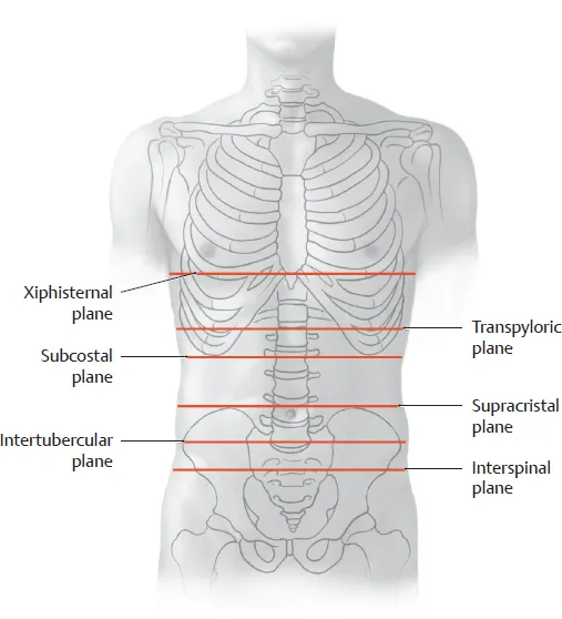
What is the subcostal plane?
It is a transverse plane that passes through the inferior borders of the costal cartilages of right and left tenth ribs
200
List the layers of the body wall in order from most superficial to the deepest.
1. Epidermis (skin)
2. Dermis
3. Superficial/subcutaneous fascia
4. Deep/internal fascia
5. Serous membrane
200
Distinguish intrinsic ligaments of a synovial joint from extrinsic ligaments of a synovial joint.
Intrinsic ligaments are a part of, or WITHIN, the fibrous joint capsule (between fibrous capsule and the synovial membrane). Extrinsic ligaments are OUTSIDE and INDEPENDENT of the fibrous capsule
200
How do arterial anastomoses and collateral circulation routes function in normal and pathological states?
Normal: typically, blood will flow between the anastomoses uninterrupted.
Pathological: Arterial anastomoses provide for the continued distribution of blood via collateral circulation routes to distal tissues should an artery become obstructed due to pathology or surgical intervention
200
Where in the body can lymph nodes be palpated?
1. Axilla
2. Cervical region (along course of internal jugular vein)
3. Pericranial region (base of the head)
4. Inguinal region
300
What is the difference between contralateral and ipsilateral? Give an example of each.
Contralateral: structures or actions that occur on opposite sides of the body
Ex: right arm, left arm
Ipsilateral: structures or actions that occur on the same side of the body
Ex: right arm, right leg
300
What are three ways that the superficial facia helps to regulate the internal temperature of the body?
1) Cutaneous nerves that innervate the arrector pili. These muscles contract to erect body hair which will trap heat and compress the sebaceous glands (releases oil)
2) Superficial blood vessels. Arterioles dilate to increase blood flow to the surface of the skin and cool it off (red flush of skin). Arterioles constrict to decrease blood flow and conserve body heat (skin appears blueish).
3) Sweat glands that release sweat to the surface of the skin that evaporates and helps to cool the body off.
300
List an example of each of these joints.
1. Fibrous
2. Cartilaginous
3. Synovial
1. Sutures of skull, ligamentum flavum of the vertebral column, interosseous membrane between the tibia and fibula
2. Intervertebral discs, pubic symphysis, epiphyseal plates
3. Most joints... hip joint, ball-in-socket joint of the shoulder, etc.
300
What is the difference between systemic circulation and pulmonary circulation?
In systemic circulation, oxygen-RICH blood is taken away from the heart via the aorta toward the body tissues to deliver that oxygen to them. The blood then returns back to the heart via the SVC and IVC and to the right atrium. (Oxygen rich blood from heart to body tissues and back)
In pulmonary circulation, oxygen-POOR blood is taken away from the heart via the pulmonary arteries toward the lungs to be reoxygenated. It then returns to the heart via pulmonary veins and enters the heart through the left atrium. (Oxygen-poor blood away from heart to lungs and back)
300
Where in the vascular system is lymph returned to the blood?
(200 bonus points: where does the lymph come from to enter these ducts?)
Lymph returns via the venous angle (place where the internal jugular vein and subclavian vein meet to form the brachiocephalic vein)
Thoracic duct receives lymph from everywhere except the right upper limb, thorax, neck, and head and drains into the left venous angle
- Right lymphatic duct receives lymph from the right upper limb, thorax, neck, and head and drains into the right venous angle
400
What are the Latin terms for these common anatomical regions?
1) Arm
2) Head
3) Leg
4) Foot
5) Ankle
6) Hand
7) Forearm
8) Thigh
1) Brachium
2) Cranium
3) Crus
4) Pes
5) Tarsus
6) Manus
7) Antebrachium
8) Femorus
400
Define bursae and describe their function.
They are closed sacs of serous membrane that secrete fluid to lubricate internal surfaces to reduce wear and tear between joints.
In their normal state, they are collapsed (potential space). When fluid accumulates or a wall is interrupted (pathological state), they will fill with serous fluid and become a real space.
400
Describe what is happening at the listed joints below in this picture
1. Right hip
2. Left elbow
3. Right shoulder
1. Flexion
2. Flexion
3. Extension
400
What is the clinical importance of end arteries?
Either do not have anastomoses with other arteries (true end arteries) or their anastomoses are ineffectual (functional end arteries). Therefore, if these vessels become blocked from a clot or surgery, the blood flow is completely restricted.
400
Explain the significance of left supraclavicular lymph nodes (signal lymph nodes) with respect to the metastasis of abdominopelvic organ carcinomas
Left supraclavicular nodes (signal lymph nodes) take part in filtering the lymph passing through the thoracic duct. Because the thoracic duct carries lymph from the abdominopelvic organs, enlarged supraclavicular nodes on the left can be the first sign of cancer of organs and tissues located deep within the thorax, abdomen, or pelvis.
500
Describe anatomical position (there are eight components).
1. Body is erect
2. Head facing forward
3. Palms face forward
4. Fingers extended
5. Upper limbs and hands lie parallel to the body
6. Thumbs point laterally
7. Feet are parallel
8. Toes facing forward
500
What are the three types of deep fascia and what are their roles?
1. Investing fascia: Covers (invests) muscles of neurovascular bundles to allow for smooth movements
2. Intermuscular septum: Thick sheets that attach to bone and separate muscles into compartments
3. Retinaculum: Thickened sheet that holds tendon into place
500
What is a joint/articulation?
Name the three types, including their composition and range of mobility
A joint is the location where two bones meet.
1. Fibrous joint: bones are PHYSICALLY connected via fibrous connective tissue. Little to no movement
2. Cartilaginous joint: bones are PHYSICALLY connected via cartilage. Little to no movement, but more movement than fibrous joints
3. Synovial joint: bones are not physically connected. They are covered in articular cartilage and held together by surrounding muscles and tissues. Highly movable. The ends of bones in these joints also contain a fibrous capsule that is composed of fibrous membrane and encloses synovial fluid.
500
Describe the order that a red blood cell would pass through starting with the SVC and IVC and ending in the ascending aorta.
SVC/IVC-> Right atrium-> tricuspid valve-> right ventricle-> pulmonary valve-> pulmonary trunk and arteries-> lungs-> pulmonary veins-> left atrium-> bicuspid valve-> left ventricle-> aortic valve-> ascending aorta
After this, the blood cell would either travel through the aortic trunk to the superior portions of the body or continue to the descending aorta to travel to the more inferior portions of the body. After traveling to the body tissues, the red blood cell would enter the veins and travel back to the SVC or IVC>.
500
Explain the significance of the umbilicus in determining the path of flow and the superficial lymph nodes encountered by lymph originating from the superficial fascia of the thoracoabdominal body wall and buttocks.
The lymph from the lower trunks as well as the lower extremities ends up congregating in the cisterna chyli, which is just below the umbilicus. The lymphatic vessels, which accompany subcutaneous veins, that are superior to the umbilicus drain to the axillary lymph nodes. Those that are inferior to the umbilicus drain to the superficial inguinal nodes.