Methods & Cells
Blood & Lymphatics
Epithelium & Connective Tissue
Muscle
Nervous Tissue
100
The indicated pale-staining region contains loosely packed, transcriptionally active DNA. What structure is being identified?
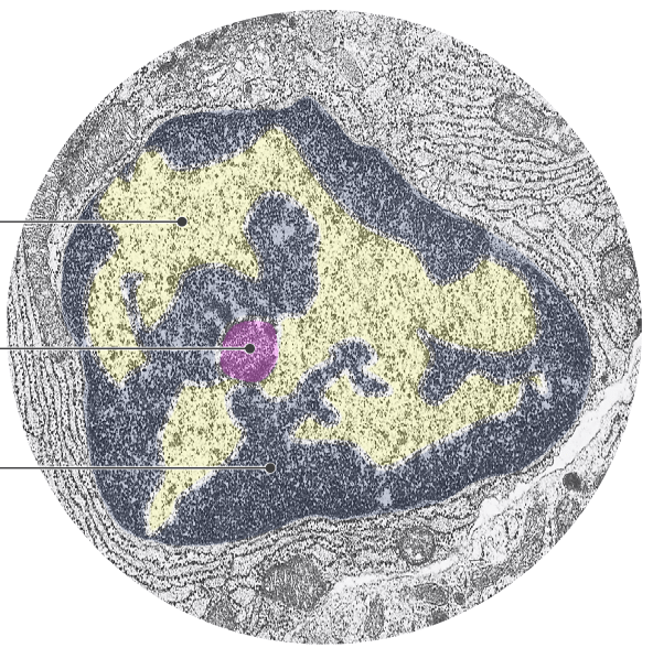
A. Heterochromatin
B. Euchromatin
C. Nucleolus
D. Golgi apparatus
E. Smooth ER
Answer: B — Euchromatin
Explanation: Abbott distinguishes euchromatin as loosely packed, transcriptionally active, and light staining. Heterochromatin is tightly coiled, inactive, and dark staining
100
Identify the indicated leukocyte.
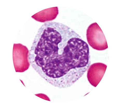
A. Lymphocyte
B. Monocyte
C. Neutrophil
D. Basophil
E. Eosinophil
Answer: B — Monocyte
Explanation: Abbott identifies the monocyte by its kidney-shaped nucleus. It is the largest WBC and differentiates into macrophages in tissues.
100
Which epithelial tissue consists of a single layer of flat cells specialized for diffusion or filtration?
A. Simple cuboidal
B. Simple columnar
C. Simple squamous
D. Stratified squamous
E. Transitional
Answer: C — Simple squamous
Explanation: Abbott defines simple squamous epithelium as a single layer of flat cells with a function of diffusion or filtration
100
Identify the muscle tissue shown.
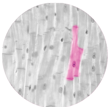
A. Skeletal muscle
B. Cardiac muscle
C. Smooth muscle
D. Dense regular CT
E. Dense irregular CT
Answer: B — Cardiac muscle
Explanation: Abbott describes cardiac muscle as having central nuclei, branched fibers, visible striations, and intercalated discs with gap junctions.
100
Which CNS glial cell acts as an immune cell and phagocytoses debris and pathogens?
A. Astrocyte
B. Ependymal cell
C. Microglia
D. Schwann cell
E. Satellite cell
Answer: C — Microglia
Explanation: Abbott describes microglia as small CNS cells with elongated dense nuclei that function as immune cells and phagocytose debris and pathogens.
200
A histologic specimen contains a structure that stains blue with H&E. Which property best explains this finding?
A. It binds the acidic dye eosin
B. It contains structures stained by hematoxylin
C. It must contain abundant lipid
D. It has a net neutral charge
E. It contains no DNA or RNA
Answer: B — It contains structures stained by hematoxylin
Explanation: Hematoxylin is a basic (+) dye that produces blue/basophilic staining and stains DNA, RNA, ribosomes, rER, and nuclei. Eosin is acidic and stains structures pink.
200
Which lymphatic organ is both a site of lymphocyte formation and the site of B-cell maturation?
A. Thymus
B. Spleen
C. Bone marrow
D. Lymph node
E. MALT
Answer: C — Bone marrow
Explanation: Abbott lists bone marrow as the site of formation of B and T cells and maturation of B cells. T cells mature in the thymus.
200
The apical modification shown consists of short, finger-like projections containing actin microfilaments. What is its primary function?
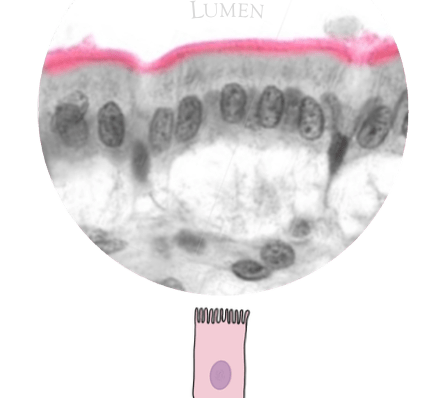
A. Movement of mucus
B. Increase surface area
C. Prevent paracellular transport
D. Attach epithelium to basement membrane
E. Connect intermediate filaments between cells
Answer: B — Increase surface area
Explanation: Microvilli are short, finger-like projections composed of actin microfilaments that increase surface area, such as for absorption. Cilia are long, hair-like microtubule-based projections involved in movement.
200
Which structure is an invagination of the skeletal muscle sarcolemma that allows rapid transmission of action potentials?
A. Myofibril
B. Terminal cisterna
C. T-tubule
D. Sarcoplasmic reticulum
E. Dense body
Answer: C — T-tubule
Explanation: T-tubules are invaginations of the sarcolemma that rapidly transmit action potentials to ensure synchronous contraction of myofibrils.
200
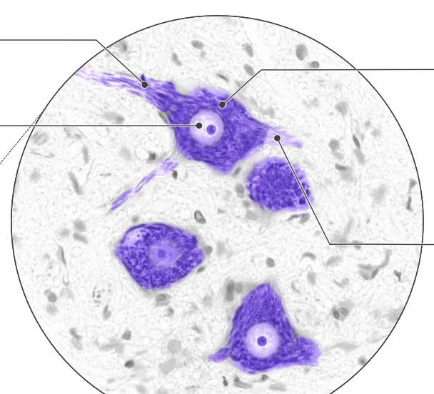
The large multipolar neuron shown contains dark clumps of rough ER in its cell body and dendrites. What are these structures called?
A. Dense bodies
B. Nissl bodies
C. Myelin sheaths
D. Satellite cells
E. Intercalated discs
Answer: B — Nissl bodies
Explanation: Abbott describes Nissl bodies as clumps of rER in the cytoplasm of somatic motor neurons. They are present in the cell body and dendrites, but not the axon.
300
A pathologist wants to visualize carbohydrate-rich mucus that appears colorless with routine H&E. Which stain from the review should be used?
A. Hematoxylin
B. Eosin
C. PAS
D. Nissl stain
E. No special stain is required
Answer: C — PAS
Explanation: Carbohydrates have a net neutral charge and appear colorless with H&E. Abbott identifies Periodic Acid-Schiff (PAS) as a magenta-colored stain used to visualize carbohydrates
300
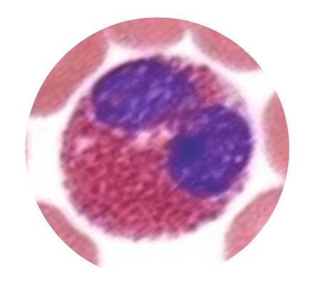
The indicated leukocyte contains a bilobed nucleus and eosinophilic granules. What is its function according to Abbott's review?
A. Phagocytosis of bacteria
B. Release of heparin and histamine
C. Release of cytotoxic granules against helminths
D. Differentiation into macrophages
E. Initiation of immune responses
Answer: C — Release of cytotoxic granules against helminths
Explanation: The cell is an eosinophil. Abbott identifies eosinophils by their bilobed nucleus and eosinophilic granules and associates them with cytotoxic granule release against helminths
300
Damage selectively destroys the junction located near the apical surface that creates an impermeable barrier between epithelial cells. Which process would be most directly affected?
A. Anchoring epithelium to basement membrane
B. Preventing paracellular transport
C. Linking intermediate filaments for structural strength
D. Connecting actin filaments for dynamic adhesion
E. Allowing passage of small molecules between cells
Answer: B — Preventing paracellular transport
Explanation: This describes the tight junction (zonula occludens). It encircles the cell near the apical surface and prevents paracellular movement of ions, water, and solutes.
300
Which structure in smooth muscle serves as an anchoring point for actin filaments and is functionally analogous to a Z-line?
A. Intercalated disc
B. Dense body
C. Terminal cisterna
D. T-tubule
E. H-band
Answer: B — Dense body
Explanation: Abbott states that smooth-muscle dense bodies anchor actin filaments and are functionally analogous to Z-lines
300
Which pairing correctly matches a myelinating glial cell with its location and capacity?
A. Schwann cell — CNS — many axons
B. Schwann cell — PNS — single axon
C. Oligodendrocyte — PNS — single axon
D. Oligodendrocyte — PNS — many axons
E. Satellite cell — CNS — many axons
Answer: B — Schwann cell — PNS — single axon
Explanation: Abbott's mnemonic is COPS: CNS contains Oligodendrocytes, PNS contains Schwann cells. A Schwann cell myelinates a single axon, whereas an oligodendrocyte can myelinate many axons.
400
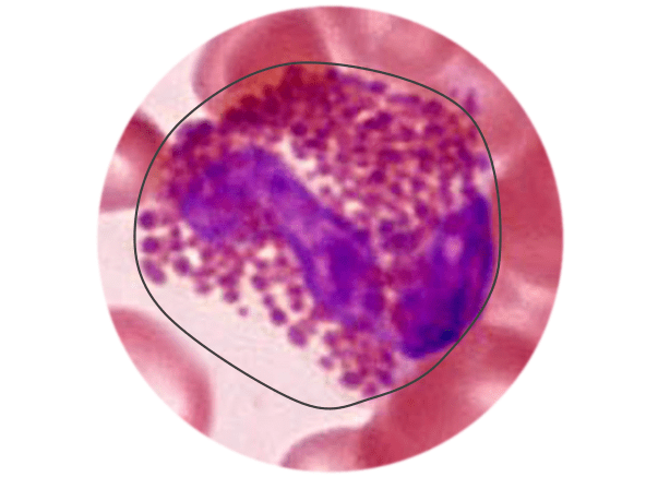
The indicated cell contains an abundance of cytoplasmic secretory granules. Which process is this cell most consistent with?
A. Constitutive secretion
B. Regulated secretion
C. Aerobic respiration
D. Protein synthesis by rER
E. Lipid detoxification
Answer: B — Regulated secretion
Explanation: Regulated secretion is signal-dependent release of stored vesicles, so these cells contain abundant secretory granules. Constitutive secretion occurs continuously and has minimal or no secretory granules
400
A patient has decreased serum albumin. According to the review, which change promotes accumulation of fluid in the interstitial space?
A. Increased oncotic pressure
B. Decreased oncotic pressure
C. Decreased hydrostatic pressure
D. Increased movement of water into capillaries
E. Decreased blood volume increasing hydrostatic pressure
Answer: B — Decreased oncotic pressure
Explanation: Abbott states that less albumin → lower osmotic/oncotic pressure → more fluid remains in the interstitial space → edema
400
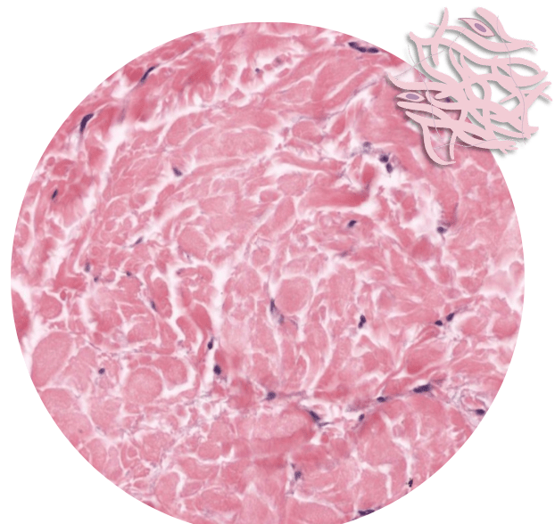
The tissue shown contains irregularly arranged fibers and relatively few fibroblasts. What mechanical property is characteristic of this tissue?
A. Rapid diffusion
B. High unidirectional stress resistance
C. Ability to withstand tension in multiple directions
D. Permits distension
E. Facilitates movement of mucus
Answer: C — Ability to withstand tension in multiple directions
Explanation: This is dense irregular connective tissue. Its irregular fiber arrangement allows it to withstand tension from multiple directions. Abbott lists the dermis of the skin as its location
400
A muscle specimen contains mononucleated cells with elongated corkscrew nuclei and no visible striations. Which additional feature should be associated with this tissue according to the review?
A. Peripheral nuclei and triads
B. Intercalated discs only
C. Dense bodies and gap junctions
D. Sarcomeres with prominent A-bands
E. Multiple peripheral nuclei
Answer: C — Dense bodies and gap junctions
Explanation: The morphology identifies smooth muscle. Abbott describes it as mononucleated with elongated/corkscrew nuclei, lacking striations and sarcomeres, and containing dense bodies and gap junctions
400
The small flattened cells forming a continuous ring around the large neuronal cell body shown have which function?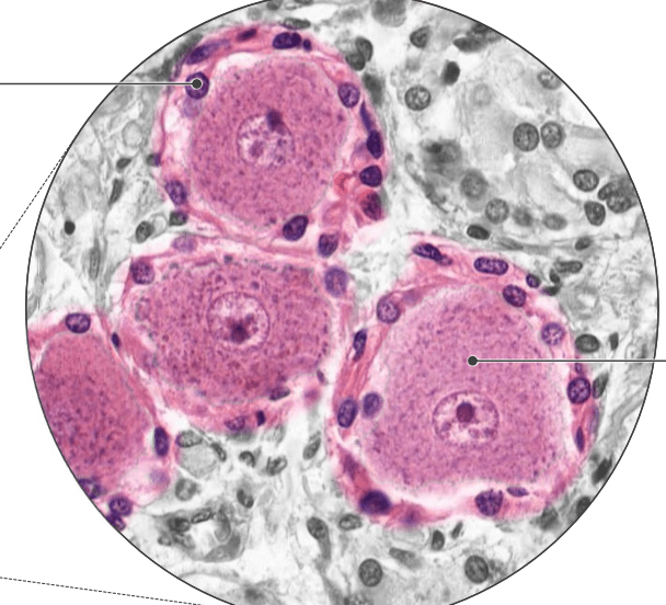
A. Produce and maintain CSF
B. Myelinate many CNS axons
C. Provide structural and metabolic support
D. Phagocytose CNS pathogens
E. Form the blood-brain barrier
Answer: C — Provide structural and metabolic support
Explanation: These are satellite cells surrounding the sensory pseudounipolar neurons of the DRG. Abbott states that they form a continuous ring around neuronal cell bodies and provide structural and metabolic support, similar to CNS astrocytes.
500
A cell has intensely basophilic cytoplasm because it contains abundant organelles responsible for protein synthesis. Which organelle most directly accounts for this appearance?
A. Smooth ER
B. Golgi apparatus
C. Mitochondria
D. Rough ER
E. Lipid droplets
Answer: D — Rough ER
Explanation: Abbott states that rER is involved in protein synthesis and stains basophilic. In contrast, sER and Golgi do not stain with H&E, while mitochondria are eosinophilic
500
An aged erythrocyte enters splenic open circulation but is unable to squeeze through the narrow slits required to re-enter circulation. What happens to it?
A. It enters white pulp and becomes a lymphocyte
B. It remains in red pulp and is phagocytosed by macrophages
C. It enters a lymph node for antigen presentation
D. It differentiates into a reticulocyte
E. It returns directly to bone marrow
Answer: B — It remains in red pulp and is phagocytosed by macrophages
Explanation: In splenic red pulp, healthy RBCs squeeze through narrow slits and re-enter circulation. Aged or damaged RBCs become trapped and are phagocytosed by macrophages. Abbott identifies red pulp as 80% of the spleen and the site of aged/damaged RBC clearance.
500
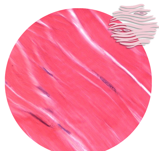
The tissue shown is subjected primarily to stress along a single axis. Which combination correctly describes it?
A. Loose fibers + abundant ground substance + beneath epithelia
B. Irregular fibers + multiple-direction resistance + dermis
C. Parallel packed fibers + unidirectional resistance + tendon/ligament
D. Flat cells + diffusion + filtration
E. Multiple epithelial layers + protection
Answer: C — Parallel packed fibers + unidirectional resistance + tendon/ligament
Explanation: Dense regular CT consists of packed, parallel rows of fibers with few fibroblasts. It provides high unidirectional stress resistance and is found in tendons and ligaments
500
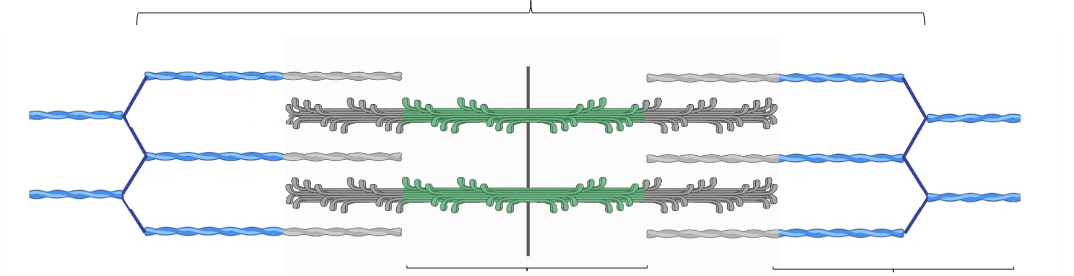
The muscle structure shown undergoes contraction. Which combination correctly describes the resulting changes?
A. H-band increases; I-band increases; A-band unchanged
B. H-band decreases; I-band decreases; A-band unchanged
C. H-band unchanged; I-band decreases; A-band decreases
D. H-band decreases; I-band unchanged; A-band decreases
E. H-band, I-band, and A-band all decrease
Answer: B — H-band ↓, I-band ↓, A-band unchanged
Explanation: Abbott gives the memory aid “During contraction, HI goes bye.” The H-band and I-band shorten, while the A-band remains the same length because it contains the entire length of myosin.
500
A peripheral nerve is examined from the level of one individual axon outward to the entire nerve. Which sequence correctly identifies the associated connective-tissue coverings?
A. Epineurium → perineurium → endoneurium
B. Endoneurium → epineurium → perineurium
C. Perineurium → endoneurium → epineurium
D. Endoneurium → perineurium → epineurium
E. Perimysium → endomysium → epimysium
Answer: D — Endoneurium → perineurium → epineurium
Explanation: Abbott organizes peripheral nerves as: individual nerve fiber/axon = endoneurium → nerve fascicle = perineurium → entire peripheral nerve = epineurium. He specifically notes that the organization parallels skeletal muscle but uses -neurium instead of -mysium