Division & Structures
Neurophysiology
Reflexes & Pathways
Support Cells & Barriers
Brain Regions & Functions
Sensory & Motor Organization
Neurons & Synapses
Plexuses & Peripheral Nerves
Integration & Communication
Diagrams
100
What are the two main divisions of the nervous system?
Central Nervous System (CNS) and Peripheral Nervous System (PNS)
100
What is an action potential and how is it generated?
Electrical signal that is generated in the neuron from ion exchange; Na⁺ (Sodium) in (depolarization), K⁺ (Potassium) out (repolarization)
100
What nerves are involved in the patellar reflex?
Quadriceps femoris, Femoral nerves, spinal segments L2, L3, L4
100
What is the function of Schwann cells?
Myelinate peripheral axons and assist in repair after injury
100
What is the function of the reticular formation?
Produces Melatonin. Controls consciousness, alertness, and sleep/wake cycles
100
What are somatic and visceral receptors?
Somatic: Monitor Skeletal Muscles
Visceral: internal organs
100
What is structural classification of neurons based on?
Number of processes that project from the cell body
100
What are the nerves of the brachial plexus?
Hint: there is 5
Axillary, radial, musculocutaneous, ULNAR, median nerve (medial & lateral cords)
100
What are the four higher functions of the nervous system?
Memory, learning, intelligence, emotion
100
Label I, O, T, & U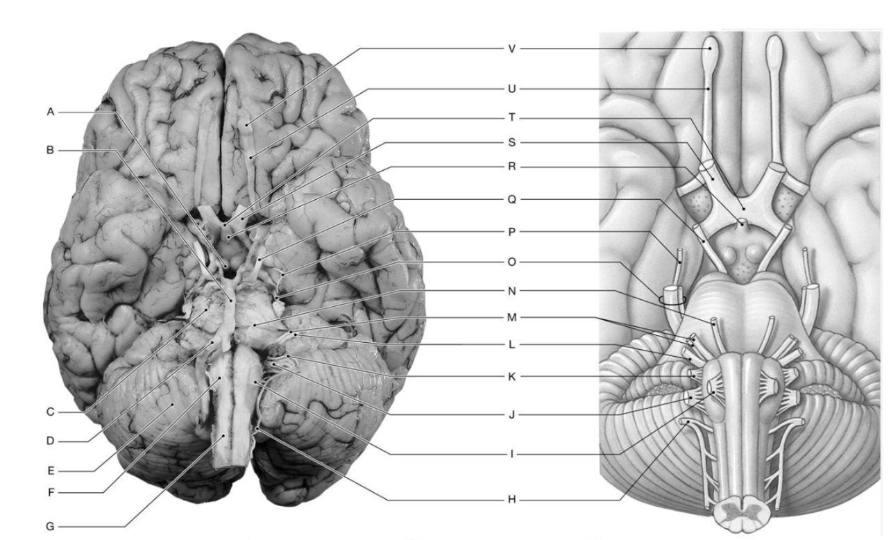
I: Hypoglossal Nerve (XII)
O: Trigeminal Nerve (V)
T: Optic Chiasm
U: Olfactory Tract
200
What is the function of the autonomic nervous system?
Regulates involuntary functions like cardiac/smooth muscle and glands
200
What is the lateral spinothalamic tract?
A pathway located in the spinal cord
200
What is a stretch reflex and its function?
Monosynaptic spinal reflex that checks the spinal cord. Maintains muscle tone and posture (body's upright position)
200
What is the function of oligodendrocytes?
Myelinate CNS axons and provide structural framework
200
What hormone is secreted by the pineal gland and what does it regulate?
Melatonin; regulates circadian rhythm
200
What are proprioceptors and what do they monitor?
Sense position of joints and muscles
200
What are the three neuron types (structural classification)?
Multipolar: most common, many dendrites (motor neurons)
Bipolar: one axon, one dendrite (rare-retina) - special sense
Unipolar: one process (sensory neurons)
200
What is the function of the femoral nerve in the patellar reflex?
Stimulates quadriceps to contract and extend knee
200
What role does the CNS play in integrating sensory and motor output?
Processes and coordinates responses
200
Label A, I, Q, & X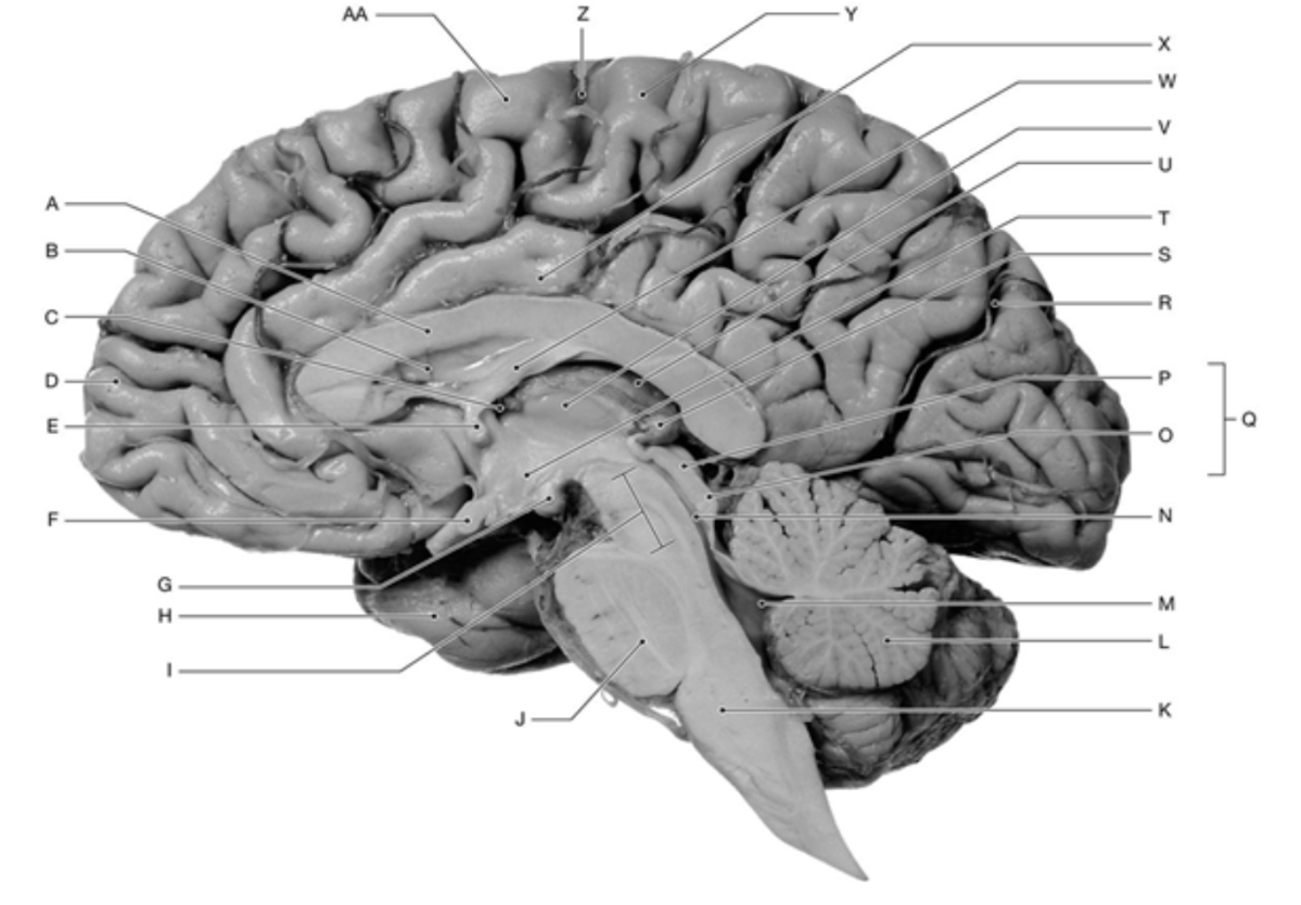
A: Corpus Callosum
I: Mesencephalon
Q: Corpora Quadrigemina
X: Cingulate Gyrus
300
What is the function of the central nervous system?
Integrates/Coordinates sensory and motor output. Also includes brain and spinal cord
300
What is the function of the axon hillock?
Site of action potential initiation
300
What are visceral (or autonomic) reflexes (long and short)?
Long: Go through CNS
Short: bypass CNS
300
What is the function of microglial cells?
Remove cell debris, waste, pathogens = phagocytosis. Also smallest glial cell.
300
What is the function of the choroid plexus?
Produces cerebrospinal fluid
300
What are effectors and how do they respond?
Muscles/glands or specialized cells that respond to motor/neuron signals
300
What is the difference between preganglionic and postganglionic neurons?
Preganglionic in CNS (First Neuron)
Postganglionic in ganglion to effector (Second Neuron)
300
What 3 cranial nerves control eye movement and What 2 cranial nerves make up the tongue?
CN III (oculomotor), IV (trochlear), VI (abducens)
Facial (VII) - 2/3 anterior of the tongue, Glossopharyngeal (IX) - 1/3 posterior of the tongue
300
Which nervous system division is associated with “rest and digest”?
Parasympathetic
300
Label K, L, M, & N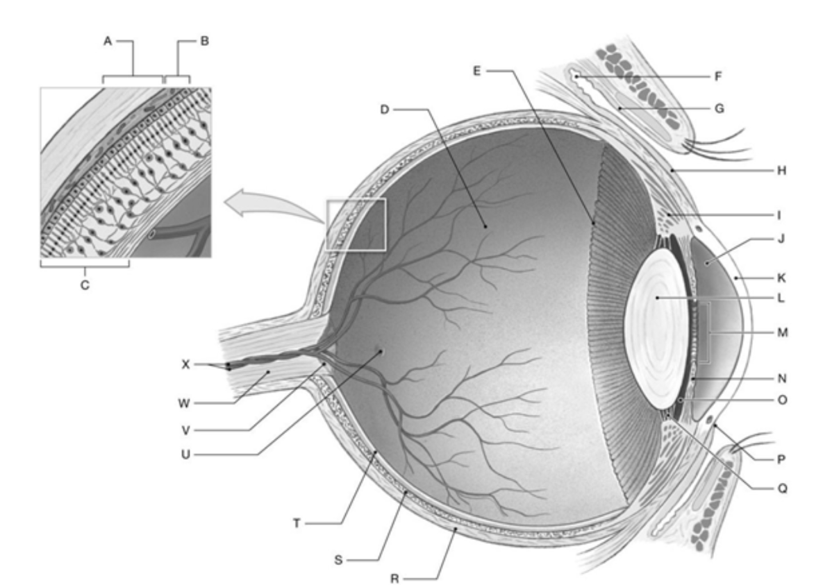
K: Cornea
L: Lens
M: Pupil
N: Iris
400
What is the origin of the sympathetic and parasympathetic nervous systems?
Sympathetic: thoracolumbar (T1-L2)
Parasympathetic: craniosacral (CN III, VII, IX, X; S2-S4)
400
What is excitability in neurons?
The ability to respond to stimuli and generate an action potential
400
What information is carried by the spinocerebellar tract?
Maintain Balance & Posture
400
What do astrocytes do?
Maintain BBB, regulate nutrients/ions, form scar tissue
400
What is the role of the inferior cerebellar peduncle?
Carries proprioceptive input to cerebellum
400
How are receptors classified?
By location
400
What is the most common neurotransmitter?
Acetylcholine (ACh)
400
What are perineurium and endoneurium?
Perineurium surrounds fascicles; around individual neurons
Endoneurium surrounds axons; layer surrounding a single axon
400
What is the purpose of the gray commissure in the spinal cord and how is gray matter organized in the spinal cord?
Connects left and right sides of gray matter in spinal cord. Allows axons to get from one side to another (cross over)
Sensory nuclei → Motor Nuclei → Posterior (dorsal) horn → lateral horn → anterior (ventral) horn → gray commissure
400
Label K, O, T, & W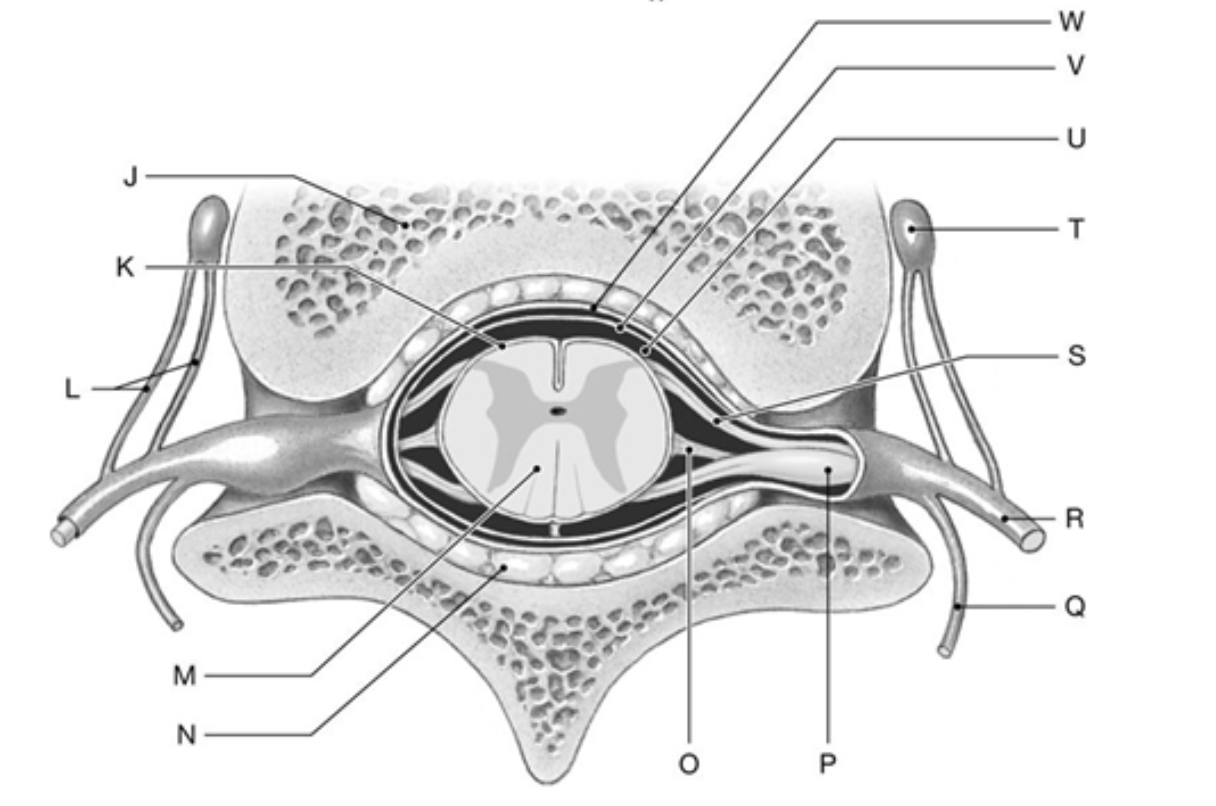
K: Pia Mater
O: Denticulate Ligament
T: Autonomic (Sympathetic) Ganglion
W: Dura Mater
500
What are the three meninges and their features?
Dura mater (tougher outermost layer), arachnoid mater (Web liked middle layer;CSF-filled), pia mater (innermost layer, attached to brain/spinal cord)
500
Name the 12 cranial nerves
Olfactory, Optic, Oculomotor, Trochlear, Trigeminal, Abducens, Facial, Vestibulocochlear, Glossopharyngeal, Vagus, Accessory, Hypoglossal
500
What information does the lateral spinothalamic tract carry?
Sensory information about pain and temperature
500
What is BBB and what are its functions?
Blood Brain Barrier. Physical/Physiological barrier, selective permeability of certain substances such as water, glucose, and ions
500
What is the function of the cerebellum in movement?
Coordinates voluntary movements and balance
500
What type of information is conducted through the sensory and motor tracts?
Sensory: to CNS
Motor: from CNS to effectors
500
What is an axon hillock?
Connects/joins axon to cell body
500
What is the function of fascicles in nerve structure?
Bundle axons into functional groups for organization
500
Which one is responsible for “fight or flight”?
Sympathetic
500
Label A, B, D, M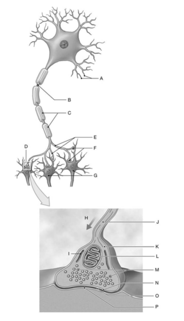
A: Dendrites
B: Axon
D: SYnapse
M: Synaptic Vesicles
600
What structures produce and absorb cerebrospinal fluid (CSF)?
Produced by choroid plexus; absorbed by arachnoid villi in superior sagittal sinus
600
What is myelin made of and how does it impact signal transmission?
Fatty insulation (phospholipids) that speeds up signal transmission (nerve impulse conduction)
600
Walking is an example of what kind of behavior?
Acquired reflex or learned motor behavior
600
What do ependymal cells do and where are they found?
Produce/circulate CSF
Line brain ventricles and central canal of spinal cord
600
What role does the brainstem play in consciousness?
Contains reticular activating system for wakefulness
600
What are the dorsal, ventral, and lateral horns associated with?
Dorsal: sensory
Ventral: motor
Lateral: autonomic
600
Which glial cell helps maintain the blood-brain barrier?
Astrocytes
600
What type of cells surround axons in the PNS?
Schwann cells
600
What is the function of interneurons in reflex pathways?
Connect sensory and motor neurons for processing
600
Label A, H, J, & Q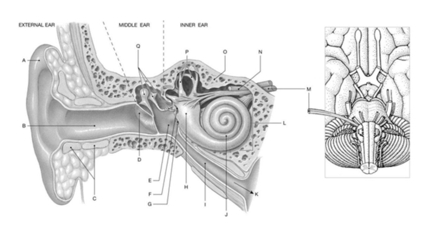
A: Auricle
H: Vestibule
J: Cochlea
Q: Auditory Ossicles