Skeletal Framework of the Head and Neck
Organization and Vascularity of the Head and Neck
Innervation of the Head and Neck
Superficial Face and Scalp
Superficial Neck
100
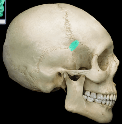
This H-shaped junction on the lateral aspect of the skull is where the frontal, parietal, sphenoid, and temporal bones meet and is a common site for clinical concern
What is the pterion?
100
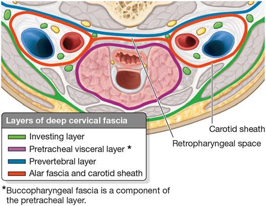
These are the three structures that are housed within the carotid sheath, formed by all three layers of the deep cervical fascia.
What are the common carotid artery, internal jugular vein, and the vagus nerve?
100
This cranial nerve carries somatic afferent fibers that provide sensory information from the face via three primary divisions: ophthalmic, maxillary, and mandibular
What is CN V (the trigeminal nerve)?
100
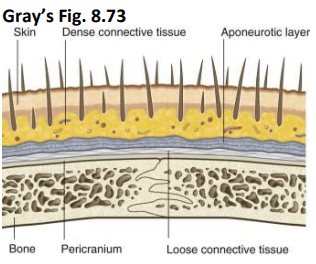
This "danger zone" of the scalp is the layer where blood and infectious material can collect and spread anteriorly to the eyelids
What is the loose connective tissue layer?
100
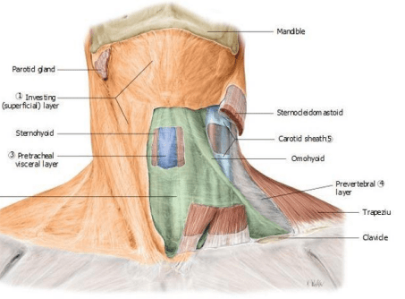
This specific layer of deep cervical fascia encloses the neck like a collar and splits to invest the sternocleidomastoid and trapezius muscles
What is the investing layer?
Bonus points: What are the three layers below the investing layer?
200
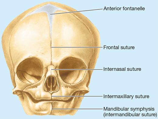
These large membranous intervals separate the flat bones of the neonatal calvaria, enabling the skull to change shape during birth
What are fontanelles?
200
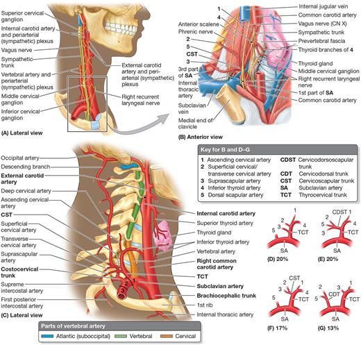
This landmark, located at the superior border of the thyroid cartilage, is where the common carotid artery divides into its internal and external branches
What is the carotid bifurcation?
Bonus points: What two important sensory structures are located here?
200
Formed by the ventral rami of C3-C5, this nerve is the most important branch of the cervical plexus and innervates the thoracic diaphragm
What is the phrenic nerve?
200
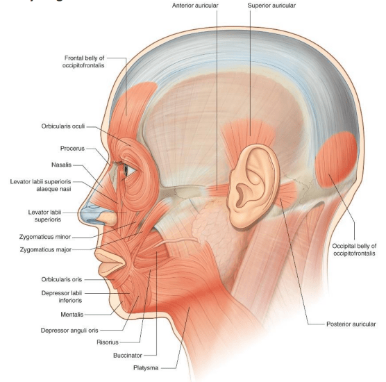
This sphincter muscle and its corresponding CN of the mouth is responsible for sealing the lips and preventing drooling.
What is the orbicularis oris (Motor Branch of CN VII)
200
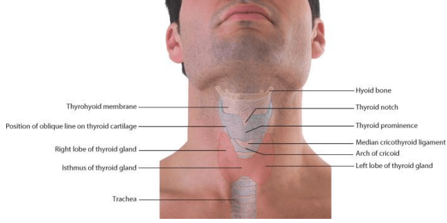
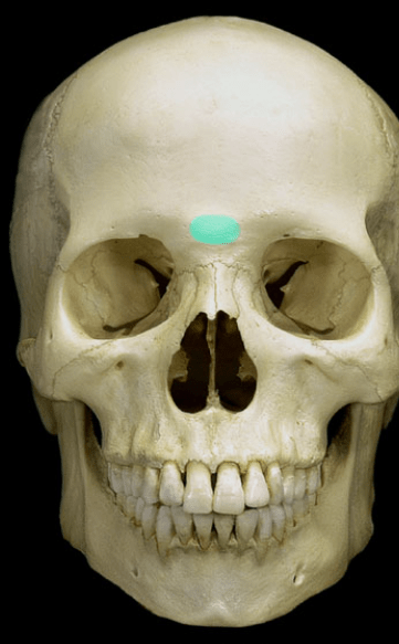
This ligament holds the hyoid in place superiorly.
What is the stylohyoid ligament?
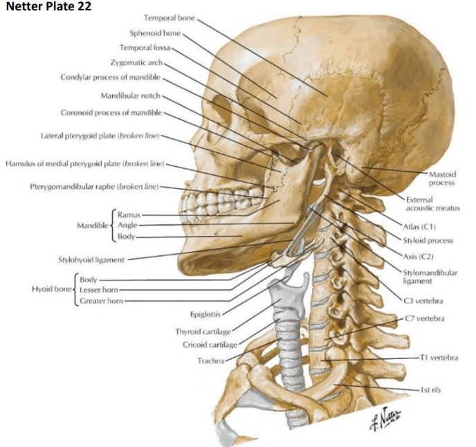
300
This midline landmark on the frontal bone is located between the supraciliary arches and is easily palpable
What is the glabella?
300
This branch of the subclavian artery enters at the C6 vertebra and exits at the C1 vertebra to provide blood supply to the brain.
What is the vertebral artery?
300
This nerve exits the jugular foramen to innervate the sternocleidomastoid and trapezius muscles; it is uniquely susceptible to injury in the posterior triangle during lymph node biopsies
What is CN XI (the spinal accessory nerve)?
300
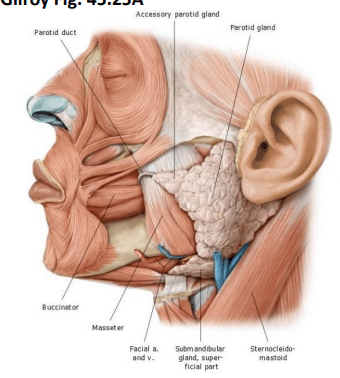
This salivary gland's duct emerges from its anterior surface, crosses the masseter muscle, and pierces the buccinator muscle to open into the oral cavity
What is the parotid gland?
Bonus Points: What nerve goes through the parotid gland, and what nerve innervates it?
300
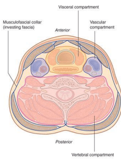
This cervical fascia layer forms the anterior boundary of the retropharyngeal space, a clinically significant route for the spread of infection
What is the buccopharyngeal fascia (part of pretracheal fascia)?
400
This clinical condition occurs when the cranial sutures fuse prematurely, potentially resulting in a wedge-shaped head known as scaphocephaly if it involves the sagittal suture
What is craniosynostosis?
Bonus Points: What will the shape of the skull be during sagittal and coronal suture craniosynostosis?
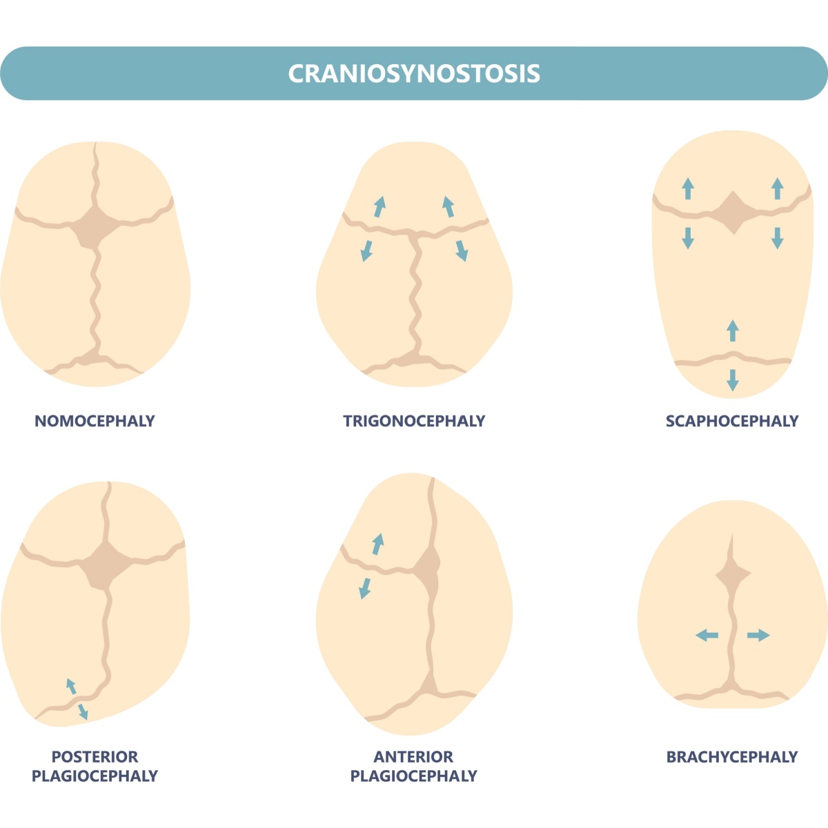
400
Located posterior to the oral cavity and the sectioned tongue, this region of the pharynx is a shared passageway for both air and ingested food
What is the oropharynx?
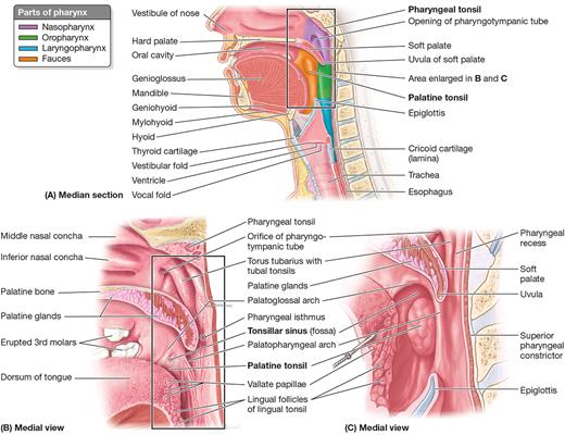
400
These sympathetic structures are formed by the fusion of paravertebral ganglia; for instance, C1-C4 fusion creates the largest of these in the neck
What are cervical sympathetic ganglia?
Bonus Points: What forms the stellate ganglia?
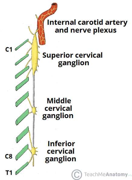
400
Unlike muscles of mastication, all muscles of facial expression receive their motor innervation from this specific cranial nerve
What is CN VII (the facial nerve)?
400
This space between the sternal and clavicular heads of the sternocleidomastoid muscle provides access to the internal jugular vein for catheterization
What is the lesser supraclavicular fossa?
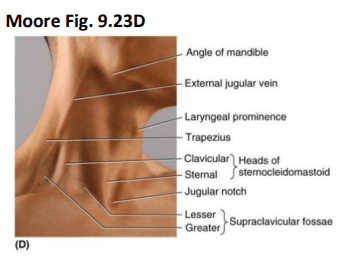
500
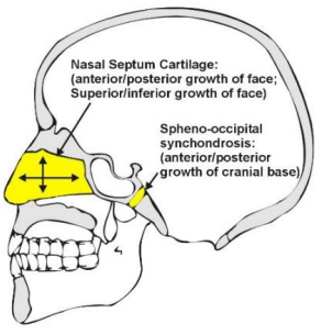
This major site of growth in the neurocranium is located at the base of the skull between the sphenoid and occipital bones
*Very hard question*
What is the spheno-occipital synchondrosis?
500
This potential anatomical variant, found in approximately 10% of individuals, must be considered during midline procedures of the trachea due to its origin from the brachiocephalic trunk
What is the thyroid ima artery?

500
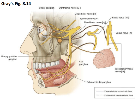
These four cranial ganglia and their corresponding cranial nerves host the postganglionic parasympathetic neuronal cell bodies that synapse for oculomotor, salivary gland, lacrimal gland, taste, motor, and general sensation of the face and tongue.
What are the ciliary ganglion (CN III), Otic ganglion (CN VII), Submandibular ganglion (CN IX), and Pterygopalatine ganglion (VII)?
500
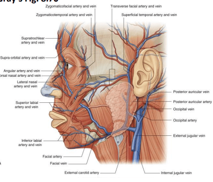
This tortuous artery supplies the face and can be palpated where it crosses the inferior border of the mandible anterior to the masseter muscle
What is the Facial artery
500
This structure is the only cartilage of the airway that completely encircles it, marking the transition between the larynx and trachea
What is the cricoid cartilage?