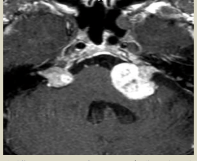
Who's that structure?!
It's classified...
Young otologists
Did you say history or histology?
MRI's are hard
100
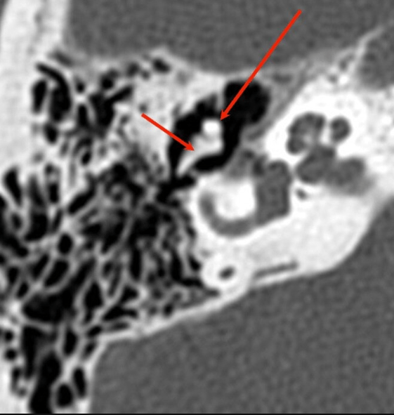
What is the head of malleus and incus body?
100
A clinical scale based on radiographic features used to determine whether a patient is a good candidate for congenital aural atresia repair
What is the Jahrsdoerfer grading scale?
100
3 yo M with history of multiple AOM. He now reports some hearing loss in the R ear. What is the next step in management?
Pneumatic Otoscopy and Tympanogram.
If OME is present, can watch for 3 mo from date of effusion onset or dx date. If 4 yoa and older then can recommend BMT and tube, adenoidectomy or both. If younger than 4 just tubes unless distinct indication exists other than OME.
100
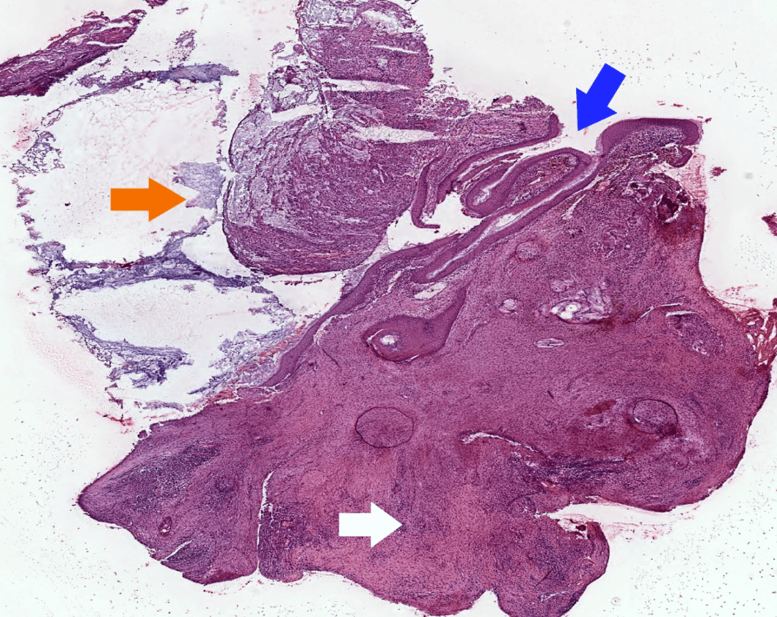 35 yo M who has a history of RAOM, ETD s/p BMT with tubes 10 years ago. Now presenting with constant drainage and fullness in the ear.
What is a cholesteatoma?
Requires 3 things on histology for dx:
1.Keratinous material
2. Keratinizing stratified squamous epithelium
3. Inflamed fibrous connective tissue stroma
100
 What weighted MRI is this?
What is a T2 weighted MRI?
(T2: H2O lights up!)
200
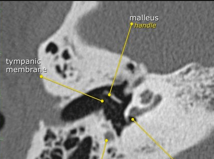 Name the two structures.
What is the facial nerve (mastoid segment) and round window?
200
Please name the 9 parameters for the Jahrsdoerfer grading scale and what parameter gets 2 points.
- Parameters (9)
- Stapes present
- Oval window
- Round window
- Malleoincudal joint
- Incudostapedial joint
- Middle ear space
- Course of facial nerve
- Mastoid air cells
- Appearance of external ear
- What is the stapes?
200
Name the 3 most common causes of AOM.
What are Strep Pneumo, Haemophilus influenzae, Moraxella Catarrhalis
200
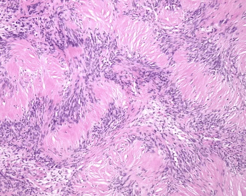 This patient most likely has this genetic disorder, that is autosomal dominant.
What is NF2?
 Spindle cell tumor (usually biphasic) and composed of compact hypercellular areas (Antoni A tissue) showing nuclear palisading (Verocay bodies) and loose microcystic areas (Antoni B tissue) with collection of lipid laden histiocytes and thick walled hyalinized blood vessels
200
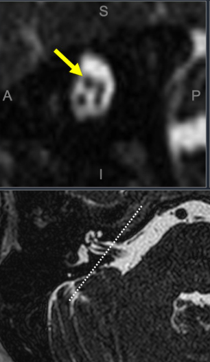 Yellow Arrow
What is the Facial Nerve?

300
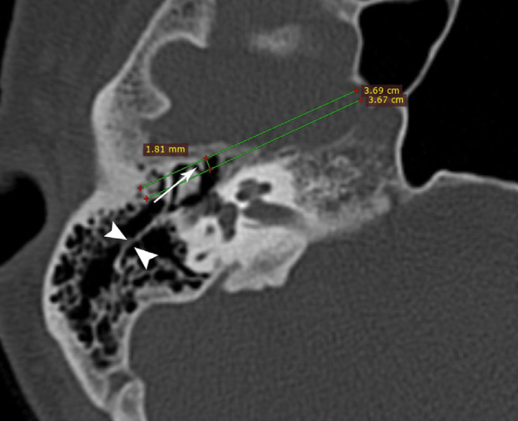
coronally oriented bony process in the middle ear cavity of the temporal bone that projects from the tegmen tympani, separating the anterior epitympanic recess from the posterior epitympanum.
What is the cog?
300
This is used to predict the likelihood of residual disease in the context of congenital cholesteatoma
What is the Potsic Staging System?
- Stage I: Disease confined to a single quadrant of the middle ear.
- Stage II: Cholesteatoma in multiple quadrants, but with no ossicular involvement and no mastoid extension.
- Stage III: Ossicular involvement without mastoid extension.
- Stage IV: Mastoid disease.
300
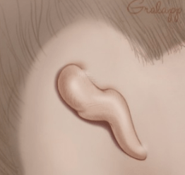 This is the most common type of microtia in which the only feature remaining is a small peanut-shaped remnant ear lobe
What is a grade 3
Grade 1: The ear is smaller than normal but the key features of the normal ear are present, though they may have minor alterations in shape or form.
Grade 2: Some of the features of the ear are missing, though usually there is still a lobule and a remnant of helix and antihelix. Grade 2 microtia is sometimes called “conchal type microtia.” The ear canals may be present, though frequently are very narrow (canal stenosis).
Grade 3: This is the most common type of microtia, in which the only feature remaining is a small peanut-shaped remnant ear lobe. Grade 3 microtia is sometimes called “lobular type microtia.” The ear canal is usually completely absent (aural atresia).
Grade 4: Complete absence of the external ear without any remnant. This is called “anotia”, and actually rarely seen
https://med.stanford.edu/ohns/OHNS-healthcare/earinstitute/conditions-we-treat/microtia.html
300

55 yo F with trouble hearing for several years from the R ear. Exam demonstrates normal appears TM and manubrium. BC>AC R ear. What is the next step in management? What is the likely diagnosis?
CT Temporal Bone and Audiogram
Otosclerosis
300
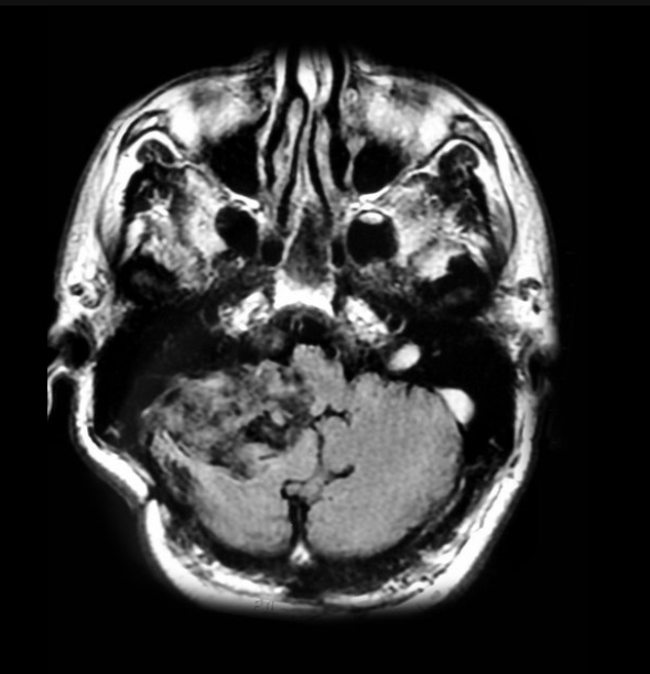 FLAIR imaging. Isodense to CSF. (Hint: heterogenous hyperintensity on FLAIR)
What is an epidermoid cyst?
400
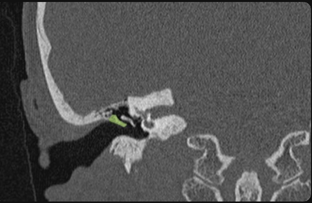 This structure can be eroded by a certain pathology. Name the structure and pathology.
Scutum.
Cholesteatoma.
400
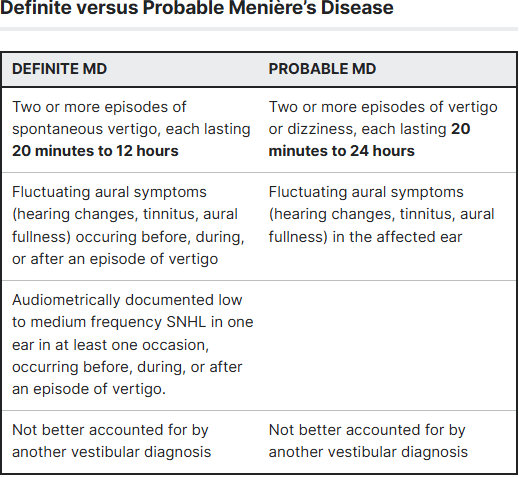
Based on Barany's Criteria, 43 yo male with symptoms of spontaneous vertigo with several episodes, some lasting more than 12 hrs without documented audiogram test, is this type of Meniere's Disease.
What is probable Meniere's disease?
400
What are the guidelines for infant hearing screening?
All infants should undergo screening before leaving the hospital, or by 1 mo of age
Rescreen/Detection of hearing loss by age 3 months
Appropriate interventions begun by age 6 months
400
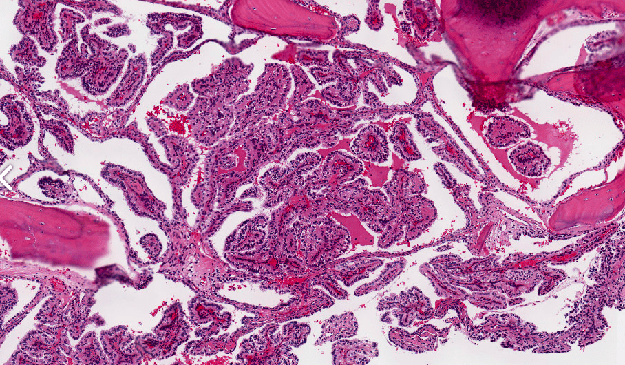 55 yo male with fluctuating, progressive single sided SNHL with tinnitus, vertigo, and ataxia. This condition is associated with a AD disease. What other tumor is this patient at risk for along the cerebellum?
Hemangioblastoma
(Histology is showing endolymphatic sac tumor which is highly associated with VHL, which is assoc. with HARP: hemangioblastoma, angiomatosis, renal cell carcinoma, pheochromocytomas)
The tumor is arranged as simple, broad, non-complex papillary projections without large cystic spaces. The spaces are often fluid filled, have extravasated erythrocytes and/or inspissated material. The cells are cuboidal, usually single layered along the papillary structures, showing indistinct cell borders. The nuclei are round and hyperchromatic
400
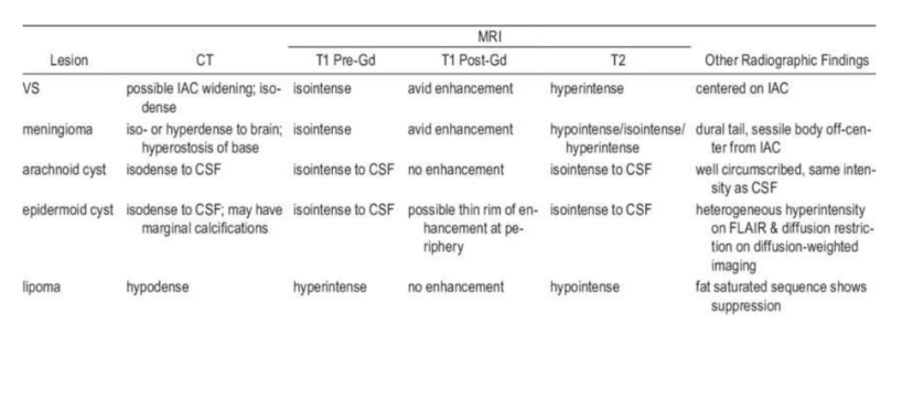
Name the intensity findings of a Vestibular Scwhannoma in the IAC on T1 pre and post Gd and T2.
What is T1 pre: isointense, T1 post: avid enhancement, T2: Hyperintense
500
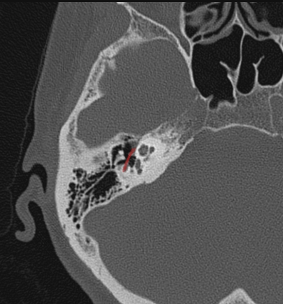
A 35-year-old patient develops acute right-sided facial paralysis. Examination reveals loss of forehead wrinkling, incomplete eye closure, and drooping of the right corner of the mouth. The patient also reports hyperacusis and decreased taste over the anterior two-thirds of the tongue. Lacrimation is preserved.
Where is the most likely location of the facial nerve lesion?
A. Distal to the stylomastoid foramen
B. Proximal to the greater petrosal nerve, at or before the geniculate region
C. Distal to the nerve to stapedius but proximal to the chorda tympani
D. Isolated marginal mandibular branch
What is B.
Hyperacusis indicates dysfunction of the nerve to stapedius, while loss of anterior tongue taste indicates chorda tympani involvement. Preserved lacrimation suggests the greater petrosal pathway is spared, so the best localization is between the greater petrosal and stapedius branches, or proximal to the stapedius branch but distal to the greater petrosal branch
500
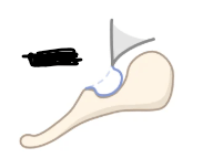 According to Tos and Poulsen Classifcation this type shows the pars flaccida adherent and retracted to the neck of the malleus with no visible air space.
What is Type II?
This classification is used to classify the retraction of the pars flaccida.
- Type 0: Normal pars flaccida.
- Type I: Retraction towards the neck of the malleus, with a visible air space behind the pocket.
- Type II: Pars flaccida is retracted and adherent to the neck of the malleus with no visible air space.
- Type III: Retraction extends beyond the bony annulus, with or without slight bone resorption of the annulus.
- Type IV: Definite bony erosion of the annulus.
500
 Name the syndrome, type of HL, and what is the defect?
Goldenhar Syndrome
Mixed HL
1st and 2nd branchial arch defect
500
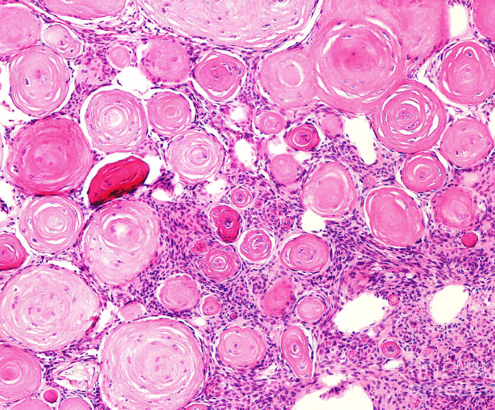
43 yo female who presents with a condition with similar histologic findings as a meningioma. This is the treatment for this patient (knowing this is high risk)
What is Total Thyroidectomy and RAI. Neck dissection if cervical neck disease present.
500
A 52-year-old patient presents with progressive unilateral sensorineural hearing loss, tinnitus, and mild imbalance. MRI of the brain with gadolinium demonstrates a 2.4-cm enhancing mass centered in the right cerebellopontine angle (CPA), extending into the internal auditory canal (IAC). The mass has an intracanalicular component and widens the porus acusticus. There is mild displacement of the adjacent pons without hydrocephalus.
Which of the following MRI findings most strongly supports the diagnosis of a vestibular schwannoma rather than a meningioma?
A. Broad-based attachment to the petrous dura with a dural tail
B. Extension into and widening of the internal auditory canal
C. Marked diffusion restriction on diffusion-weighted imaging
D. T2-hypointense signal with extensive calcification on CT
E. Intra-axial location with surrounding vasogenic edema
What is B?
Vestibular schwannomas typically arise from Schwann cells of the vestibular division of cranial nerve VIII and commonly extend from the IAC into the CPA, producing the characteristic ice cream cone appearance. Smooth widening of the IAC supports this diagnosis.