Column 1
Column 2
Column 3
Column 4
Column 5
Column 6
Column 7
100
What are the types of muscular tissue?
skeletal
smooth
cardiac
100
What is the name of this structure

Fascicle
100
attaches bone to bone
ligaments
100
Type of muscle that is found in the limbs
skeletal
100
This muscle type is only found in the heart
cardiac
100
attaches muscle to bone
tendon
100
Gap between the neuron and motor end plate of a muscle
Synapse / Synaptic Cleft 
200
What is the name of this layer of connective tissue?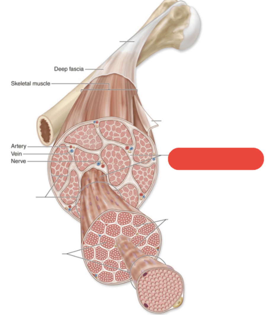
Perimysium
200
Individual muscles are separated by ________
Fascia
200
Muscle tissue type that is connected to the bones
skeletal
200
Muscle cells found within fibers are called ________
myocytes
200
Name the 2 types of muscle tissue that are striated
cardiac and skeletal
200
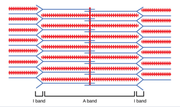
Myofibrils are made of myofilaments called ______ and _______
actin (thin) and myosin (thick)
200
Protein that binds to calcium and moves tropomyosin
Troponin 
300
Thin filaments in the muscle are made up _______
Actin
300
Name 3 functions of the Muscular System
Movement of the body (uses bones as anchors and levers)
Maintains Posture
Generates Body Heat
Plays a role in other body systems
- Respiration, Digestion, Urination
300
What is this layer of connective tissue named?
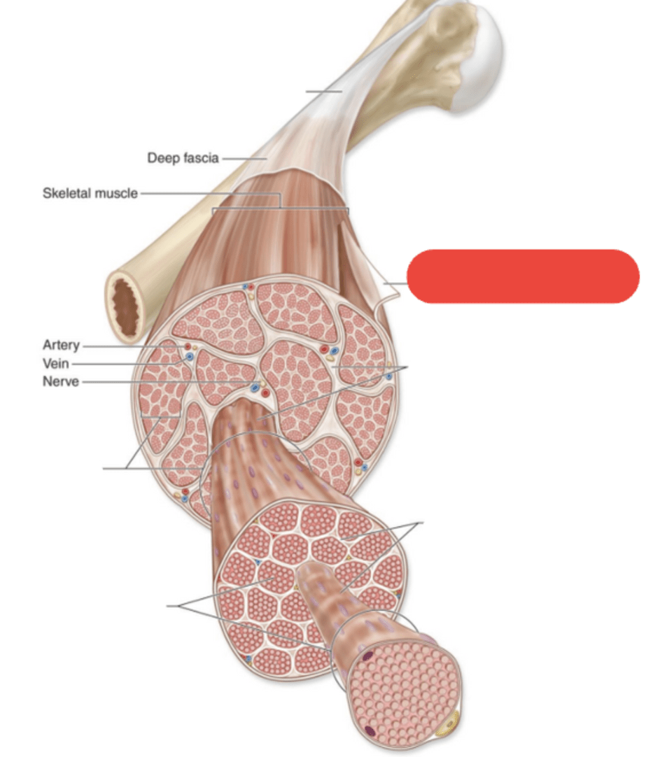
Epimysium
300
Thick filaments in the muscle are made up ________
Myosin
300
Name each type of muscle tissue and the type of control we have over them
Skeletal = voluntary
Cardiac = involuntary
Smooth = involunatry
300
Layer of connective tissue that separates and surrounds fascicles (bundles of muscle fibers)
Perimysium
300
Connection formed when myosin binds to actin during a muscle contraction
Cross Bridge
400
This tissue is mainly found in the walls of hollow organs
smooth muscle
400
What is the name of this structure: the smallest unit of contraction in a muscle
Sarcomere
400
Layer of connective tissue that surrounds each individual muscle fiber
Endomysium
400
Dark bands in the muscle are known as the _________ and are made up of the protein ________
A band
Myosin (thick filament)
400
Is this sarcomere contracted or relaxed?
Contracted
Compare it to the sarcomere below that is relaxed.

400
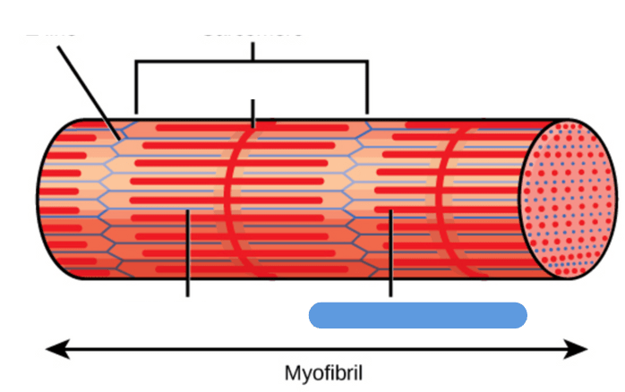
Name of this structure: holds myosin filaments in place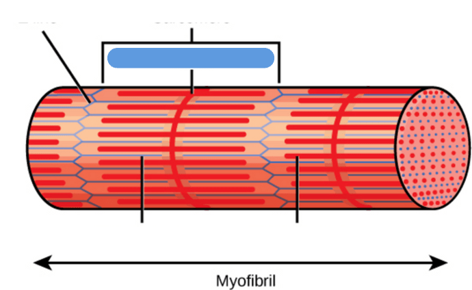
M-Line
Middle of the sarcomere; holds myosin filaments in place.
400
Protein that blocks actin’s binding sites until calcium signals the muscle to contract
Tropomyosin 
500
Outermost layer of connective tissue that surrounds entire muscle
Epimysium
500
Type of muscle tissue that is not striated
smooth
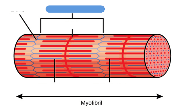
500
Muscle fibers are made up of ___________
myofibrils
500
What is the name of this struture: Ends of the sarcomere; connects actin filaments.

Z-line
500
The __________ is the smallest unit of contraction in a muscle fiber
Sarcomere
500
Energy molecule that allows myosin to release from actin and reset during a muscle contraction
ATP (Adenosine Triphosphate)
500
Put the following in order from most superficial to deep
Myofibril
Endomysium
Perimysium
Skeletal muscle
Myofilaments (actin and myosin)
Fascicle
Muscle fiber
Epimysium
Epimysium → Skeletal muscle → Perimysium → Fascicle → Endomysium → Muscle fiber → Myofibril → Myofilaments (actin and myosin)
600
What is this structure called? 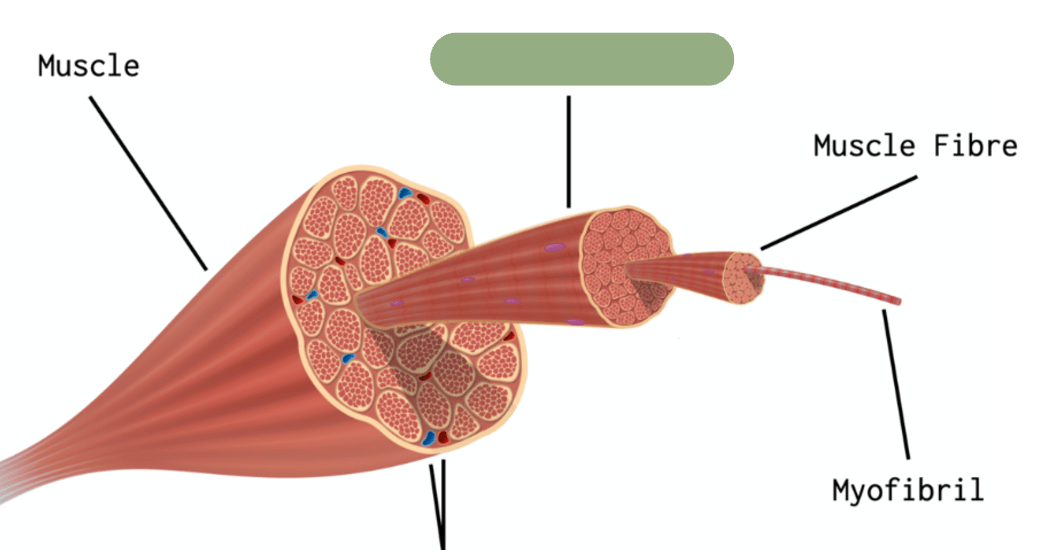
Fascicle
600
What is the name of this structure? 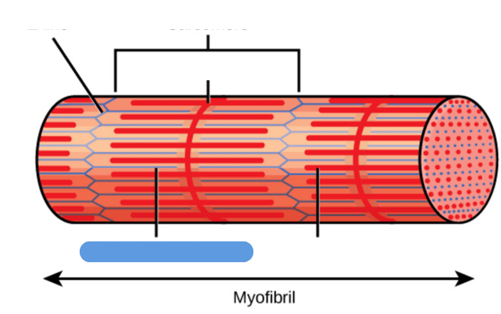
Actin (thin) filament
600
Explains how muscles contract by the sliding of actin (thin filaments) past myosin (thick filaments) within the sarcomere, shortening the muscle fiber and generating force.
Sliding Filament Theory
600
What is the name of this connective tissue?

Endomysium
600
What is the name of this structure?
Myosin (thick) filament
600
What is the name of this structure? 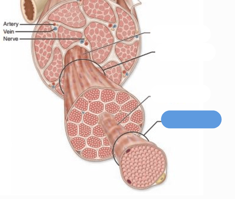
Muscle Fiber
600
Put the numbers in the correct order for the steps of a muscle contraction
1. Myosin pulls actin toward the center of the sarcomere, shortening the muscle.
2. Calcium binds to troponin which moves tropomyosin away from actin, allowing myosin to attach to actin, forming cross-bridges for contraction.
3. Acetylcholine (ACh) binds to muscle receptors, creating an electrical signal in the muscle fiber.
4. ATP binds to myosin, helping it detach from actin and reset.
5. An electrical signal releases calcium from the sarcoplasmic reticulum into the muscle.
6. A nerve signal reaches the neuromuscular junction (NMJ) to begin muscle contraction.
7. When the nerve signal stops, calcium is stored again (in the sarcoplasmic reticulum), the tropomyosin covers the actin binding sites again, and the muscle relaxes.
8. The nerve releases acetylcholine (ACh) into the synapse, signaling the muscle to contract.
1️⃣ → 6. A nerve signal reaches the neuromuscular junction (NMJ) to begin muscle contraction.
2️⃣ → 8. The nerve releases acetylcholine (ACh) into the synapse, signaling the muscle to contract.
3️⃣ → 3. Acetylcholine (ACh) binds to muscle receptors, creating an electrical signal in the muscle fiber.
4️⃣ → 5. An electrical signal releases calcium from the sarcoplasmic reticulum into the muscle.
5️⃣ → 2. Calcium binds to troponin which moves tropomyosin away from actin, allowing myosin to attach to actin, forming cross-bridges for contraction.
6️⃣ → 1. Myosin pulls actin toward the center of the sarcomere, shortening the muscle.
7️⃣ → 4. ATP binds to myosin, helping it detach from actin and reset.
8️⃣ → 7. When the nerve signal stops, calcium is stored again (in the sarcoplasmic reticulum), the