A&P
Priority !
Screening Tools
You want me to remember things?
DRUGS!
100
Name the responsibilities of the brain stem?
Life functioning: heart beat, breathing, blood pressure and swallowing
100
You're patient is having a seizure! What do you do immediately!
- Protect the airway, turn on side, ensure nothing is in the mouth, get suction for emesis
100
What does BEFAST stand for?
Balance
Eyes
Face
Arms
Speech
Time to call 911
100
What does troponin measure and at what interval do we draw troponin levels for cardiac patients?
Troponin is a protein complex found in cardiac muscle fibers. When cardiac myocytes are damaged or die (as in an MI), these proteins are released into the bloodstream. It is the gold-standard biomarker for myocardial necrosis (cell death).
- Initial (0-hour): Taken immediately upon arrival for patients presenting with suspected Acute Coronary Syndrome (ACS).
- Serial (typically 1 to 3 hours later): Depending on your hospital’s specific protocol (High-Sensitivity Troponin protocols usually use 0hr/1hr or 0hr/2hr).
100
What medication is first line treatment to administer to a patient when they hare having a seizure?
Benzo's!
- Ativan
- Diazepam
- Midazolam
200
Name and describe a type of abnormal breathing pattern and why its happening?
The specific breathing patterns you mentioned occur because the brainstem—the control center for involuntary life functions—is being damaged, causing a breakdown in the complex "respiratory rhythm generator."
Here is why each pattern occurs from a physiological perspective:
1. Cheyne-Stokes (Diencephalon/Hemispheric involvement)
This pattern is caused by a lag in the feedback loop between the brain and the body’s chemistry.
The Mechanism: When there is damage to the cerebral hemispheres or the diencephalon, the brain becomes "overly sensitive" to carbon dioxide levels. The patient hyperventilates, causing CO2 levels to drop too low; the brain then detects this and shuts off breathing (apnea). Once CO2 levels rise again, the brain overreacts, and the cycle repeats. It is essentially an "oscillation" caused by a damaged thermostat.
2. Central Neurogenic Hyperventilation (Midbrain/Upper Pons)
This pattern is caused by the loss of inhibitory signals that normally regulate the respiratory rate.
The Mechanism: In a healthy brain, the respiratory centers are constantly modulated by inhibitory signals coming from the forebrain and higher brainstem. When the midbrain or upper pons is damaged, these "brakes" are severed. The respiratory center in the brainstem essentially runs "unlocked" and at full speed, resulting in rapid, deep breaths that do not respond to changes in the body’s blood gas levels.
3. Apneusis (Mid-to-Lower Pons)
This pattern is caused by the disconnection of the "off-switch" for inspiration.
The Mechanism: Normal breathing is regulated by two centers in the pons: the apneustic center (which promotes inspiration) and the pneumotaxic center (which inhibits inspiration). The pneumotaxic center acts as the "off-switch" that tells the lungs to stop inhaling and start exhaling. If the mid-to-lower pons is severely damaged, the "off-switch" is destroyed, meaning the brain is told to "inhale" but never receives the signal to stop, resulting in those long, gasping, sustained inspirations.
4. Cluster breathing: Cluster breathing occurs when the rhythmic integration center—which coordinates the transition between breathing and resting—becomes unstable due to localized brainstem injury.
The Mechanism: Unlike the sustained "locked" rate of neurogenic hyperventilation or the "failed off-switch" of apneusis, cluster breathing represents a decoupling of respiratory drive. The brainstem is still attempting to trigger breathing, but the neural circuits responsible for maintaining a smooth, rhythmic cadence are malfunctioning. It is essentially a "stutter" in the brainstem's respiratory rhythm generator, signaling that the lesion is moving further down toward the medulla, which is the final bridge between the brain and the autonomic control of life-sustaining functions.
5. Ataxic breathing: Ataxic breathing is a completely irregular and chaotic pattern of shallow and deep breaths with unpredictable periods of apnea, signaling imminent failure of the respiratory centers in the medulla oblongata.
Why it happens:
Ataxic breathing is the most ominous of these patterns because it represents the final stage of brainstem decompensation before total respiratory arrest.
The Mechanism: The medulla oblongata contains the primary respiratory rhythm generators (the Pre-Bötzinger complex). When a lesion—usually caused by severe intracranial pressure or herniation—reaches the medulla, the fundamental "clock" that dictates the timing and rhythm of breathing is destroyed. Because the neural circuitry responsible for maintaining any semblance of order is essentially fragmented, the lungs receive random, disorganized electrical signals rather than a coherent command to breathe.
Clinical significance: Ataxic breathing is often the last stage of "rostral-to-caudal" deterioration before the patient transitions into agonal breathing and permanent apnea (brain death).
In summary: These patterns are diagnostic "map markers" for neurologists. Because the respiratory center is located in the brainstem, the specific type of abnormal breathing tells the clinician exactly how far down the brainstem the injury or pressure (herniation) has progressed.
200
Your patient suddenly starts to have slurred speech, you notice a L sided facial droop and she stopped using her L arm. Her airway is intact and she is breathing spontaneously, what is your next priority intervention besides calling the code stroke?
CT stat! Also try and grab a BGL on the way
200
What components make up a GCS?
Eye Opening
Verbal ResponseMotor Response
200
In regards to ESI resources, are oral medications considered a resource?
NO!
200
Name one second line medication we administer to patients for seizure management in the ED?
- Valproic acid
- Phenytoin
- Fosphenytoin
Levetiracetam (Keppra)
300
How many cranial nerves are there?
12
300
Upon initial evaluation of your patient they state they have a headache and "it's the worst headache of [their] life" - what is your initial priority?
Worst headache of life is a red flag for a stroke! We need to get MD evaluation immediately or consider calling a code stroke and getting straight to CT.
300
In the NIH exam, we evaluate ataxia. What does that mean and what do we ask patients to do to evaluate this?
Ataxia- is a neurological sign or symptom that means a lack of voluntary coordination of muscle movements.
- We ask patients to do the finger-to-nose and heel-shin tests
300
A patient with a change in mental status off of their baseline should always be an ESI....
2!!
300
Name 3 medications we would administer if we suspected a patient had bacterial meningitis?
- Antibiotics
- Antipyretics
- Steroids (Dexamethasone)
- Pain management
- Osmotic agents if concerned about increased ICP
400
Name the 4 lobes of the Cerebrum.
Frontal Lobe, parietal lobe, occipital love and temporal lobes
400
You're performing you triage for a waiting room patient and you start to suspect your patient could have meningitis - what is your priority?
Isolate! If patient has bacterial meningitis, very dangerous and contagious.
400
NIHSS- how do we evaluate for dysarthria in the NIHSS Exam?
We ask them to repeat words : MAMA, TIP TOP, FIFTY FIFTY, THANKS HUCKLEBERRY, BASEBAL PLAYER
or phrases: You know how. Down to earth. I got home from work. Near the table in the dining room. They heard him speak on the radio last night.
Dysarthria is a motor speech disorder caused by damage to the nervous system, which weakens or paralyzes the muscles needed to produce speec
400
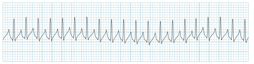 Name the rhythm and intervention?
SVT!
- Vagal maneuvers
- Adenosine!
- Unstable? Synchronized cardioversion
400
What is the dosing for TNK for strokes and what is the MAX dose?
.25 mg/kg with a max dose of 25 mg TOTAL
500
Put these in order of least to most superficial:
gray/white matter, skin, dura mater, pia mater and arachnoid mater, bone
Skin -> bone->dura mater -> arachnoid mater -> pia mater -> gray/white mater (brain)
500
You just pushed TNK and 1 minute later your patient starts experiencing tachypnea and swelling in the lips. What are your priority interventions?
1. Notify the doctor 2. Protect the patient airway 3. Get allergic reaction medications
500
Demonstrate how you would evaluate a patient's extinction and inattention.
500
[BLANK] is characterized by an accumulation of fluid in the lungs air sacks, called alveoli. This accumulation of fluid makes breathing difficult as it physically prevents gas exchange. Causes can be cardiogenic or non cardiogenic. Some cardiogenic causes include heart failure and MI, due to impaired heart function allowing for fluid buildup which leaks to the lungs. Non cardiogenic causes can include things such as fluid overload or severe infection.
Pulmonary Edema!
500
What 3 medications traditionally make up the "migraine cocktail"?
- Benadryl
- Reglan
- Toradol