Uterus Pathology
Functional Cysts
Ovarian Pathology
1st Trimester GYN
PID/Endometriosis
100
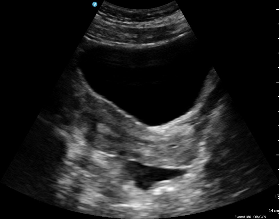
What pathology is represented in this image?
Nabothian cysts
100
What functional cyst is known as the cyst of pregnancy?
Corpus luteum cyst
100
What is the most common cause of ovarian enlargement in young women?
simple ovarian cysts
100
This signifies the beginning of gestation.
first day of LMP
100
The most common place for PID to start
Fallopian tubes
200
A 42-year-old female complains of intermittent spotting between menstrual cycles. Her patient history is: Regular menstrual cycles and no pelvic pain, no prior abnormal transvaginal exams. What pathology is seen in this image?
Cervical Polyp
200
27-year-old female with a positive qualitative pregnancy test, no fever, and no prior ovarian surgery. Uterus appears within normal limits. Right ovary measures 3.8 x 2.6 x 3.1 cm and shows a round cystic structure measuring 2.7 x 2.2 x 2.4 cm, presenting a ring of fire appearance.
Corpus luteum cyst
200
This pathology is most commonly seen in patients with trophoblastic disease or pregnant patients.
theca lutein cysts
200
Initial fetal heartbeat occurs between ____ weeks.
5.5-6
200
the most common cause of PID
untreated STDS
300
Describe what's being seen in this image and its location.
Posterior cul-de-sac fluid
300
A 29-year-old female complains of severe nausea and vomiting. Patient has Gestational Trophoblastic Disease. No pelvic pain. On ultrasound multiple large multiloculated cysts were present on both ovaries.
Theca Lutein cyst
300
This pathology is a benign epithelial tumor lined by mutinous elements of the endocervix and bowel.
mucinous cyst adenoma
300
This germ cell layer becomes the musculoskeletal and circulatory systems.
mesoderm
300
when the infection from PID spreads up towards the abdomen, specifically the liver and surrounding peritoneum
fitz-hugh-curtis syndrome
400
30-year-old female expressing persistent heavy vaginal bleeding. She had a dilation and curettage performed 5 weeks ago after a miscarriage. Beta hCG is negative. On ultrasound, snake-like anechoic structures are seen within the myometrium, providing a mosaic/intramural pattern with color Doppler because of areas with aliasing.
Arteriovenous Malformation (AVMS)
400
22-year-old female complains of mild intermittent lower pelvic discomfort. The ovary measures 4.2 x 2.8 x 3.5 cm and contains a thin-walled anechoic cyst with well-defined margins.
Follicular cyst
400
This ovarian pathology can mimic stage 2-3 ovarian cancer. It displays a moth eaten, cystic pattern.
ovarian metastases
400
a pregnancy with low hCG, and a visible embryo but no cardiac activity
missed abortion
400
the name for an adnexal mass caused by the ovaries and fallopian tubes fusing together but still being distinct structures
tubo-ovarian complex
500
A 52-year-old female complains of progressive lower abdominal pain and increased abnormal girth for 4 months. She also explains that she had had intermittent pelvic pressure, urinary frequency, and unexplained weight loss. She was previously diagnosed with uterine fibroids 6 years ago. Ultrasound image shows a heterogeneous solid hyper vascular mass.
Leiomyosarcoma
500
A 24-year-old patient presented with sudden right-sided pelvic pain. Paain began abruptly yesterday. Reports mild nausea. Last menstrual period 3 weeks ago. Negative pregnancy test.
![]()
Hemorrhagic cyst
500
This pathology, which commonly occurs in 40-70 yr old patients, is malignant, bilateral, and prone to rupture. A common complication is pseudomyxoma peritoneum.
mucinous cystadenocarcinoma
500
what are the parameters for a confirmed pregnancy loss?
embryo with a CRL of 7 mm but no heartbeat, or a MSD that is greater than 25 mm with no visible embryo
500
the ovary is the most common location for this to be found
endometriosis