Tibia Landmarks
Fibula Facts
Articulations & Joints
Muscle & Ligament Attachments
Odds & Ends Of Ossification
100
This ridge begins at the tibial tuberosity and runs down the front of the tibia
Anterior Border
100
The fibula does not articulate with this part of the knee
The Femur
100
The tibia and fibula are connected along their lengths by this structure
Interosseous Membrane
100
This muscle inserts at the tibial tuberosity via the patellar tendon
Quadriceps Femoris
100
The tibia does not articulate with this sesamoid bone
Patella
200
These two tubercles form the intercondylar eminence
Medial and Lateral Intercondylar Tubercles
200
This structure at the distal end forms the outer bump of the ankle
Lateral Malleolus
200
The proximal tibiofibular joint is this type of joint
Synovial Joint
200
Gerdy's Tubercle is the insertion point for this structure
Iliotibial Band (IT Band)
200
This structure lies between the medial and lateral condyles of the tibia
Intercondylar Eminence
300
This surface articulates with the talus at the ankle
Tibial Plafond (inferior surface)
300
This nerve wraps around the neck of the fibula and is vulnerable to injury
Common Fibular (Peroneal) Nerve
300
The distal tibiofibular joint is classified as this type of joint
Fibrous (Syndesmosis) Joint
300
The semimembranosus muscle attaches here on the posterior tibia
Medial Condyle
300
The tibia and fibular begin ossifying in this stage of life
Fetal Life
6th - 7th month
400
This structure provides an attachment site for the patellar ligament
Tibial Tuberosity
400
These are the three boarders of the fibular shaft
Anterior, Posterior & Interosseous Borders
400
This tibial feature helps form the ankle mortise
Medial Malleolus
400
The pes anserinus is formed by the insertion of these THREE muscles
Sartorius, Gracilis & SemiTendinosus
400
The distal fibular projection is also known as this
Lateral Malleolus
500
Name the pathology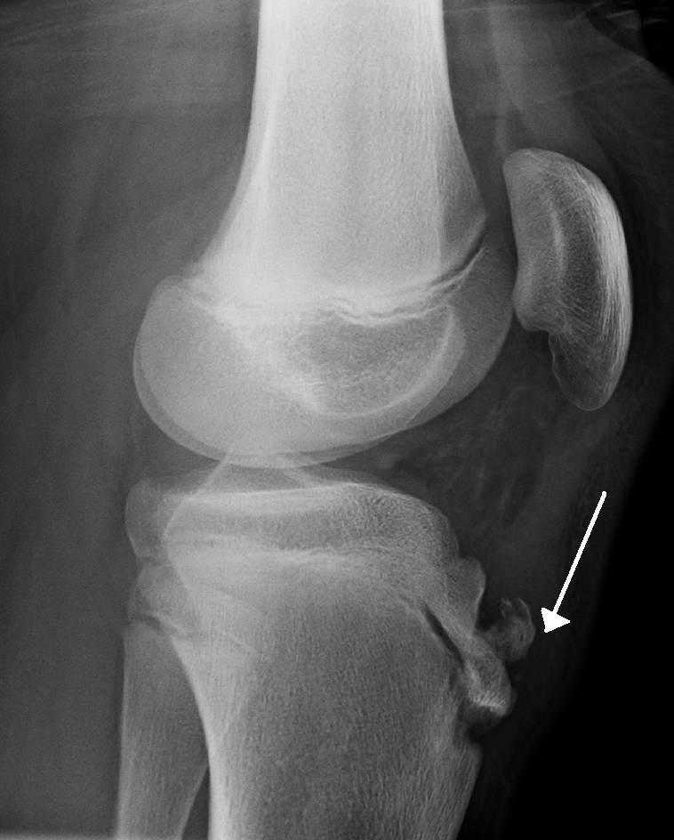
Osgood-Schlatter Disease
500
Name the structure indicated by the red block
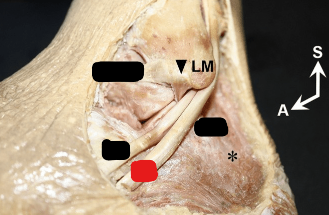
Peroneus Longus
500
Name this structure of the talus

Trochlea
500
Name the orange/yellow block structure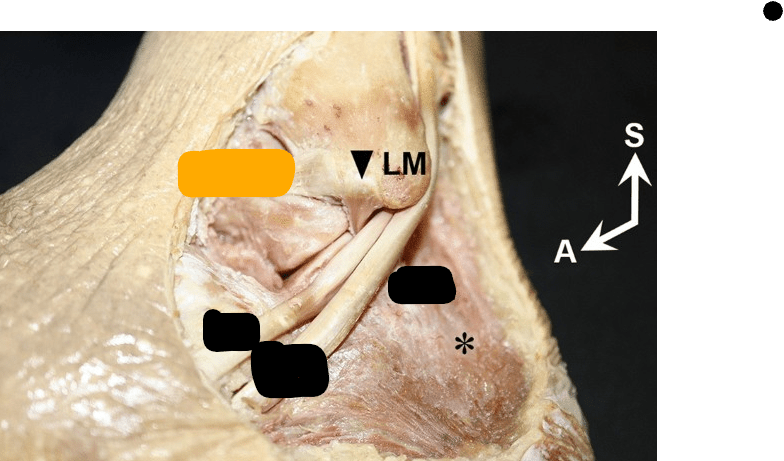
ATFL
500
Approximately how old is this child
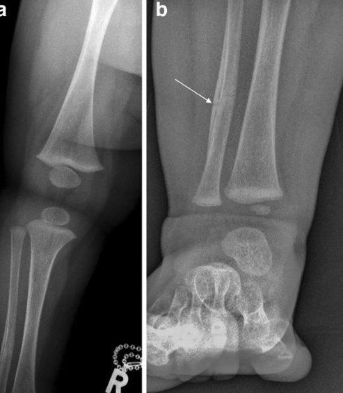
Pre-Ambulatory (<18 months)