Cardio 2
Cardio Dx
Abdomen/Renal
More Signs
Likely dx
Final Jeopardy
100
Name this view:
Apical 2 chamber
100
Recurrent fevers and this on PLAX:
Mitral valve Endocarditis
100
Prominent Finding(s) seen in this RUQ clip:
Ascites and Pleural Effusion with Jellyfish sign
100
Whats this sign called:
D Sign
100
Cause of Dyspnea in patient with these findings: (See both clips):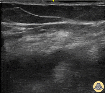

Acute PE
200
Name this view and level: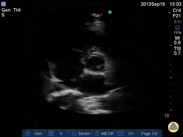
Parasternal Short (SAX) Aortic valve
200
Hypotension and the following PLAX and PSSX view:
Cardiac tamponade
200
Cause of hematuria in this patient:
Bladder cancer
200
What "mammalian" sign is shown here:
Bat-wing sign
200
Cause of Dyspnea in a patient with these findings:
CHF exacerbation
300
Based on this image, most likely type of cardiomyopathy: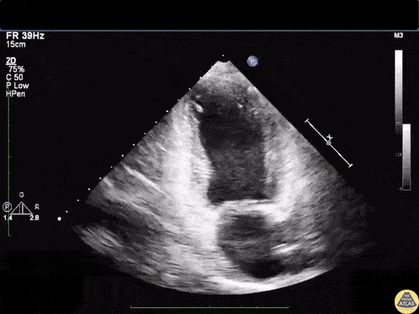
Stress/takotsubo
300
Chest pain and this image on PLAX: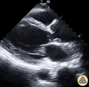
Aortic dissection
300
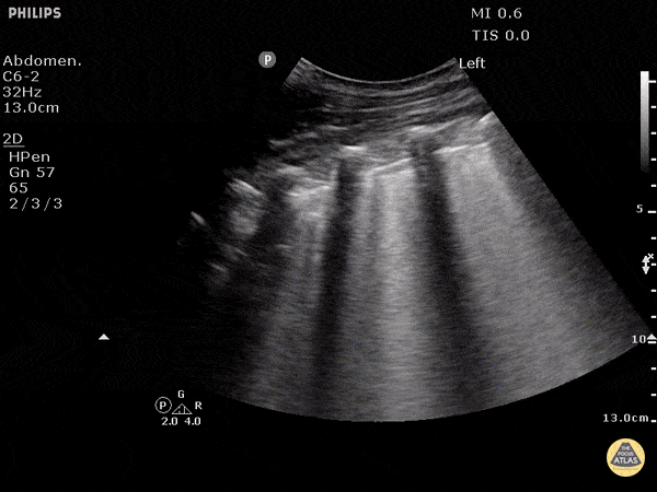
Cause of abdominal distention in this patient:
Ascites
300
What is the sign shown in this lung scan:
Lung Point
300
Patient with severe chest pain, Scan of the right neck, what is the Dx:
Aortic dissection extending to the carotid
400
Name the view and the prominent finding: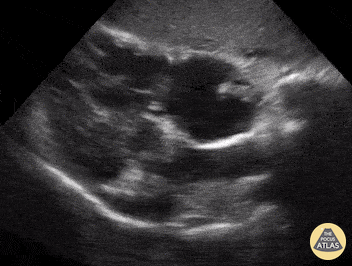
Subcostal 4 chamber, clot in transit
400
Fevers and this image: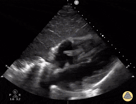
Tricuspid valve endocarditis
400
What disease does this patient have:

Polycystic kidney disease
400
Renal Sign shown here:
Bear paw sign/Bear claw sign (severe Hydro)
400
Cause of dyspnea ina patient with this lung scan: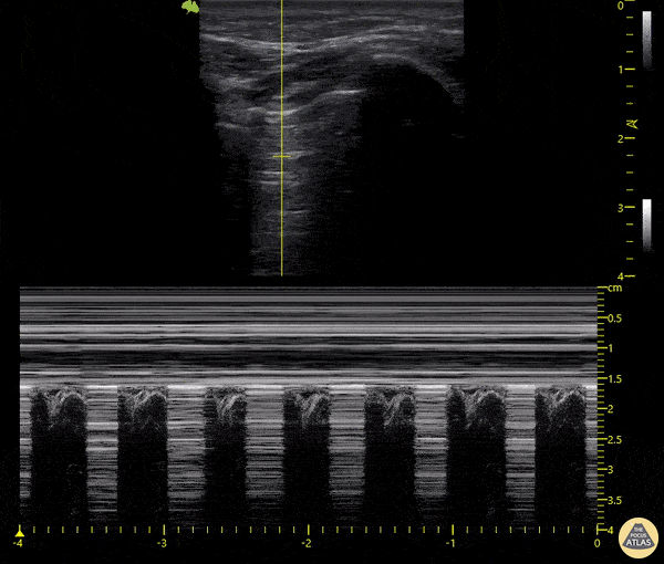
Pneumothorax
500
Finding shown here:
VSD
500
Mitral stenosis with mitral regurgitation with the classic MV "hockey stick" sign seen classically in which disease?

Rheumatic mitral valve disease
500
Why was this foley not draining: (Watch full clip)
Lodged in bladder diverticulum
500
Cardiac sign seen on this PLAX view:
McConnell's sign
500
Whats wrong with this patient: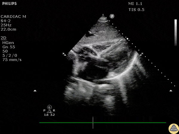
Vfib Cardiac Arrest/ Accept death
500
This ultrasonographic finding, first described in the 1990s, can be seen in both severe tricuspid regurgitation and cardiac tamponade. It refers to abnormal diastolic movement of this structure due to elevated right-sided pressures and may be missed unless you freeze-frame during ventricular diastole.
What is RV trampolining of the lateral wall and or intra-ventricular septum.