ANATOMY
PATHOLOGY
NAME THAT VIEW
ARTIFACTS/KNOBOLOGY
100
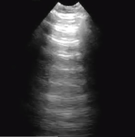
This butterfly-shaped gland is located anterior to the trachea and lateral to the esophagus
What is Thyroid?
100
Acute dyspnea with right heart strain and absent color flow in a pulmonary artery branch on Doppler suggests this diagnosis

What is Pulmonary Embolism?
100
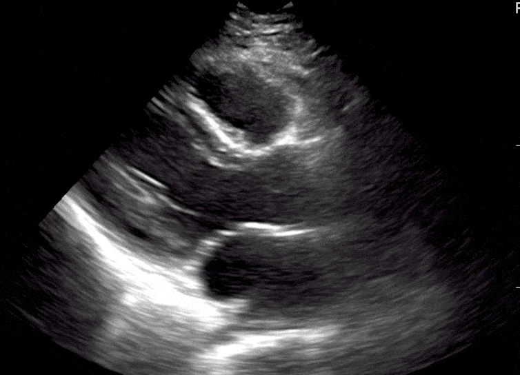
This cardiac ultrasound view displays the left ventricle, left atrium, right atrium, and right ventricle.

What is Apical 4 Chamber View?
100
Often represented by an acronym, this fundamental safety principle dictates that output power and exposure time should be kept to the minimum necessary to achieve a diagnostic image.
What is ALARA?
200
This organ is identified by a hypoechoic cortex surrounding an echogenic central sinus and demonstrates corticomedullary differentiation.

What is Kidney?
200
On ultrasound, this thyroid finding may appear hypoechoic with irregular margins and punctate echogenic foci, prompting further risk stratification.
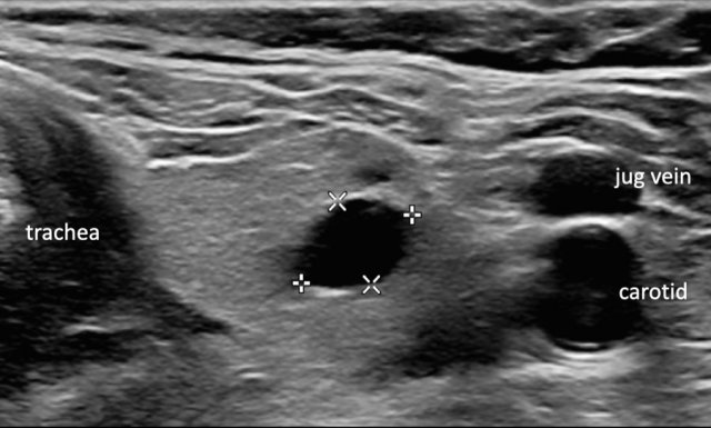
What is Thyroid Nodule?
200
Characterized by the 'bird's beak' appearance of the supraspinatus tendon tapering onto the humerus, this transducer orientation aligns parallel to the muscle fibers."
.png)
What is Long-Axis View?
(or Longitudinal view)
200
Increasing this setting allows visualization of deeper structures, but requires the machine to 'listen' longer, lowering the frame rate.
What is Depth?
300
This cardiac chamber’s systolic function is commonly estimated by visual assessment of wall thickening in the parasternal long-axis view.
What is Left Ventricle?
300
This pathology appears as echogenic intraluminal foci that cast posterior acoustic shadowing and demonstrate mobility with patient repositioning.
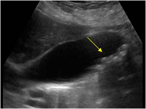
What is Gallstones?
300
This cardiac view is used to visualize the left ventricle and right ventricle.

What is Parasternal Short Axis View?
300
This artifact causes increased echogenicity deep to fluid-filled structures.

What is Posterior Acoustic Enhancement?
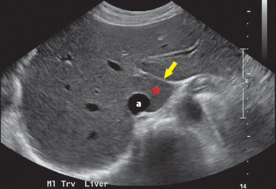
400
In trauma ultrasound, free fluid often accumulates between this organ and the left kidney, and its parenchyma should be less echogenic than the liver.
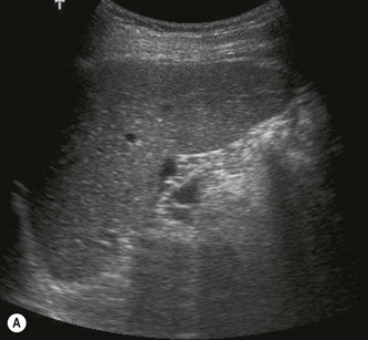
What is Spleen?
400
Ultrasound showing chamber collapse during diastole with impaired ventricular filling indicates this cause of obstructive shock.

What is Cardiac Tamponade?
400
From the standard Apical 4-Chamber window, rotating the transducer 60 to 90 degrees counter-clockwise eliminates the right heart chambers to reveal this view.

What is Apical 2 Chamber View?
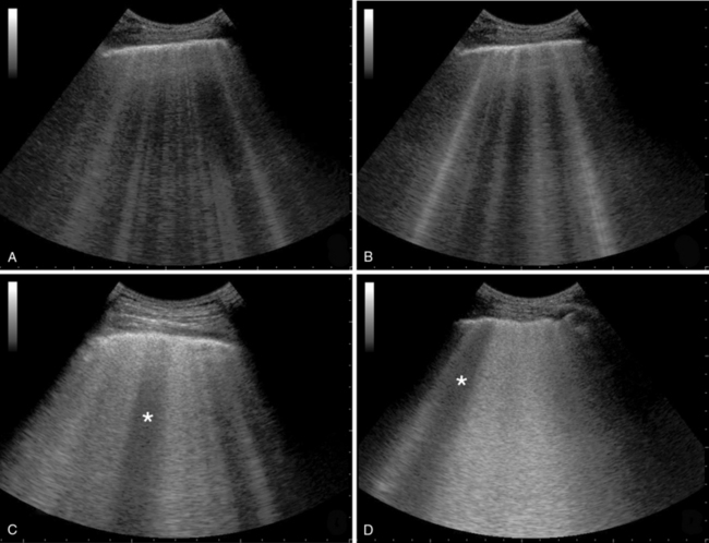
400
These horizontal, equidistant reverberation artifacts repeat deep into the image and are a classic sign of air, indicating either a normal 'dry' lung or a pneumothorax.
What are A Lines?
500
This distinct echogenic line separates the Caudate Lobe from the Left Lobe of the liver and is the fibrous remnant of the fetal Ductus Venosus.
What is Ligamentum Venosum?
500
This pathology has diffuse bilateral B-lines across multiple lung zones with preserved left ventricular systolic function, no focal consolidation, and a collapsible inferior vena cava in a hypoxic patient.
What is Acute Respiratory Distress Syndrome?
500
Often described as looking like a 'candy cane,' this view is obtained by placing the transducer in the notch above the sternum to visualize the aortic arch.

What is Suprasternal Notch View?
500
When using M-Mode on the lungs, normal lung sliding produces a granular appearance below the pleural line, commonly referred to as this 'vacation-themed' sign.
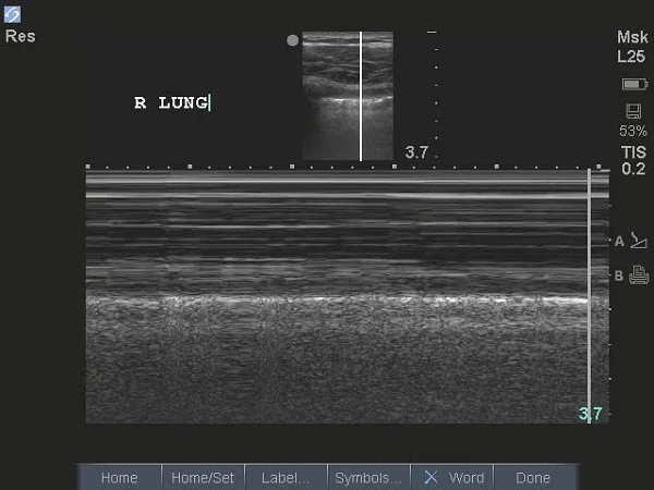
What is Seashore Sign?