Terminology
Chest
Abdomen
Upper Extremity
Lower Extremity
100
Involuntary motion can be caused by:
heartbeat, chills, peristalsis, tremor, spasm, pain
100
The thoracic cavity is separated from the abdominal cavity by the:
diaphragm
100
The _________ attaches the small intestine to the abdominal wall.
mesentary
100
Which carpal is located on the lateral side of the proximal row?
scaphoid
100
Second largest tarsal that occupies the highest position in the foot:
tarsal
200
A specific plane that passes through the midline and divides the body into equal anterior and posterior halves
Midcoronal
200
The _______ separates the right and left pleural cavities.
mediastinum
200
Where is the central ray directed for an AP projection of the abdomen in the supine position?
level of iliac crests
200
A lateral projection of the hand in full extension is best for visualizing:
foreign bodies and fracture displacement
200
An oblique of the foot requires medial rotation to place plantar surface of foot _____ degrees to the IR
30
300
What position does the image display?
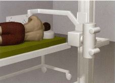
right lateral decubitus
300
Chest procedures should always be performed in upright or decubitus positions to demonstrate:
air/fluid levels
300
Where is the central ray directed for projections of the abdomen when the diaphragm is of interest?
2" above iliac crest
300
Which projection of the elbow demonstrates the radial head free of superimposition?
AP oblique - lateral/external rotation
300
A Jones fracture is best demonstrated in what foot projection?
oblique
400
A body position where the patient's right anterior surface is in contact with the IR and the left anterior surface is elevated:
Right anterior oblique (RAO)
400
If less than 10 ribs are visible above the diaphragm, what error was performed?
insufficient inspiration or image taken upon expiration
400
What position was the patient?
left lateral decubitus
400
A radiograph of an AP projection of the shoulder with external rotation will demonstrate the _________ in profile laterally.
greater tubercle
400
The fibula is _____ and ______ to the tibia.
inferior and posterior
500
Name a fibrous syndesmosis joint:
distal tibiofibular joint; cuboidonavicular joint;
500
Define pneumothorax and describe the radiographic appearance:
free air in the pleural cavity - increased density with absence of lung markings
500
An AP abdomen radiograph reveals elongation of the right iliac wing and foreshortening of the left iliac wing. Which direction was the patient rotated?
right
500
Hypersthenic patients require a ______ in CR angulation for AP axial projections of the clavicle.
decrease
500
What error is present?

over rotation