Structurally Shown
Anatomy in Action
Positioning Proficiency
Joints
Methodology
100
The Stetcher method is performed to demonstrate was structure?
What is the scaphoid?
100

What is the intervertebral disc space of C5-C6?
100
This position is used when inserting an enema tip.
What is the Sim's position?
100
These two articulating structures form the zygapophyseal joint.
What are the superior and inferior articulating processes?
100
Another name for the froglegs position.
What is the modified Cleaves method?
200
The Fuchs method is performed to demonstrate this structure.
What is the odontoid process (dens)?
200
These three bones make up the pelvis.
What are the ilium, ischium, and pubis.
200
The tip of this tube resides in the stomach.
What is a nasogastric tube?
200
The atlanto-occipital joint is the articulation between which two bones.
What are the occipital bone and C1(atlas)?
200
This method is used to demonstrate an axiolateral view of the hip when the patient cannot flex his/her unaffected hip.
What is the Clements-Nakayama method?
300
The Settegast method will demonstrate this view.
What is an axial view of the patella?
300
When performing the a lateral projection of the scapula, the arm should be positioned this way to best demonstrate the scapular body.
What is across the chest?
300
On the image below, this structure on the proximal humerus is visualized in profile laterally.

What is the greater tubercle?
300
The pubic symphysis and an example of this type of joint.
What is amphiarthrodial.
300
When performing this method, the femur makes a 20° angle to vertical.
What is the Holmblad method?
400
The patient was placed in this position in the below radiograph.

What is the RAO position?
400
For a lateral projection of the thumb, the central ray should be directed perpendicular to this landmark.
What is the first MCP joint?
400
This could be done to improve the positioning of the patient in the below radiograph.
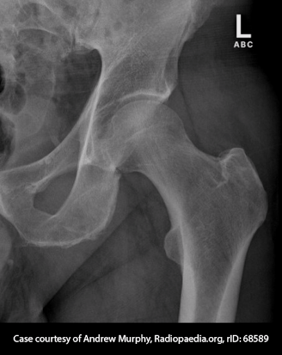
What is internally rotate the affected leg?
400
The pubic symphysis and sacroiliac joints are this type of joint.
What is amphiarthrodial?
400
This is demonstrated on the Grashey method.
What is a profile view of the glenoid cavity.
500
For a lateral projection of the elbow, this humeral structure must be perpendicular to the image receptor.
What are the humeral epicondyles?
500
This organ is responsible for the metabolism of glucose.
What is the pancreas?
500
For an AP oblique projection of the ribs, the patient should be positioned this way to best visualize the left axillary ribs.
What is in the 45 degree LPO position?
500
The joint located between C1 and C2 is called this.
What is the atlantoepistropheal joint?
500
This method is performed to see C7-T1 in the dorsal decubitus position.
What is the Pawlow method?