Anatomy
Pathology
Perio
Miscellaneous
100
Identify two anatomical landmarks visible on this radiograph: Maxillary Right Pre-molar PA, Mandibular Anterior PA.
Inverted Y
Genial tubercles
100
Identify any possible carious lesions present.
#5D, 31M
100
What is the stage and grade of the patient?
Stage II Localized III
Grade B
100
Identify restorative dental materials visible in this radiograph.
Composite, Porcelain fused to metal crown, Endo gutta percha, Endo posts (metal and fiber reinforced),
200
Are there any anomalies or variations from normal anatomy present in this case?
Pulpal stones (#18, 21 & 28)
Fused roots (#2, 15)
200
List pathological findings in the CIS
Internal resorption on #6
External resorption #19
Peiapical abscess #19 & 30
200
What type of bony wall defect is present in the CIS?
one-wall defect
two-wall defect
2, 3, 6, 30, 31
200
Is there evidence of previous endodontic treatment?
Yes.
#4, 29 & 30.
300
How might the anomalies impact the patient’s dental health?
Pulp stones: no symptoms
Fused roots: less periodontal attachment
300
What clinical findings would you expect to be associated with tooth #19 or #30?
Any of the following: Supuration, Fistula, Abscess, Mobility, Edematous, Bleeding, supra eruption.
300
What would be the first phase of treatment planning?
Treating the Endo Abscess
300
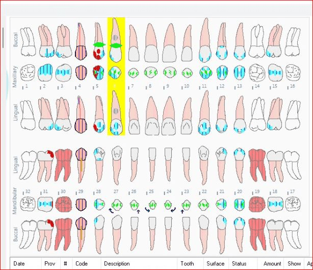
Dentition charting of Quad 1 and 4
400
Which condition could have caused the radiolucency on tooth #19?
Caries.
400
what are the radiographic indicators of a successful or failed endodontic treatment?
Successful: Root canal filling has no visible voids or gaps and is within 0.5 to 2mm of the radiographic apex
Failed: Vsible gaps or voids in the filling, Periapical radiolucencies