CR Placement
Collimation
Positioning
Corrective Actions
Anatomy
Image Evaluation
100
T -7
What is the longitudinal centering for a chest exam?
100
Maximum collimated light field for a lateral elbow projection.
What is 10 x 12 portrait?
100
The positioning error on the internal oblique ankle projection?
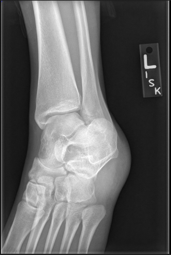
What is the foot is not dorsiflexed?
100
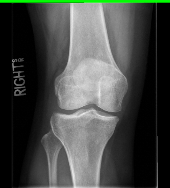
To correct the positioning error of the following internal knee projection, the knee should be ________.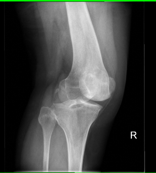
What is extend the knee?
100
Pertinent anatomy not properly demonstrated.
What is the tibiotalar joint?
100
The right SC joint is superimposed over the sternum and the left SC joint is open and projected away from the sternum on a routine PA chest projection.
What is a RAO rotation?
200
Transverse CR placement for a lateral chest projection
What is mid-thorax
200
Maximum collimated light field for a PA scapular Y projection.
What is 10x12, portrait?
200
The movement that projects the clavicles to the level of T-3 & T-4 on a PA chest projection.
What is leaning the patient anteriorly towards the IR?
200
The following lateral chest projection is nicely positioned except for __________?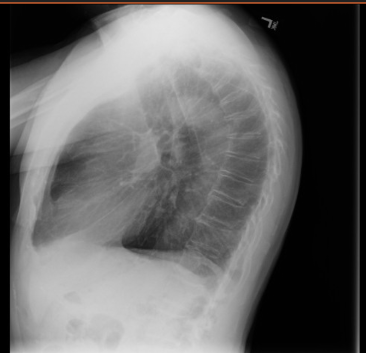
What is the patient's arms are not elevated out of the lung field?
200
The three concentric rings demonstrated on a lateral elbow projection.
What are the trochlear sulcus, ridges of the capitulum and trochlea, and the trochlear notch?
200
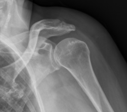
A closed glenohumeral joint with more than 1/3 of the coracoid superimposed over the humeral head on a Grashey projection.
What is over rotated?
300
Grashey transverse & longitudinal CR placement
What is the glenohumeral joint?
300
Maximum collimated light field for a Grashey projection.
What is 8x10, landscape?
300
Demonstrates an opened glenohumeral joint space.
What is a Grashey projection?
300
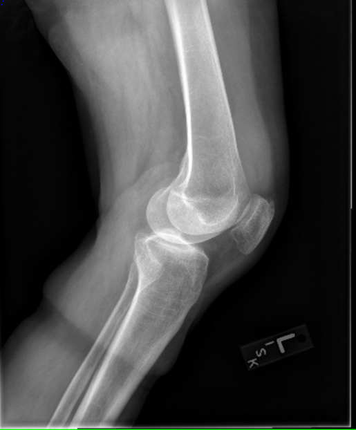
The lateral femoral condyle is demonstrated anteriorly to the medial condyle.
What is medially rotate the knee?
300
Has a thick border extending from the glenoid cavity to the inferior angle of the scapula.
What is the lateral border?
300
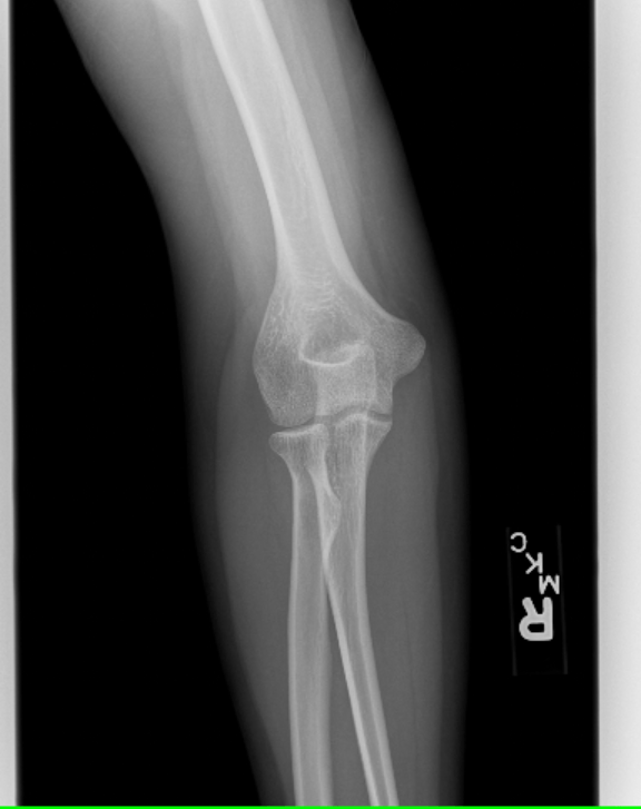
Opened capitulum radial joint space and a closed trochlea ulnar joint space on an AP elbow projection.
What is elevated humerus?
400
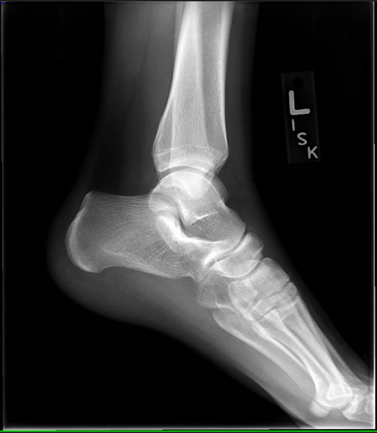
Evaluate the transverse & longitudinal CR placements.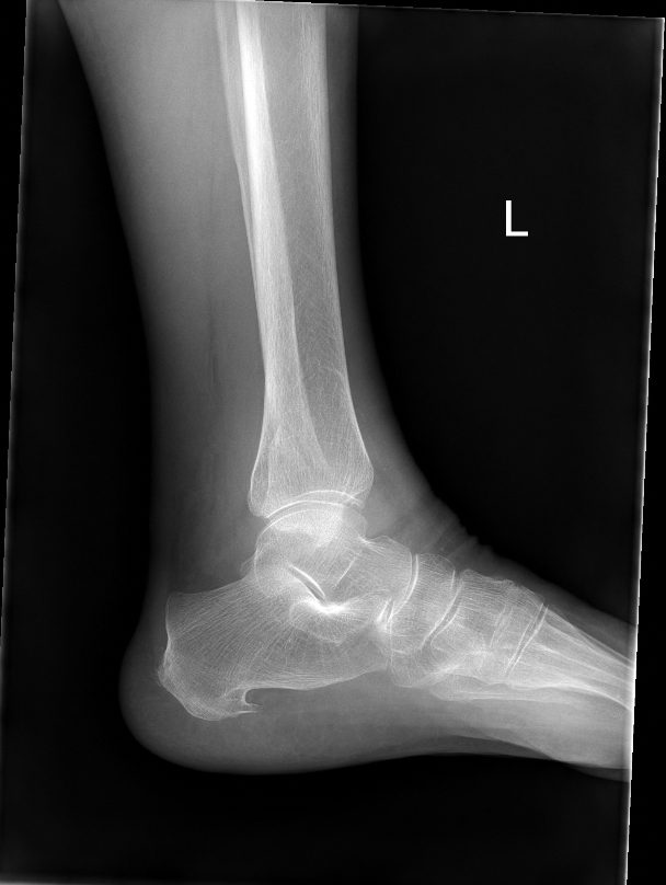
What is longitudinally centered proximal to the medial malleolus and transversely centered anterior to the medial malleolus?
400
The following image is _______ collimation.
What is under collimated?
400
Demonstrates the distal fibula free of superimposition.
What is an internal angle oblique, 45 degree rotation?
400
The medial femoral condyle is demonstrated inferior to the lateral condyle.
What is apply a 5 to 7 degree cephalic tube angel?
400
Pertinent anatomy not accurately demonstrated. 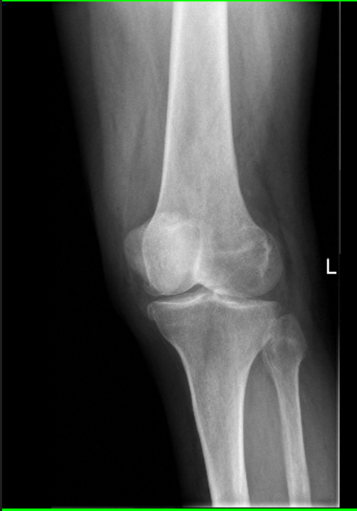
What is the proximal tib-fib joint?
400
The radial tuberosity seen in profile anteriorly on a lateral elbow projection.
What is an externally rotated wrist?
500
Transverse & Longitudinal CR placement for a Knee projection.
What is a 1/2 inch distal to apex of the patella?
500
The following image is ______ collimated.
What is accurately collimated?
500
Evaluate the rotation.
What is under rotated?
500
The glenohumeral joint is not demonstrated.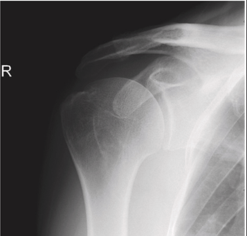
What is decrease the obliquity? The part is over rotated.
500
Anatomy demonstrated as a Y on a scapular Y projection.
What is the coracoid, acromion, and scapular body?
500
The olecranon process is not demonstrated in profile and more than half of the radial head is superimposed by the ulna (coracoid process).
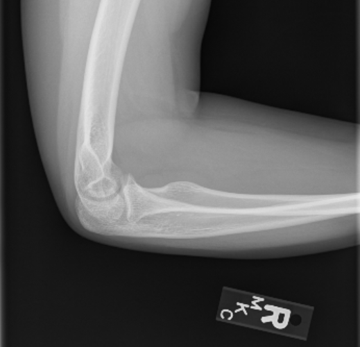
What is an elevated humerus?