The femoral triangle is bordered by the sartorious, adductor longus and this ligament
Inguinal Ligament
This bone lies within the quadriceps tendon and is easily palpable
Patella
This anterior muscle is best palpated by dorsiflexing and inverting the foot
Tibialis Anterior
This muscle attaches to the bony prominence at the base of the fifth metatarsal and is a key palpable landmark
Peroneus Brevis to the Styloid Process
This joint complex lies between the tarsal and metatarsals
LisFranc Joint
Nerve, Artery, Vein & Lymphatics
These bony landmarks of the femur and tibia are easier to palpate when the knee is FLEXED
Femoral Condyles & Tibial Plateaus
These two large posterior legs muscles form the calf
Medial and lateral heads of gastrocnemius
These three ligaments of the lateral ankle are responsible for most people's pain after an inversion ankle sprain. Name them
ATFL
CFL
PTFL
This muscle mass is located on the dorsal lateral foot and helps extend toes 2-4
Extensor Digitorum Brevis (EDB)
This large nerve in the gluteal region is NOT palpable
Sciatic Nerve
This tubercle is just below the lateral tibial plateau and is the insertion point for the IT band
Gerry's Tubercle
This muscle on the lateral leg is easily palpated and everts the foot as it inserts on the fifth metatarsal base.
Peroneus Brevis
This bony landmark is palpable just distal to the talus and supports it
Sustentaculum Tali
Plantar Fascia (Plantar Fasciitis)
This bony prominence is considered the "sit bone" and is palpable at the inferior pelvis
Ischial Tuberosity
This nerve wraps around the neck of the fibula and divides into superficial and deep branches
Common Peroneal Nerve
This sharp prominence of the shin bone becomes described as a saber in the presence of late stage syphillis
Tibial Crest
This ligament complex, deep and superficial, supports the medial ankle and is estimated, not palpated
Deltoid Ligament
These bones are located plantar to the 1st metatarsal head
Tibial and Fibular Sesamoids
Name the structure that is being palpated:
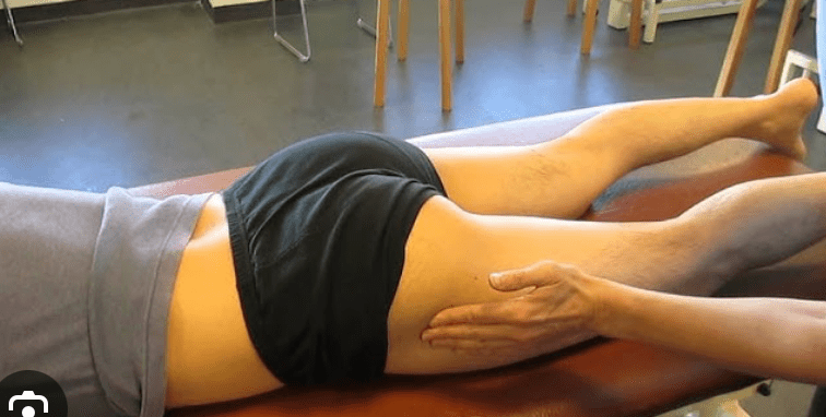
Greater Trochanter
Name the structure indicated by the red arrow
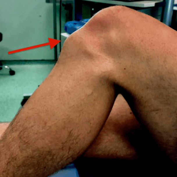
Tibial Tuberosity
Name the structure marked in purple and with the arrow 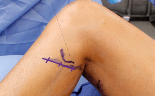
Fibular Head or Neck (Both Accepted)
Name the structure the person is pointing to with his left index finger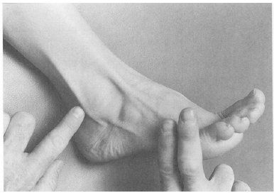
Tendon of the Peroneus Longus
Name the structure being indicated by the person's index finger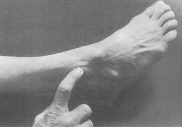
Superficial Peroneal Nerve