Bones
Muscles
Joints and Ligaments
Distal Equine Forelimb
100
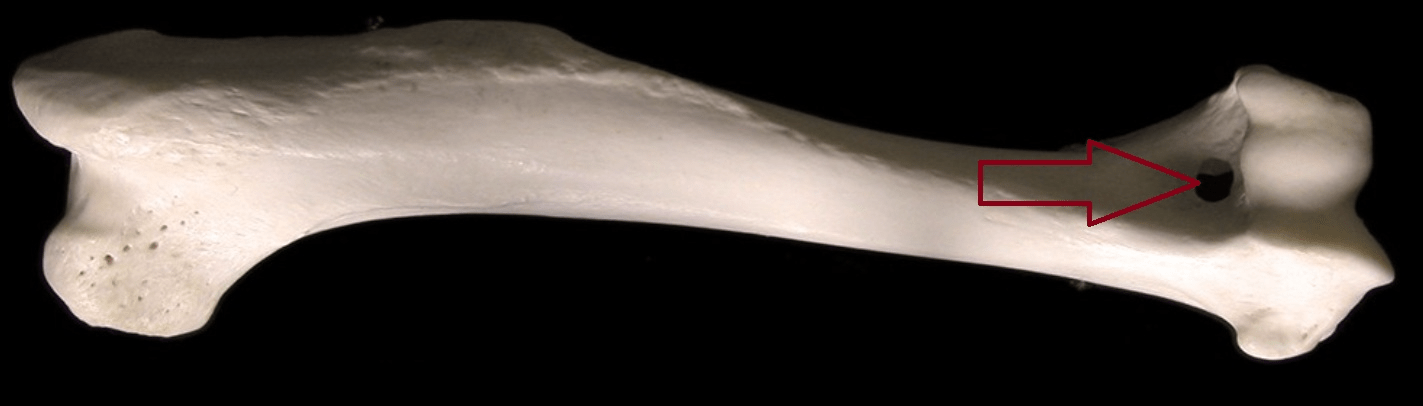
What is the arrow pointing to?
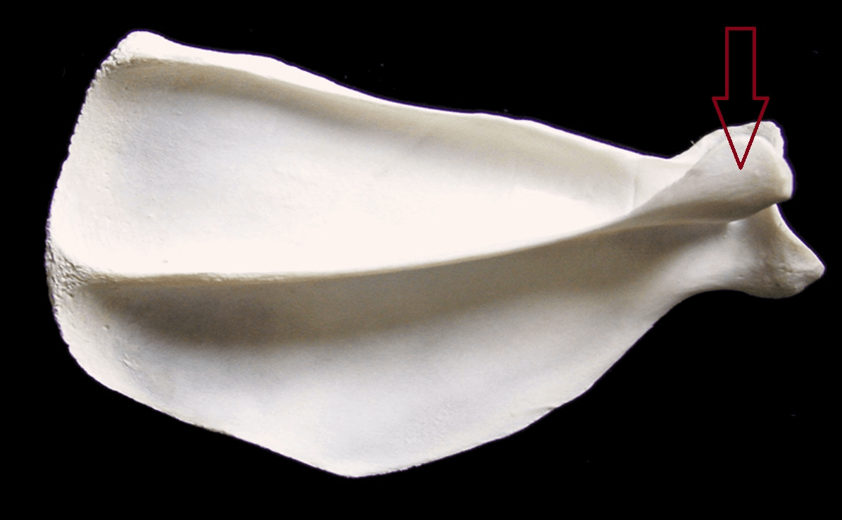
Acromion Process
100
What is the attachment of a limb to the trunk through muscles instead of bones called?
Synsarcosis
100
Which range of motion is greater passive or active?
Passive
100
Name each joint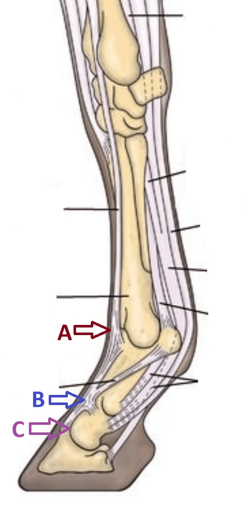
A: Fetlock
B: Pastern
C: Coffin
200
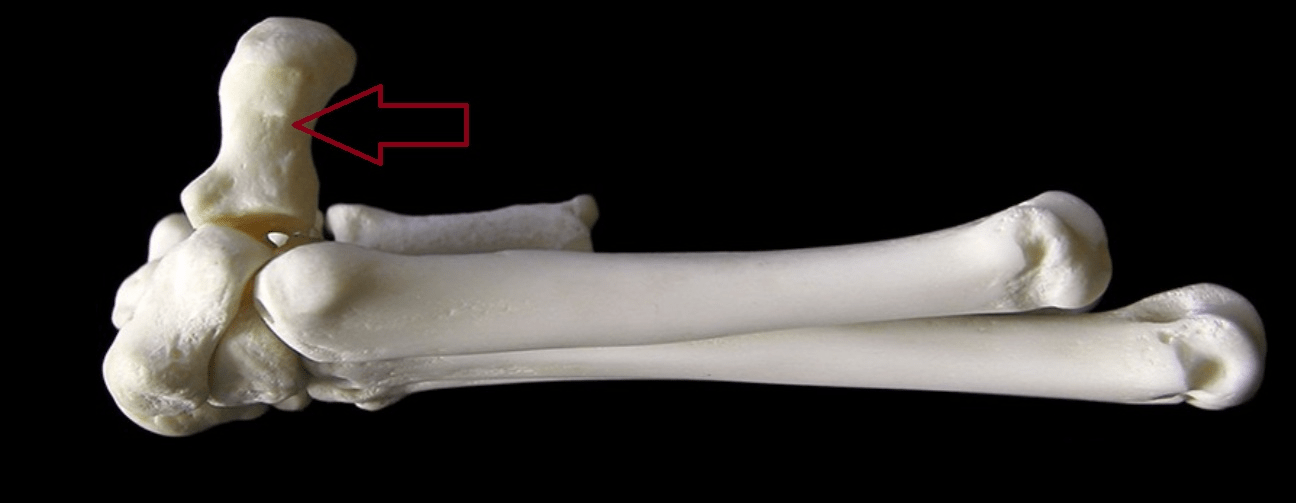
Accessory carpal bone
200
What muscles adduct the forelimb when non-weight bearing?
Superficial and Deep Pectoral m.
200
What ligaments limit the joint to moslty extened and flex and what joint doesn't have them?
The lateral and medial collateral lig.
The shoulder joint
200
What is the other name for the distal sesamoid bone in horses?
The Navicular bone
300
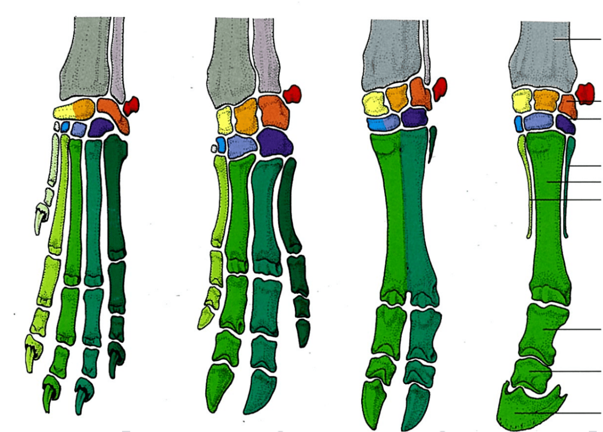
What bone is Orange
Intermediate Carpal bone
300
What are the 4 major muscles on the cariolateral aspect of the forelimb and what is their function?
1. Extensor carpi radialis
2. Common digital extensor
3. Lateral digital extensor
4. Ulnaris lateralis
Extensors of the carpus and digits
300
What is the purpose of the Transverse humeral ligament?
To hold the tendon of the biceps brachii in the intertubercular groove
300
When giving intra-articular injections into the the carpal joint where would you give them?
Antebrachiocarpal joint
Middle Carpal joint
400
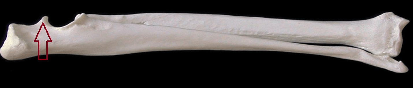
What is the arrow pointing to?
The Supratrochlear foramen
400
A horse presents with atrophy and instability to the shoulder muscles on the right side. What nerve is most likely affected and what is this condition commonly called?
Suprascaplular nerve
Sweeny Shoulder
400
What tendon allows cats to retract their claws?
Deep digital flexor tendon
400
What ligaments/tendons prevent the buckling forward of the pastern joint when the foot hits the ground?
Oblique Sesamoiden lig
Superficial digital flexor tendon
500
Anconeal process
500
A dog presents with a lower radial nerve injury what clinical signs would be present?
Knuckling of the digits and toe dragging due to being unable to extend them.
500
What are the three types of elbow dysplasia?
1. Ununited anconal process
2. Fragmentation of the medial coronoid process
3. Osteochondrosis of the humeral condyle
500
Where does the interosseous lig originate and where does it insert?
Origin: Proximal canon bone
Divides into 2 at distal canon bone
Insertion: abaxial surface of the proximal sesamoid bones and joins with common digital extensor tendon on the dorsal aspect of the proximal phalanx.