Sheep Heart
Heart
Blood vessels
Nervous System Histology
Brain
Special Senses
Nerves
100
What valve is shown here?
Pulmonary semilunar valve
100
This photo represents cardiac muscle. The large, red bands represent what?
Intercalated discs
100
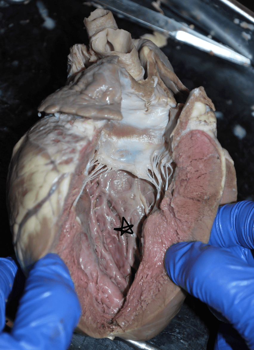
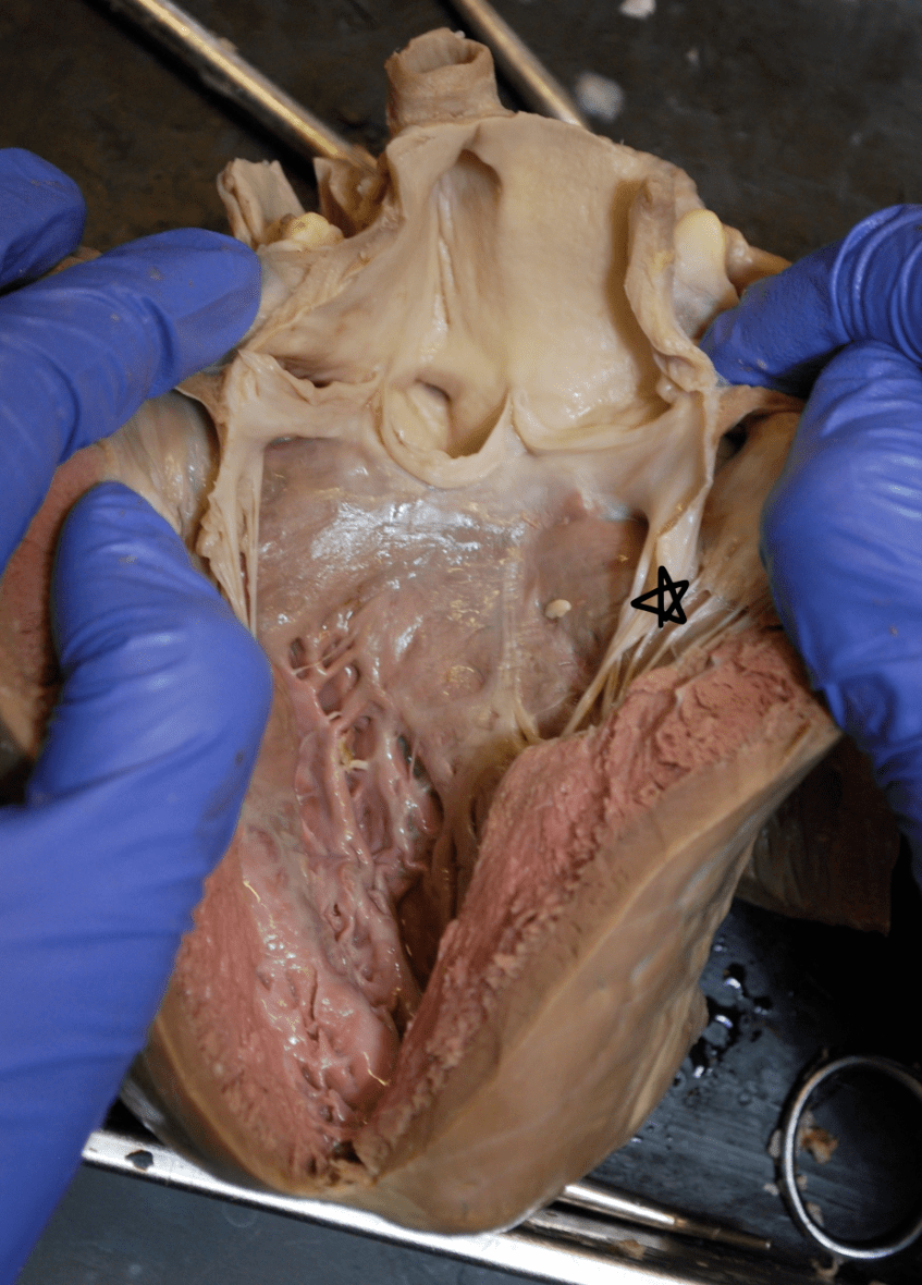
List the structures with a star.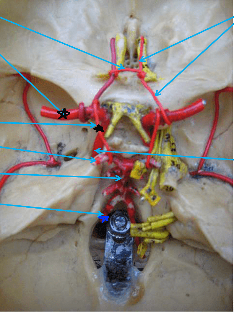
Middle cerebral artery
Posterior communicating artery
vertebral artery
100
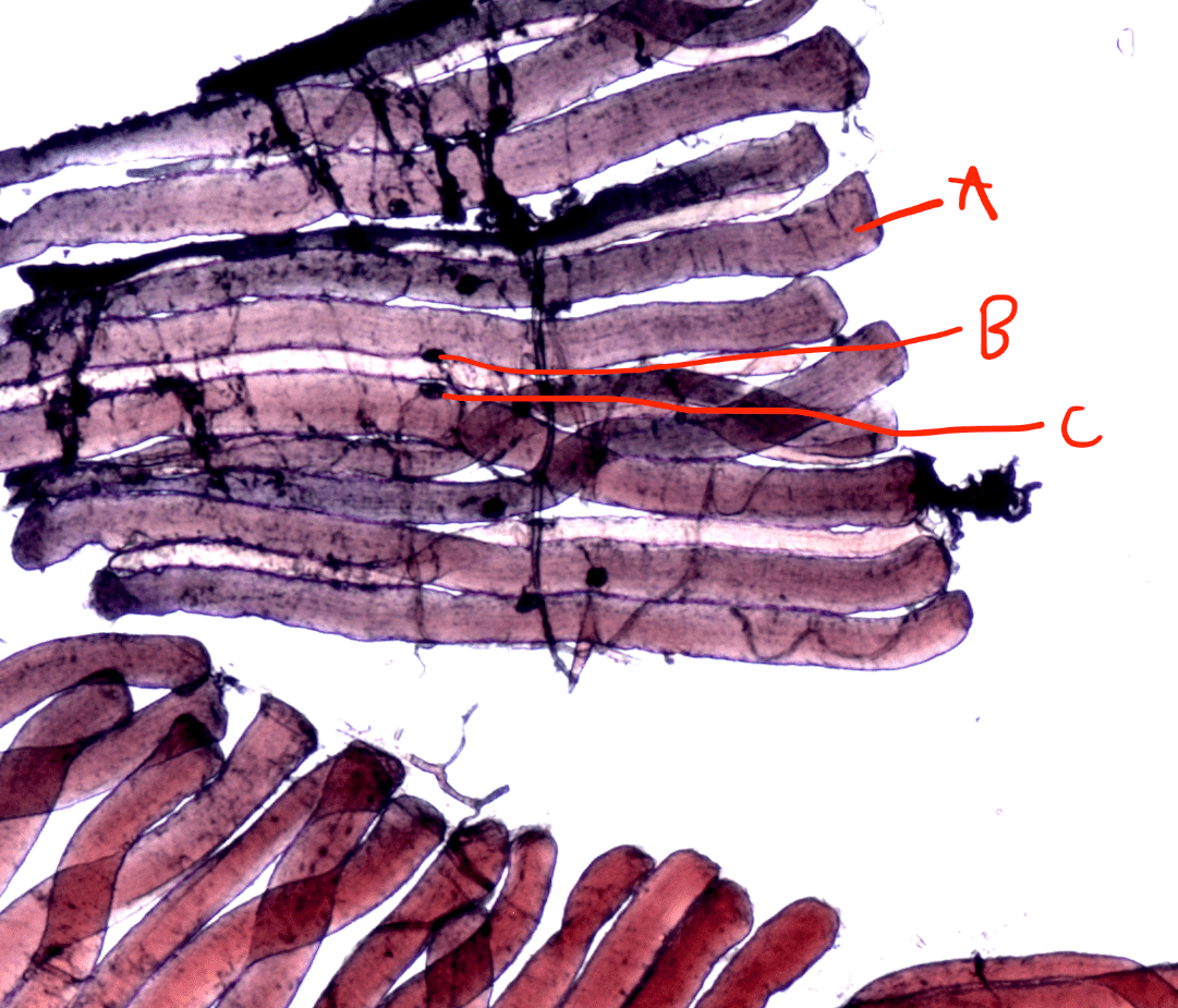
Label A.
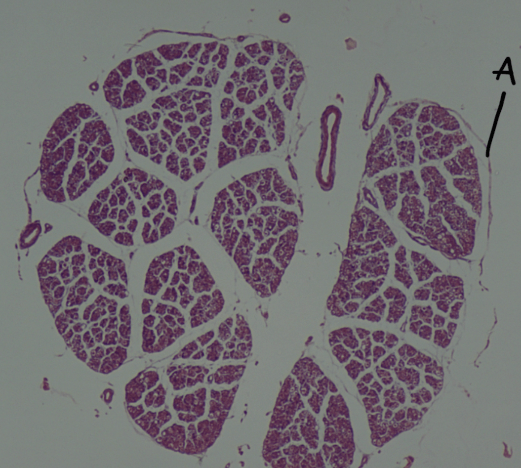
Epineurium
100
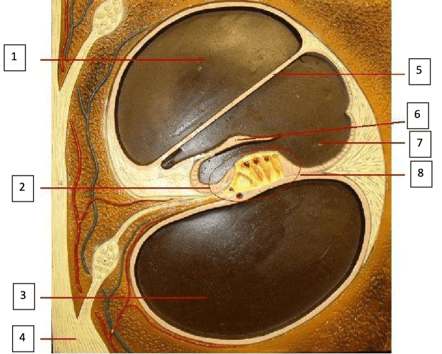
Label 2, 3, 7, 12
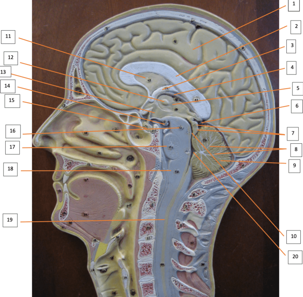
2: Corpus callosum
3: Fornix
7: Corpora quadrigemina
12: Intermediate mass of Thalamus
100
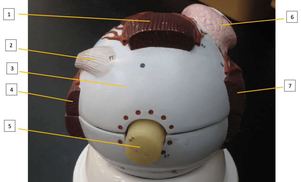
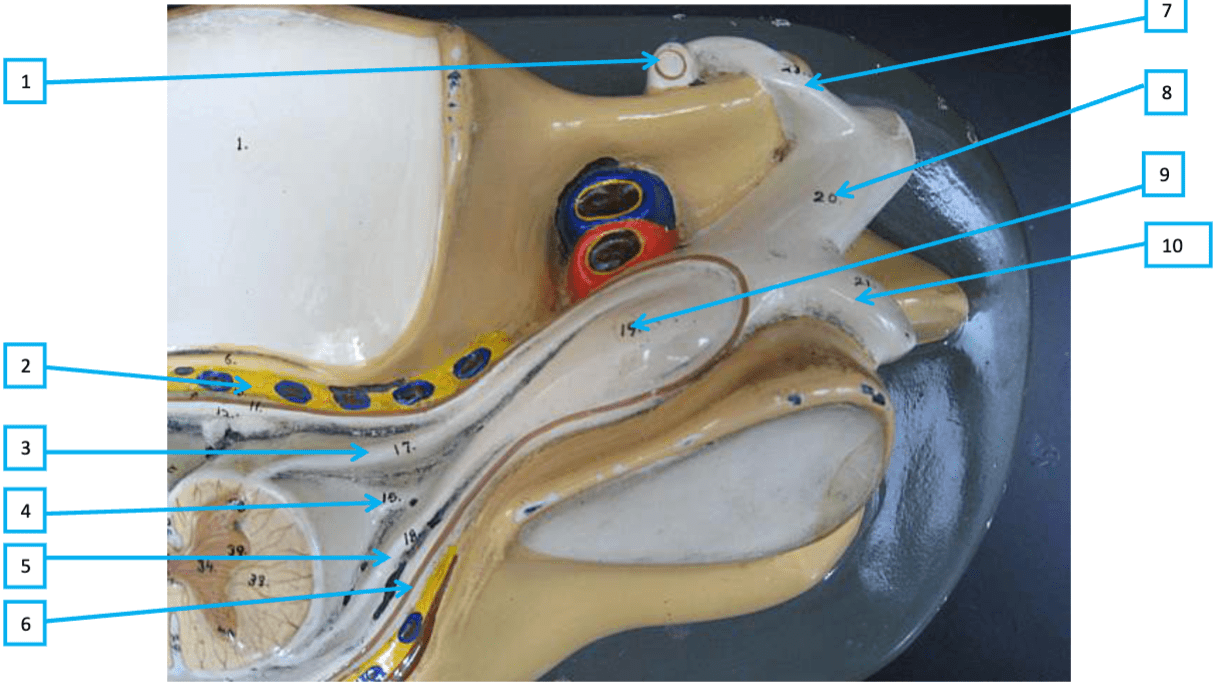
Label 1, 3, 5
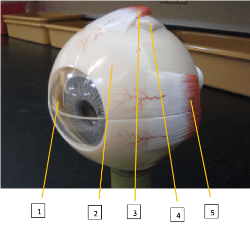
1: Cornea
3: Superior rectus muscle
5: Medial rectus muscle
100
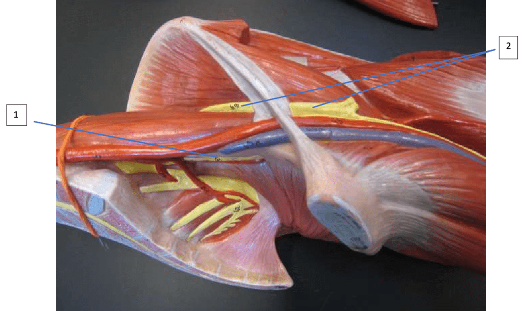
Label 1, 2, 3, 4
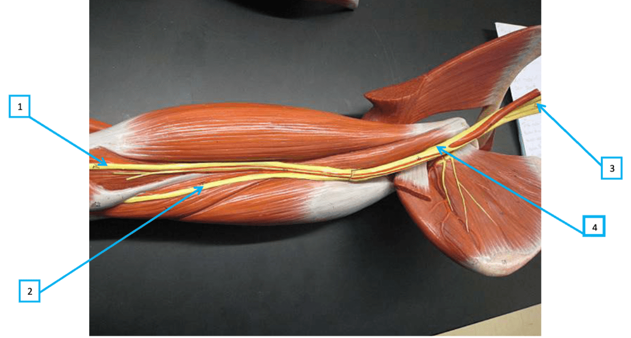
1: Median nerve
2: Ulnar nerve
3: Brachial plexus
4: Musculotaneous nerve
200
What vessel has been dissected in this image?

Pulmonary Trunk
200
List all of the structures that are starred.
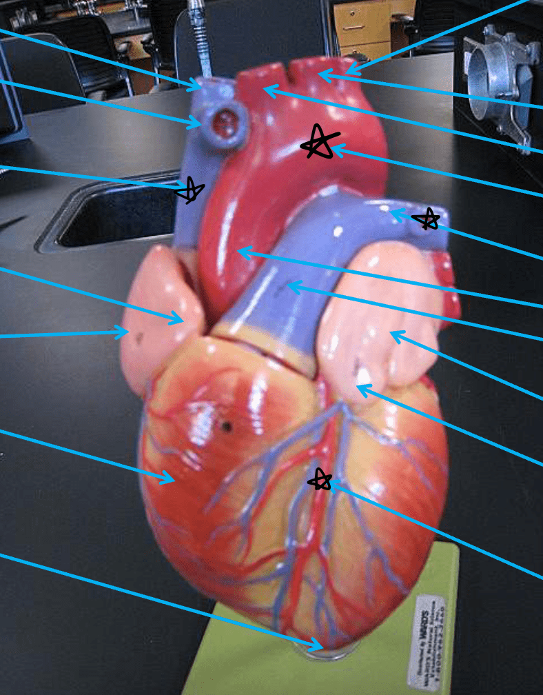
Aortic arch
Superior Vena Cava
Pulmonary artery
Anterior interventricular branch of the left coronary artery
200
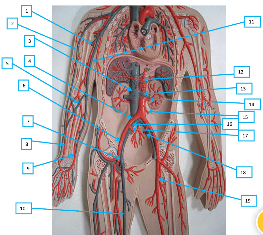
Label structures 2, 3, 4, and 5
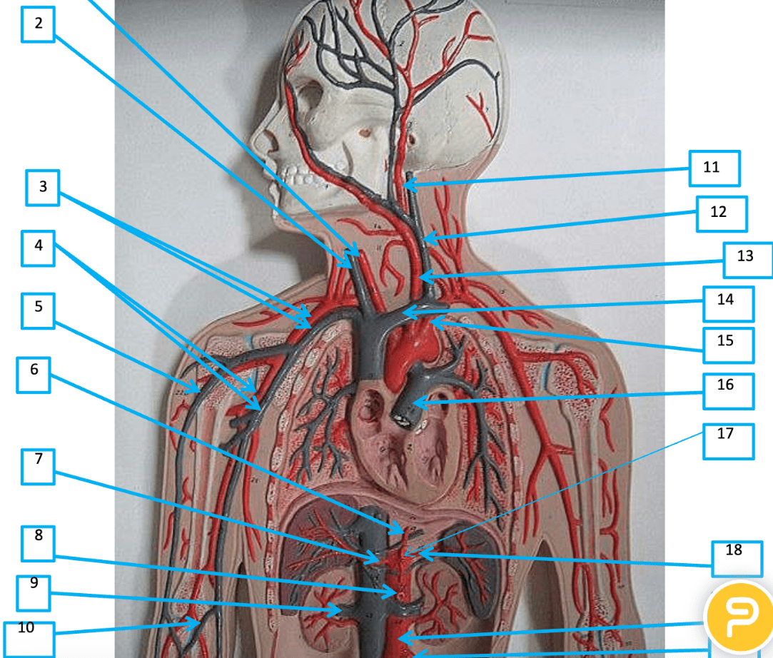
2: Right internal jugular vein
3: Right subclavian artery & vein
4: Right axillary artery & vein
5: Cephalic vein
200
Label A, B, C, D.
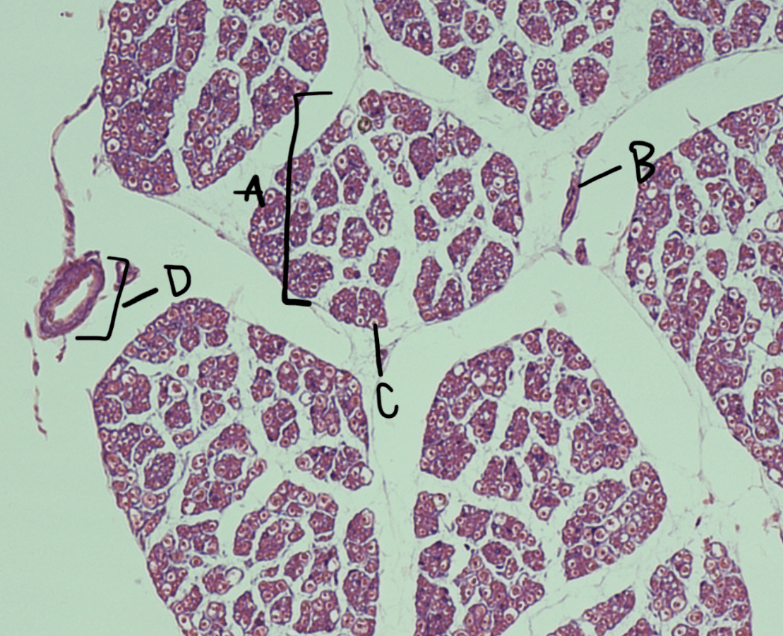
A: Nerve fascicle
B: Perineurium
C: Myelinated axon
D: Blood vessel
200
Label 3, 4, 6, 15, and 17
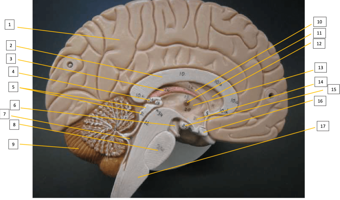
3: Choroid plexus – 3rd ventricle
4: Pineal gland
6: Cerebral peduncle
15: Mammillary body
17: Medulla Oblongata
200
Label 1, 4, 6, 10
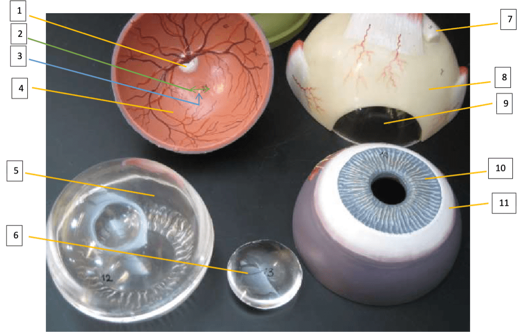
1: Optic disc
4: Retina
6: Lens
10: Iris
200
Label 1 and 2.
1: Obturator nerve
2: Femoral nerve
300
What vessel is denoted by the star?
Left common carotid artery
300
Label the structures that are starred.
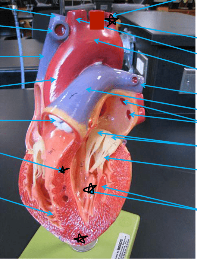
Left subclavian artery
Interventricular septum
Papillary muscle
Apex of the heart
300
Label 1, 11, 18, and 19
1: Right brachial vein
11: R basilic vein
18: L internal iliac vein
19: L femoral artery
300
This is a motor neuron. Label A, B, C.

A: Cell body
B: Nucleus
C: Dendrite/Axon (some sort of projection)
300
Label 5, 8, and 13.
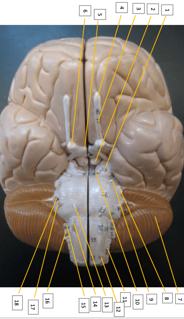
5: Optic chiasm
8: Trigeminal nerve
13: Abducens nerve
300
Label 4, 5, 6, 7
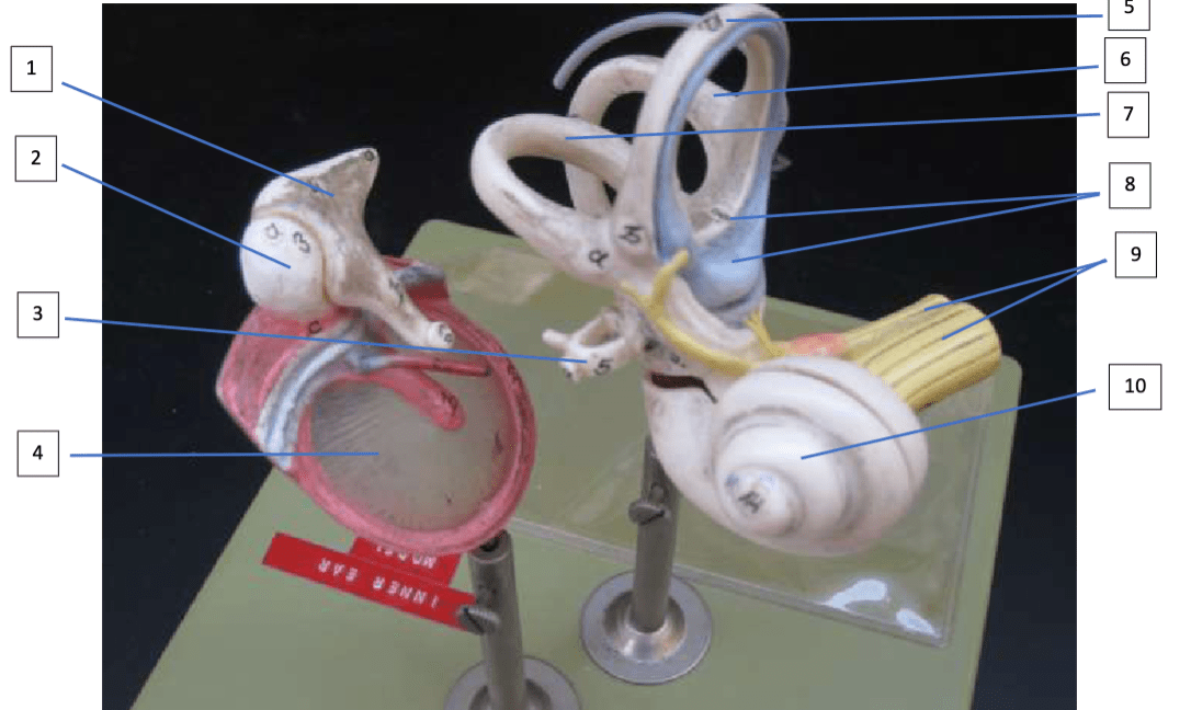
4: Tympanic membrane
5: Anterior semicircular canal
6: Posterior semicircular canal
7: Lateral semicircular canal
300
Label 10, 13, 19

10: Ventral nerve root
13: Dorsal nerve root
19: Epidural space
400
What is this on the ventricle wall?
Trabeculae carneae
400
Label the structures that are starred.

Descending aorta
Azygos vein
Pulmonary vein
Coronary sinus
Inferior Vena Cava
400
Label 1, 2, 9, and 10
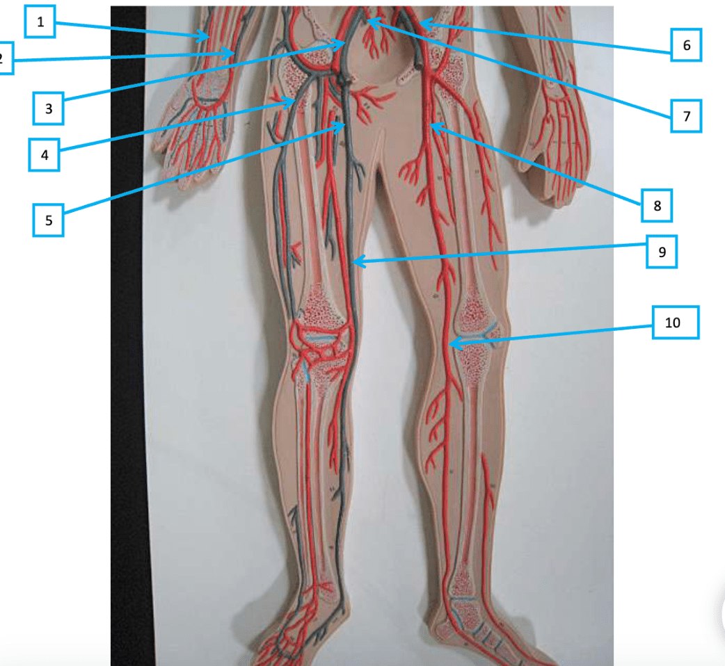
1: R radial artery
2: R ulnar artery
9: R great saphenous vein
10: L popliteal artery
400
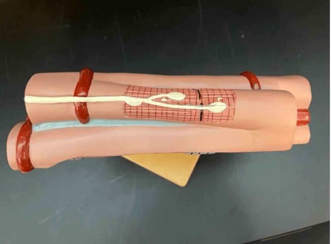
What is being shown in this photo? Also, label A and B. What specialized tissue connects with the synaptic end bulb at C?
Neuromuscular junction
A: muscle fiber
B: Synaptic end bulb
C: Motor end plate
400
Label 6, 8, 12, 14

6: Olfactory tract
8: Oculomotor nerve
12: Vestibulocochlear nerve
14: Hypoglossal nerve
400
Label 1, 3, 5, 6
1: Scala vestibuli
3: Scala tympani
5: Vestibular membrane
6: Tectorial membrane
400
Label 2, 4, 8
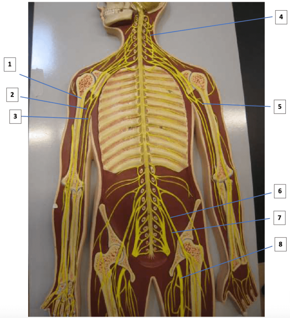
2: Median nerve
4: Phrenic nerve
8: Sciatic nerve
500
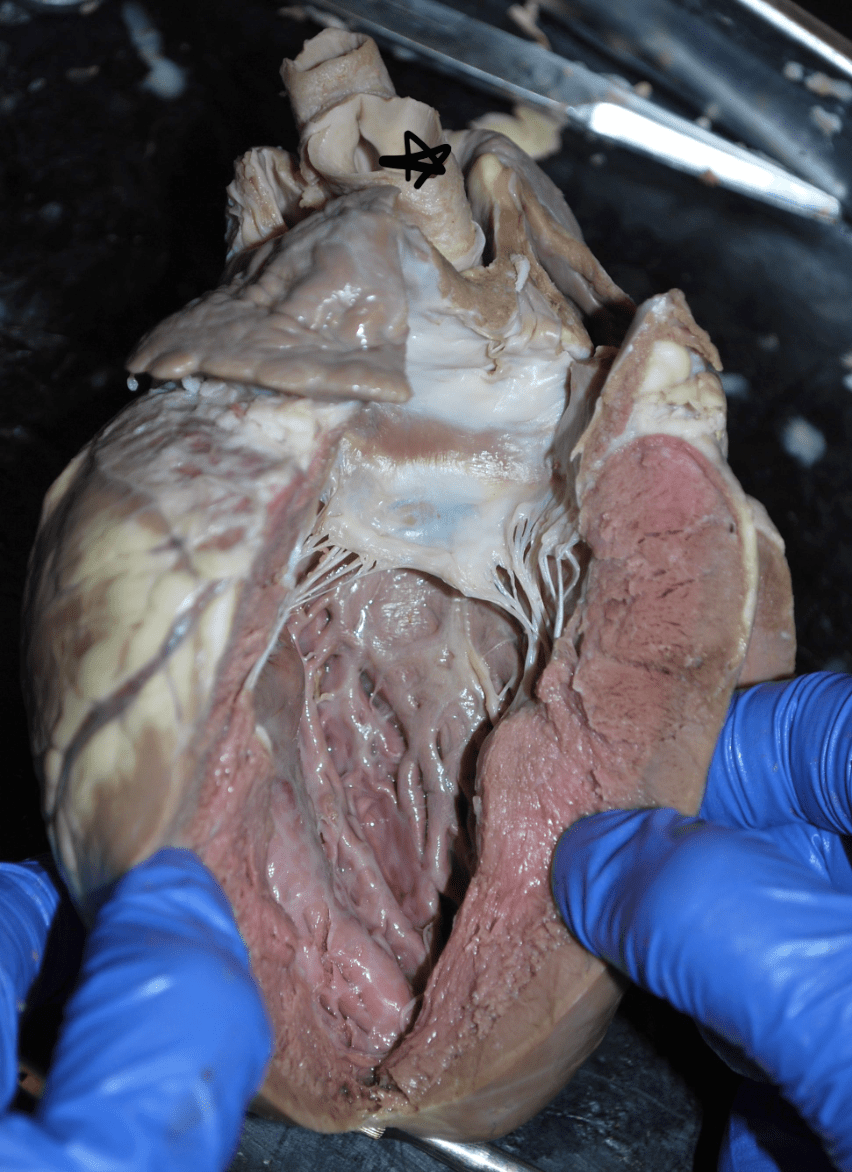
What valve has been dissected here?
Mitral Valve
500
List the structures with a star.
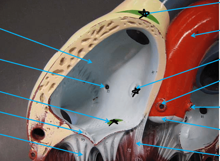
SA node
Fossa ovalis
AV node
500
Label 1 and 2.
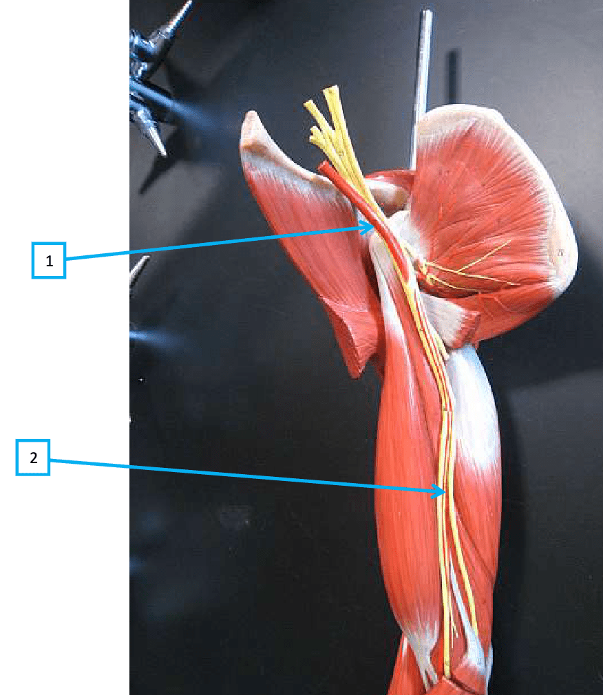
1: Axillary artery
2: Brachial artery
500
Label A.
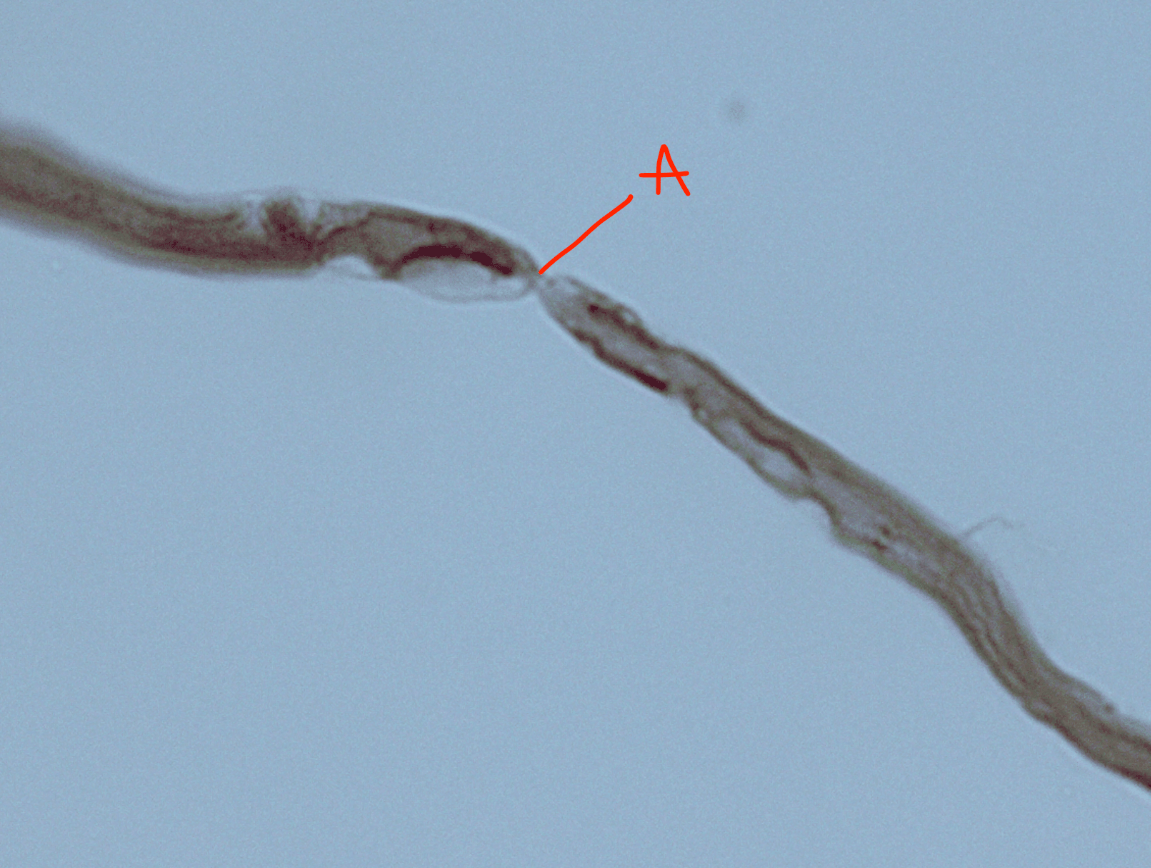
A: Node of Ranvier
500
What skull foramina allow the passage of the Trigeminal (V) nerve?
Superior orbital fissure
Foramen rotundum
Foramen ovale
500
Label 2, 3, 6, 7
2: Tendon of superior oblique muscle
3: Sclera
6: Lacrimal gland
7: Lateral rectus muscle
500
Label 4 and 5.
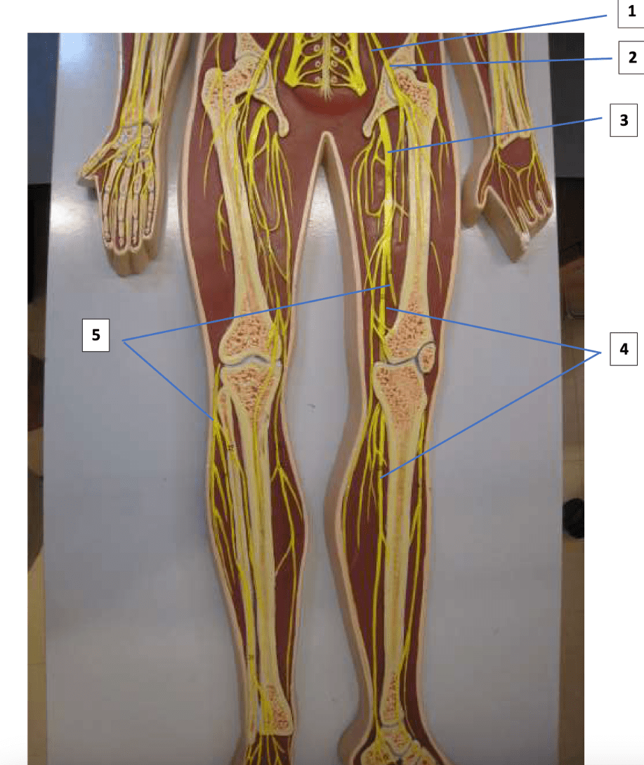
4: Tibial nerve
5: Fibular nerve
600
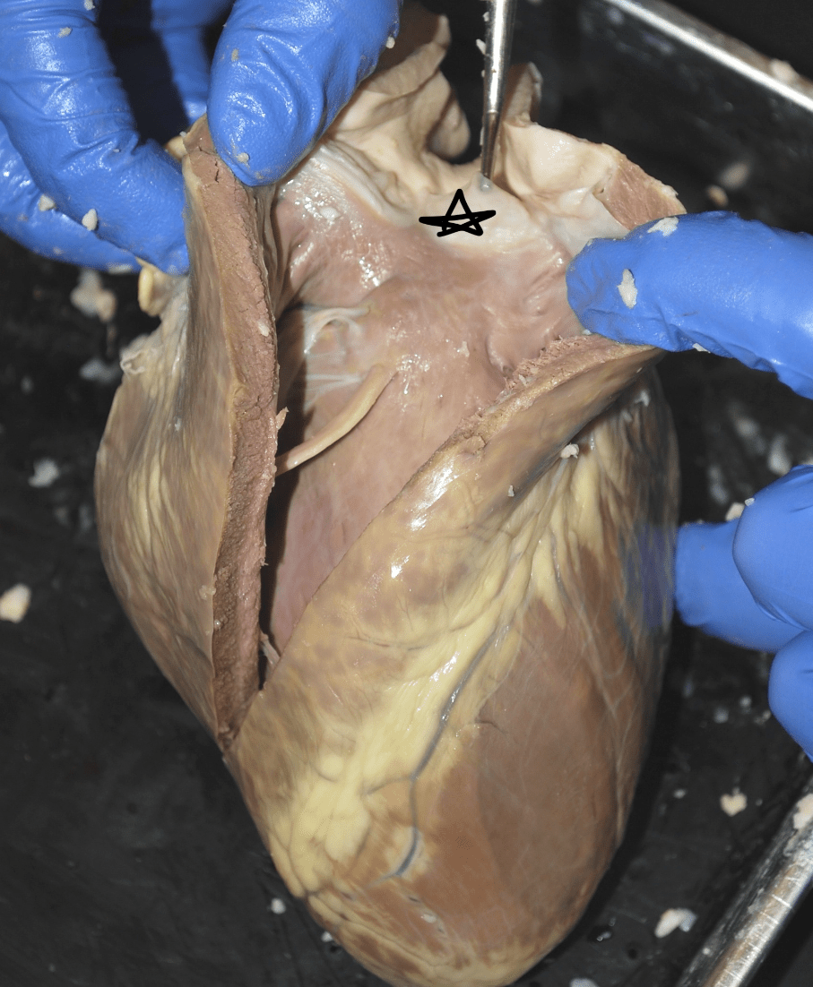
What structure is denoted by the purple star?

Anterior Interventricular sulcus
600
List the structures that have a star.
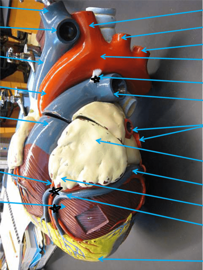
Ligamentum arteriosum
Great cardiac vein
Middle cardiac vein
600
Label 3, 6, 8, 16
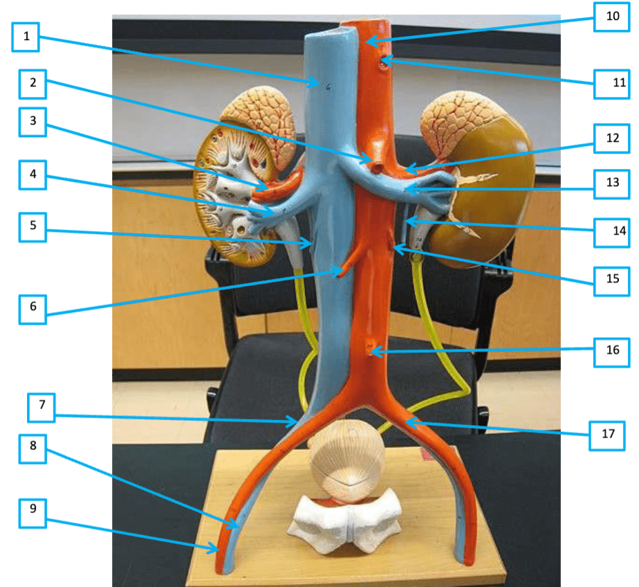
3: R renal artery
6: R gonadal artery
8: R femoral vein
16: Inferior mesenteric artery
600
Label A, B, C, F, and I
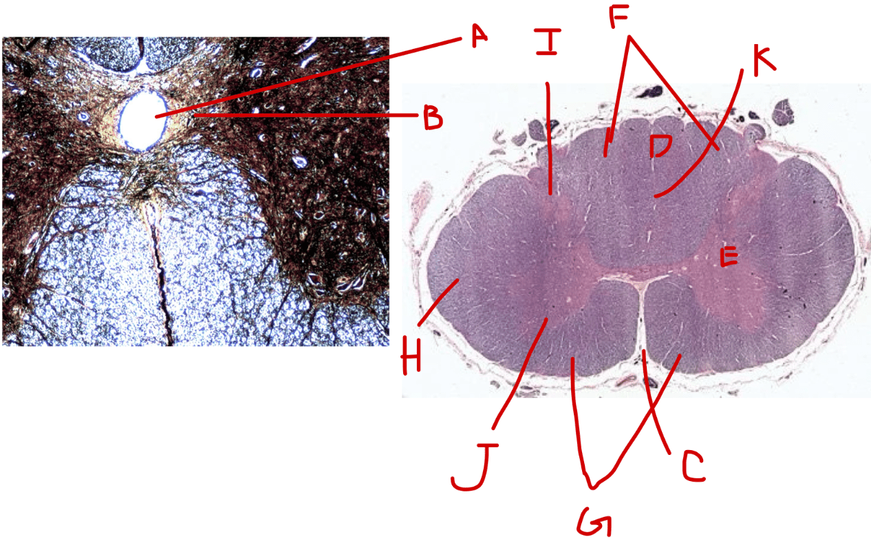
A: Central canal
B: Gray commissure
C: Anterior median fissure
F: Posterior white column
I: Posterior gray horn
600
Which cranial nerves pass through the jugular foramen?
Glossopharyngeal (IX)
Vagus (X)
Accessory (XI)
600
Label C, D, E, F, G
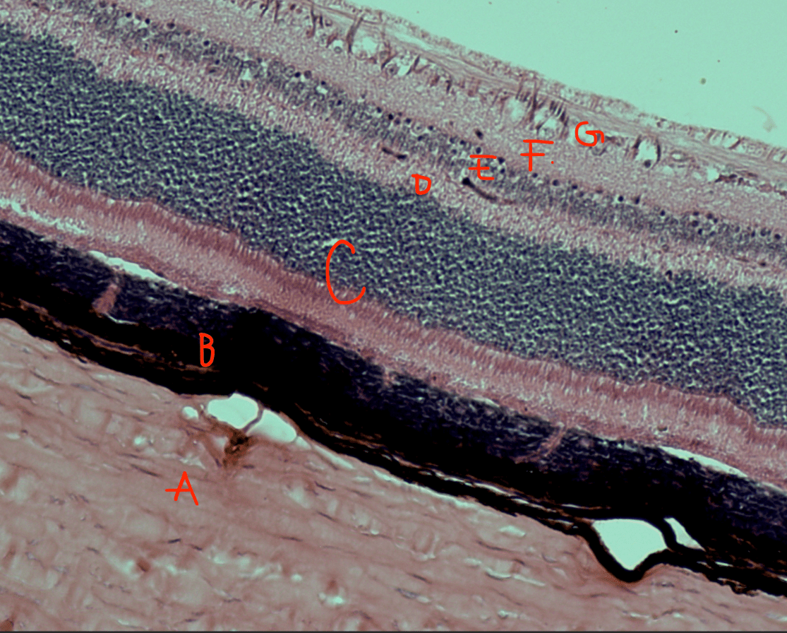
C: Photoreceptor layer
D: Outer synaptic layer
E: Bipolar cell layer
F: Inner synaptic layer
G: Ganglion cell layer
600
Label 7, 8, 9, 10
7: Rami communicantes
8: Ventral ramus
9: Dorsal root ganglion
10: Dorsal ramus