Hernias
Hernias
Pharm and Genetics
Rando
Review
100
These are the boundaries of the inferior lumbar triangle (Petit Triangle)
Superior to Iliac crest
Lateral edge Lat Dorsi
Posterior edge External oblique
Cause: Weakness in the lumbodorsal fascia
Best repaired with mesh anchored to the fascia over the bone.
100
What are the Boarders of th hernia
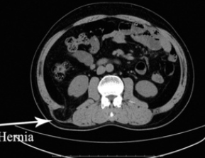
12th rib
Paraspinal muscles (lat edge of quadratus lumborum)
Posteruir edge of Internal oblique
Lumbar hernias are more common in this space than in the Petit Triangle.
Cause: Weakness in the lumbodorsal fascia
Best repaired with mesh anchored to the fascia over the b
100
What is the expected gene mutation seen with FAP?
APC
FAP review:
Associated Tumors to Monitor/ think of:
1. Thyroid
2. Duodenal Adenomas
3. Colorectal Cancer
4. Desmoids
Surgical options:
1. Total colectomy with ileorectal anastomosis
2. Total proctocolectomy w/ or without Jpouch creation.
100
During a total thyroidectomy, the left inferior pole is being dissected out, and the artery is encountered. What is the vessel and its origin?
Inferior thyroid artery from the thyrocervical trunk
100
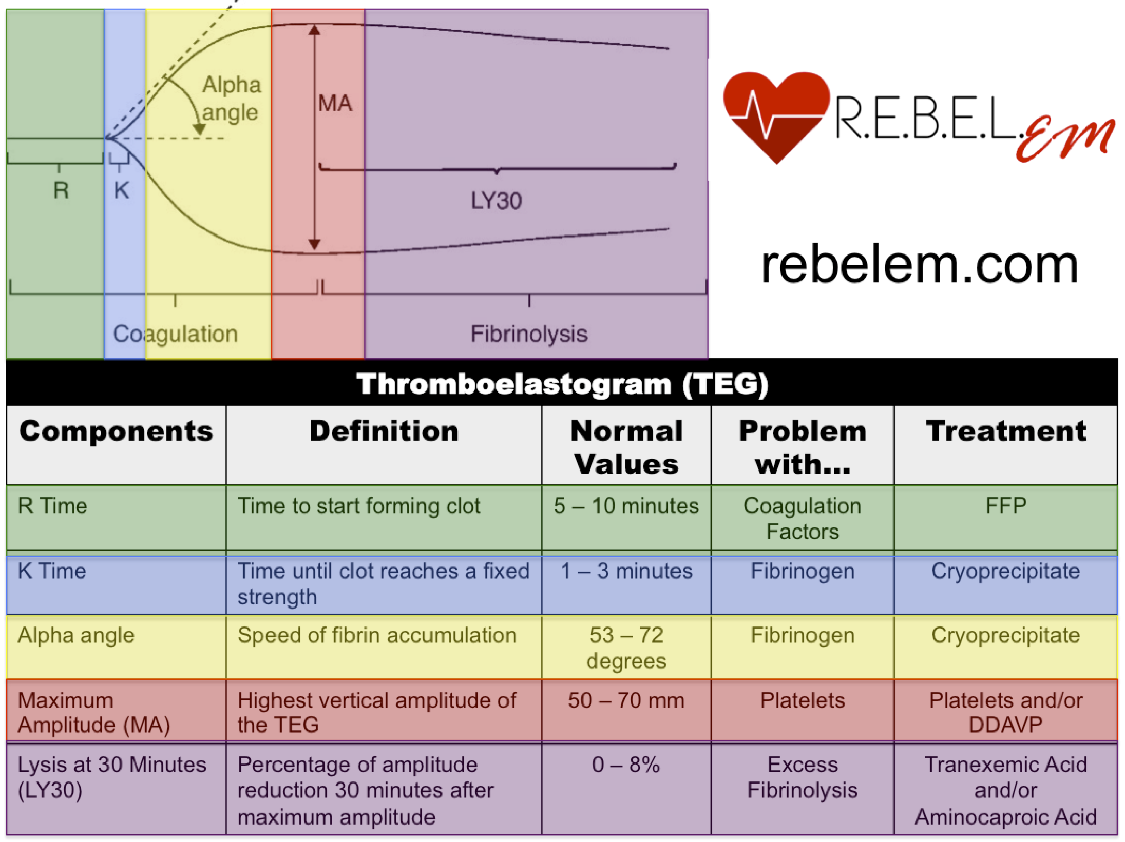
What product (s) do you give a patient with low MA on a TEG?
200
You are performing an uncomplicated open ventral hernia repair using a retromuscular technique in a 45-year-old man and are selecting the mesh to use. There has been no contamination during the case. Which type of mesh is most appropriate in this case?
A. Macroporous midweight polypropylene mesh
B. Polyglycolic acid (Vicryl) mesh
C. Polytetrafluoroethylene (PTFE) mesh
D. Absorbable synthetic mesh
E. Biologic mesh
A. Macroporous midweight polypropylene mesh
Polypropylene mesh is a permanent synthetic mesh that becomes incorporated into native tissue and has a low rate of recurrence. Polyglycolic acid mesh, an absorbable mesh, is often used in contaminated fields because there is no prosthetic material remaining over the long term; however, it does not provide long-term tissue support. Polytetrafluoroethylene, or PTFE, mesh does not become incorporated into native tissue. Synthetic absorbable mesh (BioA, Phasix) is associated with worse long-term outcomes than synthetic nonabsorbable mesh, although the lack of a long-term foreign body may be an advantage in an infected field. Biologic mesh is typically composed of an acellular collagen matrix that theoretically promotes neovascularization and native collagen deposition; thus, it can be used in contaminated fields. However, the high cost of biologic or absorbable synthetic mesh makes it less appropriate for routine repair in clean operative fields.
200
What is the advantage of a posterior component separation vs an anterior?
Less wound complications. Don't need to create large skin flaps.
- There is no difference in the amount of fascial release.
200
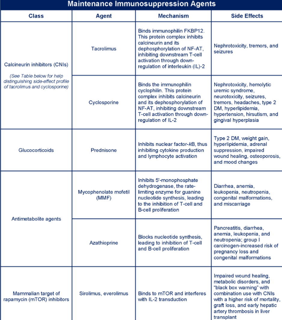
What class of transplant immunology medications causes nephrotoxicity? Give one example.
200
Name at least 6 guidelines for surgical intervention in asymptomatic primary hyperparathyroidism. (As in, when would you operate? There are 8 answers)
- Age younger than 50
- Calcium higher than 1mg/dl obove normal (8.88-10.4)
- Nephrolithiasis
- Nephrocalcinosis
- Osteoporosis w. T-score less than -2.5
- Compression fracture
- Urinary Ca greater than 250ug per 24 hours (Women) or 300up (men)
- Neuropsychiatric symptoms
200
You performed an excisional biopsy for a 44-year-old female who came to you with a breast mass with discordant imaging and pathology findings. Pathology shows lobular carcinoma in situ, Pleomorphic Type with positive margins. How do you counsel the patient?
She will need re-excision for negative margins because it is pleomorphic
If it was the Classic type of LCIS, ADH, ALH, do not need negative margins
DCIS - 2cm Margins
300
A healthy 70-year-old man with a BMI of 30 kg/m² presents to the clinic with a painful bulge at the site of a previous vertical midline incision. On examination, he has an easily reducible mass with a 4-cm fascial defect. What is the best management of this problem?
A. Repair with component separation
B. Reevaluation after weight loss
C. Rechecking monthly to assess change in size
D. Primary fascial repair
E. Repair with mesh placement
E. Repair with mesh placement
Incisional hernias do not improve with time and have a substantial risk of incarceration, so watchful waiting is not optimal in a patient who is a good surgical candidate.
The risk of recurrence with primary closure is as high as 50%, and therefore mesh should be used, especially with a defect larger than 3 cm.
Component separation should not be necessary for this size defect.
A patient with a BMI of 30 does not require weight loss—expert hernia consensus guidelines suggest a BMI <30 is sufficient for hernia repair. BMI >50 should not undergo a hernia repair. For any BMI 30-50, consideration of the size of the defect, ability to repair, and other patient risk factors are evaluated, with the final decision between the patient and surgeon.
300
A 58-year-old woman with hepatitis C cirrhosis (Child-Pugh class B) presents with a long-standing 5-cm umbilical hernia that has been irreducible for over a year. She reports intermittent dull discomfort but no acute pain, no nausea or vomiting, and no obstipation. Examination shows a firm, nontender, chronically incarcerated hernia with normal overlying skin. She has moderate ascites on diuretics. Labs show albumin 3.0 g/dL, total bilirubin 2.6 mg/dL, and INR 1.5. Under what conditions would you repair her hernia? And how would you repair it?
- new tenderness, erythema, or dusky/thinned/taut overlying skin (impending rupture)
- signs of strangulation (severe pain, leukocytosis, lactate elevation)
-bowel obstruction (nausea/vomiting, obstipation, distension, transition point on imaging).
Any of these shifts management from watchful waiting to urgent repair despite the cirrhosis.
Repair: Avoid mesh in the setting of ascites because of markedly increased infection risk; perform a primary, multilayer tissue closure to minimize ascitic leak. Intraperitoneal drain. Judicious volume management.
300
This is the genetic mutation in a patient presenting with midline neck swelling and numerous painless nodules on the lips or tongue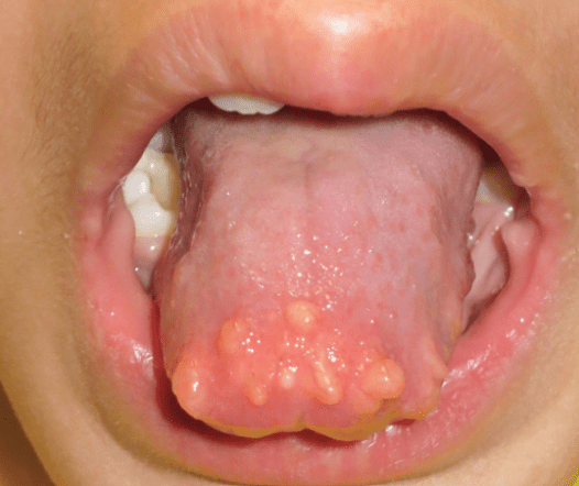
RET
MEN2 is an AD disorder.
MEN2A: Medullary thyroid carcinoma, pheochromocytoma, and hyperparathyroidism
MEN2B: Medullary thyroid carcinoma, pheochromocytoma, Mucosal neuromas, Marfanoid habitus
300
What is the "rule of sixes" for assessing readiness to use an AV fistula?
No deeper than 6mm from the skin
At least 6 mm in diameter
6cm in length
600ml/min flow rate
6 week to mature
300
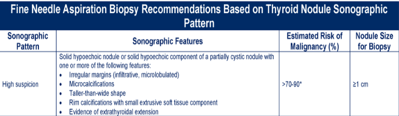
What are the high-risk/suspicious sonographic features seen on imaging for a thyroid nodule? (6)
400
An 82-year-old woman (BMI 18) presents to the emergency department with two days of colicky abdominal pain, nausea, bilious emesis, and obstipation. She has lost 15 lb over the past few months and reports intermittent right medial thigh pain that worsens when she extends and internally rotates the hip and eases when she flexes it. Vitals show a heart rate of 108 and a soft, distended abdomen. CT is shown below. You take her to the OR and are having trouble reducing the hernia what 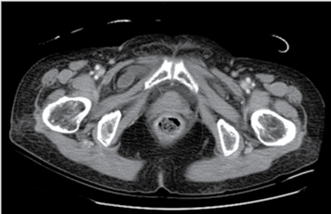
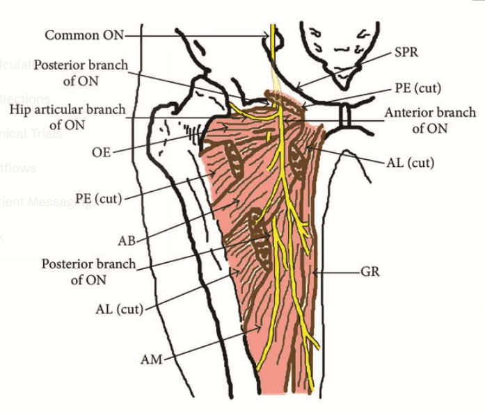
These hernias occur through a weakness in the obturator membrane and are often bilateral. The membrane may need to be incised to enlarge the defect and allow for safe reduction of the incarcerated contents without need for an enterotomy. When incising the membrane, care should be taken to avoid injury to the obturator nerve, vein, and artery, which pierce the membrane at the superolateral border. Incision should be directed inferiomedially.
400
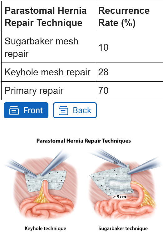
A 61-year-old man with a BMI of 38 kg/m², poorly controlled type II diabetes (HbA1c 9.2%), and an active 20-pack-year smoking history underwent an abdominoperineal resection with end colostomy for rectal adenocarcinoma four years ago. He presents to the surgery clinic with a gradually enlarging bulge around his colostomy and increasing difficulty maintaining an appliance seal, with intermittent leakage and skin excoriation. He denies obstructive symptoms. On examination there is a large, easily reducible bulge encircling the stoma that enlarges with Valsalva. A CT scan confirms a loop of colon and omentum herniating through the parastomal fascial defect, without evidence of obstruction or strangulation. He is highly motivated to proceed with definitive repair.
What is the most appropriate management for this patient?
A. Primary suture (fascial) repair of the parastomal defect
B. Open onlay biologic mesh repair
C. Stoma relocation to the contralateral abdominal wall without mesh
D. Minimally invasive (laparoscopic or robotic) mesh repair using a Sugarbaker technique
E. Continued nonoperative management with a hernia support belt indefinitely
D. Minimally invasive (laparoscopic or robotic) mesh repair using a Sugarbaker technique
Patients with a symptomatic parastomal hernia and risk factors for wound complications and recurrence (obesity, diabetes, smoking) are preferentially managed with a minimally invasive, mesh-reinforced repair. Mesh reinforcement markedly reduces recurrence versus primary suture repair, and the minimally invasive approach reduces wound-related morbidity compared with open repair in high-risk patients.
400
A 72-year-old man presents with a recurrence of non–small cell lung cancer following neoadjuvant chemotherapy and resection. On further evaluation, tissue from a biopsy is found to express significant levels of programmed death ligand-1, making this tumor amenable to treatment with which agent?
A. High-dose interleukin-2
B. Nivolumab
C. Ipilimumab
D. Rituximab
E. Infliximab
B. Nivolumab
Nivolumab, pembrolizumab and atezolizumab=monoclonal antibodies disrupting the interaction between the T-cell protein programmed death-1 and its ligand programmed death ligand-1.
These drugs are finding a role in the treatment of melanoma, non–small cell lung cancer, renal cell carcinoma, and urothelial carcinoma.
- Interleukin 2 (IL-2)= T-cell activation and proliferation.
IL-2 can result in durable responses in a small percentage of patients with renal cell carcinoma or melanoma.
- Ipilimumab = monoclonal antibody targets CTLA-4.
Downregulating T-cell responses. This drug plays a role in the management of melanoma.
- Rituximab = monoclonal antibody against B-cell protein CD20.
The drug has an important role in treatment of B-cell leukemias and lymphomas as well the B-cell–mediated autoimmune disease rheumatoid arthritis.
- Infliximab = Blocks Tumor Necrosis Factor (TNF)
Can be used in the management of immune-mediated diseases such as rheumatoid arthritis, ankylosing spondylitis, inflammatory bowel disease, psoriasis, hidradenitis suppurativa, and refractory asthma.
400
These are the indications for parathyroidectomy in primary hyperparathyroidism. (7)
- Age less than 50 years
- Laboratory studies: serum calcium greater than 1 mg/dL above the upper limit of normal
- Bony fracture
- Osteoporosis as measured with a bone density T-score less than or equal to –2.5 on DEXA at any site
- GFR less than 60 mL/min
- Nephrolithiasis or nephrocalcinosis
- Hypercalciuria (> 250 mg/d for females and > 300 mg/d for males)
400
A 69-year-old woman presents with several months of gradual abdominal distention and a 30-lb weight gain. Her history is notable for 15 years of work in a shipyard, coronary artery disease, prior tobacco use, and stage II breast cancer treated with surgery and chemotherapy 12 years ago. A screening colonoscopy 3 years ago was normal. She has no changes in bowel function. On examination, she has a distended, mildly tender abdomen with a fluid wave; there is no palpable pelvic or breast mass and no lymphadenopathy. CT of the chest, abdomen, and pelvis shows moderate ascites, diffuse smooth and nodular peritoneal thickening with an "omental cake," mesenteric infiltration, small bilateral pleural plaques, and scattered sub-centimeter bilateral lung nodules.
What is the most likely diagnosis?
A. Peritoneal carcinomatosis from metastatic lung cancer
B. Peritoneal carcinomatosis from breast cancer
C. Peritoneal carcinomatosis from metastatic colon cancer
D. Peritoneal carcinomatosis from gastric cancer
E. Primary peritoneal mesothelioma
E. Primary peritoneal mesothelioma
In a patient with a documented history of asbestos exposure who presents with isolated peritoneal disease — ascites, peritoneal and mesenteric thickening, omental caking, and accompanying pleural plaques — without an identifiable dominant primary tumor, primary peritoneal mesothelioma should be strongly suspected. Sx typically include abd pain, weight gain, and increased abd girth.
A. Metastatic lung cancer — Peritoneal metastasis from lung cancer is uncommon and almost always occurs in the setting of a dominant lung primary and widespread systemic (brain, bone, liver, adrenal) disease rather than isolated peritoneal disease.B. Breast cancer — Peritoneal spread from breast cancer typically occurs late, in the context of established metastatic disease elsewhere (bone, liver, nodes), and most often with lobular histology. Isolated peritoneal recurrence 12 years out from stage II disease with no locoregional or systemic recurrence would be distinctly unusual
C. Colon cancer — A normal colonoscopy 3 years ago and the absence of GI symptoms, obstruction, or a colonic mass make colorectal carcinomatosis unlikely.
D. Gastric cancer — Gastric carcinomatosis usually presents with GI symptoms (early satiety, weight loss, obstruction) and often gastric wall thickening/linitis plastica; none are present here
.
500
You are performing an open inguinal hernia repair with mesh (Lichtenstein). How do you do this? Oral boards style.
1. Make an oblique incision 1- 2 cm above the inguinal ligament
2. Expose and incise the external oblique aponeurosis in the direction of its fibers, exposing the spermatic cord and protecting the ilioinguinal nerve
3. Mobilize and isolate the spermatic cord with a Penrose drain
4. Reduce the hernia contents (If indirect, may choose to open, inspect, reduce, and ligate the sac at the level of the internal ring)
5. Perform a tension-free repair w/ permanent mesh secured to the periosteum of the pubic tubercle with 2cm overlap, conjoint tendon, and shelving edge of external oblique
6. Slit the mesh to accommodate the spermatic cord and recreate the internal ring by suturing the tails together
7. Ensure hemostasis and close the external oblique fascia, Scarpa's, and skin.
500
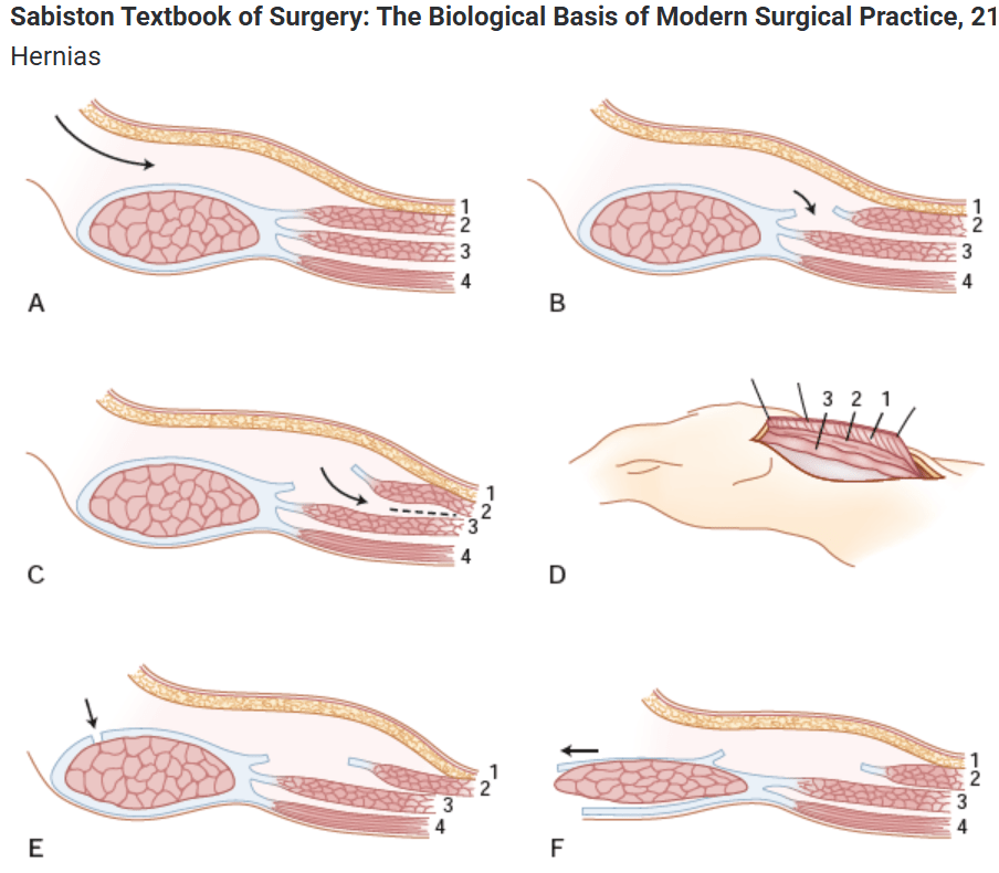
You are taking a patient to the OR for a complex midline ventral hernia with loss of domain. Assuming it is large enough to require component separation, how will you perform an open posterior OR anterior component separation? Oral Boards Style
Predictive rule of thumb Carbonell Rule: If the defect width is ≥ twice the width of the retromuscular space on one side, a component separation will most likely be required to close the defect and set the mesh without tension
POSTERIOR
(Consider saying DVT PPX and Abx)
1. Open midline, isolate hernia sac and fascial defect
2. Incise the retrorectus spaces 1cm off midline to the linea semilunaris, preserving the neurovascular bundles.
3. Incise the retrorectuc sheath just medial to the perforating NV bundles, exposing the transversus abdominis muscle
4. Divide the transversus abdominis muscle
5. Continue the dissection out to the psoas
6. Perform the same dissection on the contralateral side.
7. Close posterior fascia and peritoneum. Place mesh in the retromuscular space.
8. Place drains over the mesh. Reapproximate the fascia and close.
ANTERIOR
1. Open midline, isolate hernia sac and fascial defect
2. Create subcutaneous flaps past the linea semilunaris, taking care to preserve NV bundles
3. Divide the external oblique aponeurosis 2cm lateral to the semilunar line
4. Develop a plane between that aponeurosis and the internal oblique muscles laterally.
5. Repeat on the contralateral side.
6. Close the fascia primarily (underlay or onlay mesh is optional)
Notes: If recurrent and mesh in place, need to excise mesh and non-absorbable sutures, lyse adhesions to gain working room and laxity in abd wall, evaluate for mesh erosion into bowel
500
A 41-year-old man received a kidney transplant 4 months ago for kidney disease secondary to DM and HTN. The patient returns for routine follow-up. He denies any urinary symptoms. Vital signs are HR 80 beats/min, respiratory rate 12 breaths/min, BP 130/60 mm Hg, and O2 saturation 98% on room air. The man is taking tacrolimus 6 mg twice daily, mycophenolate 720 mg twice daily, and prednisone 5 mg daily for immunosuppression. His latest blood work shows BK viremia, with a level of 20,000 copies/mL. What is the initial treatment for the BK viremia?
A. Leflunomide
B. Cidofovir
C. Levofloxacin
D. Discontinuation of mycophenolate
E. Admission for IV steroids
D. Discontinuation of mycophenolate
First-line treatment of BK viremia in the setting of transplantation is to reduce immunosuppression, and mycophenolate should be discontinued.
There is no indication for IV steroid treatment; this is administered for rejection and will increase the patient’s level of immunosuppression.
Leflunomide, cidofovir, and levofloxacin are second-line options for BK viremia if the viral level remains high despite reduced immunosuppression.
500
A 48-year-old patient with a history of DM, HTN, and chronic kidney disease is now 2 weeks status-post–kidney transplant (deceased donor). She comes into the office for a visit because of a low-grade fever and oliguria at home. She is found to have elevated creatinine and low-grade fever. She is admitted, and a workup, including a kidney biopsy, is performed. The biopsy demonstrates evidence of rejection. The pathophysiology of this patient’s rejection can be best described as?
A. Antibody-mediated
B. Interleukin-6–mediated
C. T-cell–mediated
D. B-cell–mediated
E. Interstitial fibrosis
C. T-cell–mediated
The patient in this question is presenting with acute rejection. Rejection is classified by timing as hyperacute, acute, or chronic. Rejection can also be differentiated based on the immune response.
- Hyperacute allograft rejection: within minutes to hours of placement of the graft and is antibody-mediated, specifically donor-specific preformed antibodies.
- Acute allograft rejection: mediated by T cells, can occur days to 6 months posttransplant.
- Chronic rejection: Months to years. Interstitial fibrosis. cause of chronic rejection is still poorly understood and variable.
500
This is how you do a Laparoscopic right (abdominal, not RP) adrenalectomy. Oral Boards style
1. Enter the abdomen (technique of choice), place ports under direct visualization, and triangulate to the lesion
2. Mobilize the liver and take down the triangular ligament to expose the retroperitoneum ( Left: mobilize the splenic flexure, spleen, and pancreas medially) to expose the adrenal gland
3. Dissect along the border of the adrenal and ligate the R adrenal V taking care not to avulse it from the IVC. (left adrenal v drains into the L renal v)
4. Completely mobilize the gland and remove it with an endoscopic retrieval bag
5. Ensure hemostasis
6. Desufflate the abdomen, close fascia and port sites