Figure 1
Figure 2
Figure 3
Figure 4
Figure 5
100
 This main technique was used to obtain the results in Fig. 1
What is fluorescence in situ hybridization (FISH)
100
This region of the RhoA mRNA was investigated in Fig. 2
What is the 3' UTR (untranslated region)
100
The authors wanted to understand this about Sema3A-mediated growth cone collapse in the experiment associated with Fig. 3
What is the ability of Sema3A to induce RhoA translation
100
This was the purpose of the experiment that produced the results in Fig. 4
What is testing whether Sema3A induces translation of a RhoA reporter
100
The authors used this technique to shut down RhoA activity in the DRG cells
What is small interfering RNA (siRNA)
200
 The authors used this secondary technique (results pictured above) to further validate their findings
What is RT-qPCR
200
The authors used this type of mechanism to deliver the heterologous mRNA to the target cells
What is the Sindhis pseudovirus
200
 When the authors added Sema3A to severed DRG axons, they made the following observation:
What is an increase in RhoA immunofluorescence in the Sema3A condition relative to the vehicle (control)
200
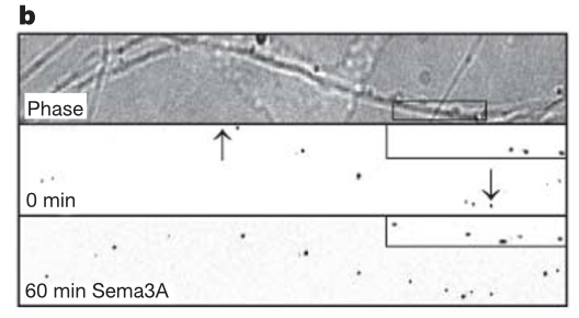 The "puncta" shown in Fig. 4 indicate these structures
What are RNA granules
200
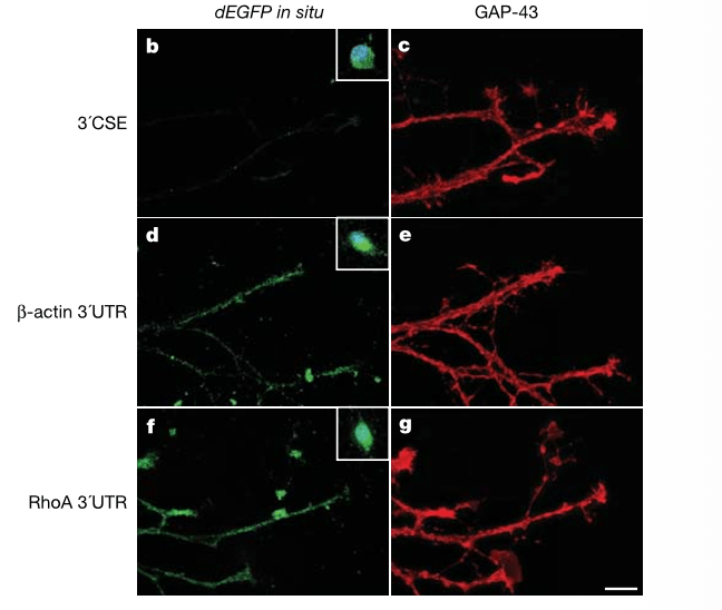
RhoA 3' UTR and CSE 3' UTR transcripts differ in this main way
What is their localization: RhoA 3' UTR transcripts can localize to the axon, while CSE 3' UTR transcripts are restricted to the soma.
300
The authors used this model organism (and specific cell type) in the study
What is the rat dorsal root ganglia (DRG)
300
 The authors used this technique to evaluate the localization of the mRNA in Fig. 2
What is fluorescence in situ hybridization (FISH)
300
The authors used these two ribosomal inhibitors to obtain the results in Fig. 3
What are anisomycin and rapamycin
300
Treatment of DRG explants (infected with the myr-dEGFP and RhoA 3' UTR viral construct) with Sema3A resulted in this observation
What is the appearance of new puncta and the increased intensity of existing puncta in the axons
300
The authors observed this after treating DRG explant cells with EGFP-RhoA3'CSE viral transcripts
What is the restoration of Sema3A-mediated growth cone collapse
400
The authors examined this other cytoskeletal component in addition to RhoA
What is β-Actin
400
The 3' CSE construct played this role in the study
What is a control (no axonal localization expected)
400
The authors made this conclusion after seeing the results pictured in Fig. 3g
What is that the increase in immunofluorescence in the presence of Sema3A was specific to RhoA (did not include GAP-43)
400
 Why are there already puncta present in the middle frame?
What is: the RNA granules exhibit baseline translational activity. Sema3A amplifies the system, but it does not create it.
400
The authors used rapamycin for this reason
What is the ability of rapamycin to block cap-dependent translation
500
These probes were used to hybridize with the mRNA in Fig. 1
What is digoxigenin-labeled (DIG) riboprobes
500
 Based on the results shown in Fig. 2, the authors concluded this about RhoA mRNA
What is the 3' UTR of RhoA is sufficient for targeting the mRNA to axons and growth cones
500
 The results of testing with ribosomal inhibitors suggests what about RhoA mRNA and Sema3A?
What is that Sema3A-mediated changes in fluorescence depend upon translation of RhoA
500
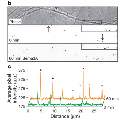 Figures B and C are related in this way
What is: Figure C is the quantification in average pixel intensity of the middle frame of B (green) and bottom frame of B (orange). Triangles denote prior existing puncta that are brighter, and asterisks denote new puncta.
500
What did the authors observe when DRG neurons infected with IRES-EGFP-RHoA were treated with Sema3A in the presence of rapamycin?
What is the restoration of Sema3A-mediated growth cone collapse (rapamycin did not block growth cone collapse)