Bad Blood
Pour some sugar on me!
Hit me with your best shot!
Stayin' Alive
Every breath you take
PEM-pourri
100
An 8-year-old previously healthy girl presents to the emergency department with bruising. She reports no recent ill symptoms or pain. She had an appendectomy earlier this year without any complications but has noted recent diffuse bleeding when she brushes her teeth and blood in her urine. She has an area of coalescing petechiae over her bilateral inner thighs that started after she rode a bicycle yesterday. Which of the following laboratory results are most likely in this patient?
- A. Abnormal direct antiglobulin test
- B. Factor V Leiden mutation
- C. Prolonged partial thromboplastin time
- D. Thrombocytopenia
D. Thrombocytopenia
Children with bleeding from low or dysfunctional platelets tend to have petechiae, excessive bleeding after minor cuts, and mucocutaneous bleeding from brushing teeth, epistaxis, or hematuria. Bleeding from clotting factor deficiencies or alloantibodies to factor deficiencies (inhibitors) presents with hemarthrosis, deep muscle hematomas, or large areas of ecchymosis and rarely presents with mucocutaneous bleeding or petechiae. Bleeding after surgeries is common with both platelet disorders and factor deficiency and, if not present in the patient’s history, suggests an acquired cause. Most congenital causes of bleeding are due to von Willebrand disease (a disorder that prevents platelet aggregation) or to factor deficiencies, such as factor VIII deficiency (hemophilia A) and factor IX deficiency (hemophilia B). While it is possible to acquire a relative factor deficiency due to alloantibodies to a specific factor, this is usually only seen in older adults and only in children already receiving factors for a known deficiency. Acquired alloantibodies to platelets is relatively common in children presenting with extreme thrombocytopenia, which presents with petechiae and mucocutaneous bleeding, as in this patient.
An abnormal direct antiglobulin test (A) suggests alloantibodies to red blood cells in circulation, as seen in hemolytic anemia, not a bleeding disorder.
A mutation in factor V Leiden (B) suggests an increased risk of developing abnormal blood clots, not bleeding.
A prolonged partial thromboplastin time (C) is sometimes seen in patients with factor VIII deficiency (hemophilia A), von Willebrand disease, or disseminated intravascular coagulation, which presents with hemarthrosis, deep muscle hematomas, or large areas of ecchymosis.
100
A 6-year-old girl with nephrotic syndrome presents to the emergency department with a fever of 103°F (39.4ºC). She has symptoms of and tests positive for influenza. She has cushingoid features from her daily prednisone prescribed by her pediatric nephrologist. Which of the following stress-dose steroids should be given in this patient?
- A. Dexamethasone 4 mg
- B. Hydrocortisone 50 mg
- C. Prednisolone 6 mg
- D. Prednisone 5 mg
B. Hydrocortisone 50
The amount of glucocorticoid the body requires increases during periods of physiologic stress. Patients with a normal-functioning pituitary-adrenal axis and functioning endocrine glands are usually able to meet the increased demand. Steroid use for more than a few weeks, such as in the above patient, causes a drop in the synthesis and release of endogenous glucocorticoids and mineralocorticoids, resulting in adrenal hypoplasia and relative adrenal insufficiency. Such patients are not able to mount the glucocorticoid surge needed during physiologic stress. Acquired adrenal insufficiency can also be caused by trauma (e.g., pituitary or adrenal hemorrhage), masses (e.g., craniopharyngioma), infiltrative processes (e.g., iron overload, sarcoidosis, Langerhans histiocytosis), and anorexia. Since patients with adrenal insufficiency cannot meet their own physiologic demands during times of physiologic stress, they need a larger dose of steroids during major trauma, illness causing fever, or a surgical procedure that requires general anesthesia. Hydrocortisone is the preferred first-line stress-dose steroid in children. It is rapid-acting and has enough mineralocorticoid action that the addition of fludrocortisone is not needed. The recommended dose of hydrocortisone is 25 mg for children 0–3 years, 50 mg for those 3–12 years, and 100 mg for those ≥ 12 years.
100
A 12-year-old boy with a history of obesity presents with groin and proximal anterior thigh pain. He has no pain below the knee. Physical examination shows a significant decrease in internal rotation of the affected hip. Radiographs show a widened growth plate. Which of the following is the most likely diagnosis?
- A. Acute transient synovitis
- B. Juvenile idiopathic arthritis
- C. Legg-Calvé-Perthes disease
- D. Osgood-Schlatter disease
- E. Slipped capital femoral epiphysis
E. Slipped Capital Femoral Epiphysis
Atraumatic pediatric hip pain or limp is usually caused by acute transient synovitis, Legg-Calvé-Perthes disease, or slipped capital femoral epiphysis (SCFE). SCFE is a fracture in the physis (growth plate) of the femoral head, which leads to slippage of the overlying epiphysis. It usually occurs during adolescent growth spurts.
100
A 2-year-old boy presents to the emergency department in cardiopulmonary arrest. The patient was found unresponsive after a few days of fevers, and the family called paramedics. Paramedics began CPR and they placed a supraglottic airway and an intraosseous line during the short transport. On arrival, CPR is in progress and the patient has the above rhythm. A length-based tape estimates a weight of 10 kg. He has no pulse. Which of the following is the most appropriate next step?
- A. Adenosine 1 mg bolus
- B. Amiodarone 50 mg bolus
- C. Defibrillation 20 J
- D. Synchronized cardioversion 5 J
C. Defibrillation 20J
Ventricular tachycardia is extremely uncommon in children but can be caused by long QT syndrome, structural heart disease, myocarditis, cardiomyopathy, or poisoning. Children without a pulse with an identified wide complex tachycardia should be treated with immediate defibrillation of 2 J/kg (20 J in this patient). If pediatric-specific pads are not available, adult pads may be placed on the front and back of the child. If unsuccessful, 4 J/kg should be tried next. Treatment with continuous cardiopulmonary resuscitation should be given between defibrillation attempts. Epinephrine 0.01 mg/kg should be given by the fastest available route (usually intraosseous) after two defibrillation attempts and every 3–5 minutes thereafter. Additional shocks can be given up to 10 J/kg or the maximum adult dose. If the patient continues to have a shockable rhythm, administration of an antidysrhythmic such as lidocaine or amiodarone can be given. In general, > 20 minutes of cardiopulmonary resuscitation is considered futile, but some children survive > 35 minutes of CPR, many of whom have a favorable neurologic outcome.
100
An 18-year-old man with sickle cell disease presents with chest pain, cough, and fever. His vital signs are a BP of 110/65 mm Hg, HR of 132 bpm, RR of 28/min, T of 101.1°F, and SpO2 of 92% on room air. His chest X-ray shows a right lower lobe infiltrate. His hemoglobin is 8.0 g/dL. Which of the following is the first-line management that is most likely indicated?
- A. Broad-spectrum antibiotics and ICU admission
- B. Exchange transfusion
- C. Nebulized bronchodilator and systemic glucocorticoids
- D. Oral antibiotics and follow-up with hematology
- E. Splenectomy and broad-spectrum antibiotics
A. Broad spectrum antibiotics
ACS is the leading cause of death in SCD and is defined as fever, chest pain, and the presence of new pulmonary infiltrates. Management consists of supportive care, supplemental oxygen if needed, broad-spectrum antibiotics, and admission to the ICU.
Exchange transfusion (B) may be required in the treatment of stroke, refractory priapism, and severe acute chest syndrome but is not first-line therapy. The priority of treatment for ACS is supportive care and initiation of antibiotics due to the risk of life-threatening infection.
Nebulized bronchodilator and systemic glucocorticoids (C) are appropriate management for an asthma exacerbation, which most commonly presents with wheezing, chest tightness, and dyspnea. The chest X-ray is typically unremarkable with asthma. Bronchodilators may be used in the management of ACS, especially for children, but systemic glucocorticoids are not routinely used.
These patients are too sick to be treated with oral antibiotics (D) and require close monitoring for decompensation in the ICU setting.
Splenectomy (E) is not necessary in SCD, as patients have likely already autoinfarcted their spleen by adulthood. Splenectomy is one of the treatments for beta-thalassemia major.
100
A 6-year-old boy with type 1 diabetes presents to the ED with vomiting and high blood sugar at home. His vital signs are within normal limits for his age. He has a blood glucose of 423 mg/dL, and additional lab tests show his pH is 7.21, sodium is 129 mEq/L, potassium is 4.1 mEq/L, and bicarbonate is 11 mEq/L. His urinalysis is notable for large ketones. An IV is obtained, and a 20 mL/kg crystalloid bolus is given. Which of the following is the best next step in this patient’s care?
- A. Administer subcutaneous insulin
- B. Give a second isotonic fluid bolus
- C. Repeat a serum blood gas
- D. Start an insulin infusion
D. Start an insulin infusion
After the first crystalloid bolus, children with DKA should be treated with a continuous insulin infusion at 0.1 units/kg/hr, along with maintenance fluids given at one-half to two times the usual rate.
Intermittent insulin injections, such as subcutaneous insulin (A), should be avoided in children with DKA due to the uncertainty of absorption in a dehydrated patient flooded with endogenous catecholamines, even when the ketoacidosis is mild. Intermittent doses frequently lead to a late and precipitous plummet in serum glucose levels as the patient’s shock is treated.
While most children with DKA have total-body fluid depletion, this is best corrected more slowly after the first fluid bolus, especially if the child is hemodynamically stable. A second isotonic fluid bolus (B) would be most appropriate if the patient is hypotensive or tachycardic.
The diagnosis of DKA is based on the initial blood gas. Repeating a serum blood gas (C) does not change the management. However, a repeat blood glucose is helpful to know the starting point when the insulin drip is initiated.
200
A 16-year-old previously healthy girl presents to the emergency department with fever, palpitations, and fatigue. Careful history reveals she has blood oozing from her gums while brushing her teeth, increased bruising, and heavy periods. On exam, she is pale and mildly tachycardic with no hepatosplenomegaly or abnormal lymphadenopathy. Her lab tests show WBC count 2,900 cells/µL, hemoglobin 9 g/dL, hematocrit 22%, platelets 75,000 cells/µL, and an absolute neutrophil count of 1,200/µL. Her reticulocyte count is extremely low. Her serum ferritin is normal. A peripheral smear shows normal cellular morphology. Which of the following is the most likely diagnosis?
A. Acute lymphocytic leukemia
B. Aplastic anemia
C. Hemophilia A
D. Immune thrombocytopenic purpura
B. Aplastic Anemia
Aplastic anemia is a rare disorder caused by autoimmune destruction of hematopoietic stem cells, resulting in an inability to create all three cell lines.
200
A 16-year-old girl with Graves disease presents to the emergency department with tremulousness, extremity paresthesias, and shortness of breath. She manages her own medications but states that she has been busy with school and forgets to take them regularly. On examination, she appears anxious. Her HR is 120 bpm, BP is 108/70 mm Hg, RR is 28/min, T is 37.6°C, and SpO2 is 96% on room air. She has mild crackles on lung exam, her extremities are warm and well-perfused, and a bounding pulse is noticed. She reports tingling in both legs but is neurologically intact otherwise. What is the most likely etiology of her symptoms?
- A. Cardiac failure
- B. Dehydration
- C. Sepsis
- D. Stroke
A. Cardiac Failure
While this patient likely has some level of dehydration (B) secondary to insensible losses, this would not cause all of the symptoms seen here, and crackles would not be expected on her lung exam.
Sepsis (C) should be considered in this patient. However, they are afebrile and do not report infectious symptoms, making this less likely than cardiac failure. You would also not expect to hear crackles on lung exam.
Patients with Graves disease are at higher risk for atrial fibrillation and subsequent thromboembolism, making ischemic stroke (D) an important consideration as well. However, simple paresthesias without focal neurologic findings on the examination do not suggest stroke.
200
A 3-year-old girl presents to the emergency department after falling into a campfire. She has partial and full-thickness burns to her hands, forearms, and abdomen that cover approximately 10% of her body surface area. She is tachycardic but alert and without signs of airway compromise. She is calm and relatively cooperative while in her parent's lap. You have arranged a transfer to a burn center. Which of the following is the preferred initial method to provide analgesia during the transfer of this patient?
- A. Midazolam 0.4 mg/kg intranasal
- B. Morphine 0.3 mg/kg intravenous
- C. Nitrous oxide 75% inhaled
- D. Sterile nonadherent dressing
D. Sterile Non-Adherent Dressing
Partial thickness burns result in exposed nerve endings. These exposed nerve endings cause hyperalgesia and significant pain even with the movement of cool air across the burned skin’s surface. This injury is complicated by the child’s anxiety, resulting in both an increased perception of pain and unreliable physiologic indicators of pain (e.g., heart rate, blood pressure). A simple sterile dressing reduces the sensation from air movement across burned skin and is a safe and remarkably effective way to control the initial pain from burns.
Intranasal midazolam (A) will not result in significant pain control for transport but may help in reducing anxiety prior to debridement or placement of the sterile dressing if necessary.
Intravenous morphine (B) is appropriate in this patient, but the initial dose is usually 0.05–0.1 mg/kg. It is also more likely to result in hypotension but is appropriate at the right dose after a sterile dressing.
Nitrous oxide (C) may be helpful for mild debridement but is not used for transport.
200
A 6-month-old boy presents to the emergency department with a head injury after a high-speed motor vehicle collision. He has decerebrate posturing, moans to pain, and needs bag-mask ventilation support. Which of the following medications is most highly recommended for administration before performing rapid sequence intubation on this patient?
- A. Atropine
- B. Fentanyl
- C. Lidocaine
- D. Ondansetron
A. Atropine
Rapid sequence intubation is the process of providing rapid sedation and paralysis for endotracheal intubation outside of the operating room. Appropriate sedation is important in most scenarios to prevent increases in intracranial pressure and laryngospasm. Commonly used medications include a combination of etomidate and rocuronium. However, ketamine and succinylcholine are frequent alternatives depending on the scenario. The addition of pretreatment before rapid sequence intubation ultimately delays the time to intubation by making the process more complex. Controversy is fueled by insufficient evidence to recommend the use of pretreatment medication in most cases. One of the few exceptions is the use of atropine, which the American Heart Association still recommends for infants < 1 year of age and those up to 5 years of age who are receiving succinylcholine. Infants < 1 year of age develop an exaggerated vagal bradycardia with the manipulation of the trachea during intubation, which worsens already poor peripheral perfusion and can lead to cardiovascular collapse. Succinylcholine also is associated with reflex bradycardia, so atropine is recommended for older patients when this medication is used for paralysis. Atropine may also be useful for intubating children with septic shock to prevent progressively unstable reflex bradycardia.
200
A 10-year-old boy with mild persistent asthma presents to the emergency department with nasal congestion and a cough. On exam, his vital signs are within normal limits for his age, but he has moderate wheezing with a prolonged expiratory phase. You treat him with an oral steroid and choose to give him albuterol using a pressurized metered dose inhaler. Which of the following is the additional use of a spacer or valved holding chamber shown to do when treating this patient?
- A. Compensates for poor technique
- B. Provides complete drug delivery
- C. Reduces particle size for better alveolar deposition
- D. Results in more symptom improvement
A. Compensates for poor technique
Pressurized MDIs are lightweight, easy to use, and suitable for almost all ages when used with a valved holding chamber and a facemask. They are shown to be at least as effective as a nebulizer system, if not superior to, in reversing acute bronchospasm. They can be titrated to effect, with 4–6 puffs being equivalent to approximately 2.5 mg of nebulized albuterol. Most problems with MDI use stem from the need to coordinate device actuation with inhalation due to the speed of aerosolized particles. Rapid inhalation can also decrease lung delivery, as medication is more likely to deposit in the larger airways at higher flow rates. The best drug delivery occurs with tidal volume breaths.
Adding a spacer allows the aerosol plume to expand, the propellant to evaporate, and large particles to settle. Depending on the spacer size, it may take two or three tidal-volume breaths to empty the spacer. A spacer is shown to correct for poor technique, significantly decreasing the amount of drug deposited in the oropharynx. It is also shown with glucocorticoids to decrease oral and systemic side effects. The use of a spacer also results in faster symptom resolution.
There is no evidence that a spacer provides complete drug delivery (B), partly because plastic spacers carry an electrostatic charge that traps some drug particles and prevents delivery to the lungs. This is seen in up to 10–20 actuations after cleaning, so when cleaning is performed, dishwashing detergent is recommended.
For proper alveolar deposition, particles must be between 8 and 20 micrometers. While larger particles can settle, a spacer itself does not reduce particle size (C).
Since a spacer does not provide better drug delivery, it also does not provide more symptom improvement (D) when compared to use without a spacer. However, it does provide more rapid symptom improvement.
200
A 12-year-old boy with acute lymphoblastic leukemia presents to the ED with a fever, anorexia, vomiting, diarrhea, and abdominal pain. His neutrophil count obtained yesterday is 200 cells/µL. On exam, he appears ill, with a T of 38.7°C (101.7°F), HR of 130 bpm, and BP of 92/52 mm Hg. He has right lower quadrant abdominal pain over the McBurney point, with guarding but no rebound tenderness. Which of the following is the preferred imaging modality to identify the source of this patient’s abdominal pain?
- A. Computed tomography
- B. Magnetic resonance imaging
- C. Ultrasound
- D. X-ray
A. Computed Tomography
Neutropenic enterocolitis (typhlitis) should be considered in severely neutropenic patients who present with fever and abdominal pain, particularly in the context of recent chemotherapy or bone marrow suppression. It often mimics conditions like appendicitis because it frequently involves the cecum, but it can affect other parts of the colon as well. Symptoms include abdominal tenderness, vomiting, cramping, abdominal distention, and bloody diarrhea. In some cases, the presentation may be more subtle, particularly in patients who are profoundly neutropenic and may not mount a strong inflammatory response.
The differential diagnosis for neutropenic enterocolitis includes several other serious gastrointestinal conditions: appendiceal abscess, Clostridioides difficile colitis, graft-versus-host disease (in post-transplant patients), and viral colitis caused by pathogens such as cytomegalovirus or norovirus. Given the overlapping clinical features, a thorough diagnostic workup is essential.
The diagnosis of neutropenic enterocolitis is typically made using abdominal CT scan, preferably with intravenous and oral contrast. The CT scan will often show thickening of the bowel wall (particularly in the cecum), as well as possible signs of perforation or pneumatosis intestinalis. While contrast helps to delineate the structures better, its use may be limited in patients with impaired kidney function or those who are unable to tolerate oral contrast due to nausea or vomiting. In addition to imaging, patients with suspected neutropenic enterocolitis should undergo a thorough infectious workup. This includes obtaining routine blood cultures due to the risk of bacteremia in febrile neutropenic patients. Stool cultures and testing for C. difficile toxins should also be performed, given the possibility of concurrent infections.
Magnetic resonance imaging (B) has been used in many centers to evaluate for appendicitis but has not been studied to evaluate for neutropenic enterocolitis.
Ultrasound (C) is sometimes used to evaluate the appendix and commonly used to evaluate the gallbladder. It can be used to show a thickened bowel wall, suggesting typhlitis, in centers without advanced imaging but has a high false-negative rate when evaluating for neutropenic enterocolitis.
X-ray (D) has been largely replaced by computed tomography when evaluating for neutropenic enterocolitis, given the high false-negative rate, but sometimes it will show a fluid-filled and distended cecum in centers without advanced imaging.
300
A 4-year-old girl presents to the emergency department with fatigue, pallor, and jaundice about 2 weeks after a viral upper respiratory tract infection. She is ill appearing and tachycardic on exam. Her white blood cell count is 19,400/µL, hemoglobin is 4 g/dL, and indirect bilirubin is 4.1 mg/dL. She has a positive direct antiglobulin test and nearly 50% reticulocytes. Which additional lab finding is expected in this patient?
- A. Decreased lactate dehydrogenase
- B, Elevated haptoglobin
- C. Low urinary urobilinogen
- D. Positive urine dipstick for blood
D. Positive Urine Dipstick for Blood
A decreased lactate dehydrogenase (A) is not found in hemolytic anemia. Lactate dehydrogenase is a nonspecific marker of tissue damage that is increased in hemolytic anemia and many other conditions.
An elevated haptoglobin (B) is not found in hemolytic anemia. Haptoglobin is created by the liver and removes hemoglobin from the bloodstream. A decreased haptoglobin level is specific to hemolytic anemia.
A low urinary urobilinogen (C) is not found in hemolytic anemia. The metabolism of hemoglobin generates bilirubin, and when there is excess hemoglobin, excess bilirubin is metabolized and excreted in all forms, including in the urine.
300
A 5-day-old infant without prenatal care presents to the emergency department after a seizure. His history and exam findings suggest DiGeorge syndrome as he has difficulty feeding, micrognathia, low-set ears, a harsh systolic murmur, no thymus on X-ray, and long QT on ECG. Measurements of his electrolytes show an ionized calcium of 0.6 mmol/L (normal 1.05–1.3 mmol/L) verified by repeat analysis. Which of the following laboratory abnormalities support the diagnosis of hypoparathyroidism in this patient?
- A. High alkaline phosphatase measurement
- B. High phosphorus measurement
- C. Low calcifediol (25-hydroxy vitamin D) measurement
- D. Low magnesium measurement
B. High Phosphorous Measurement
Laboratory findings of hypoparathyroidism include a very low calcium level with a low or inappropriately normal parathyroid hormone level. Most patients have an extremely elevated serum phosphorus level as parathyroid hormone is also responsible for inhibiting kidney proximal tubule phosphate reabsorption.
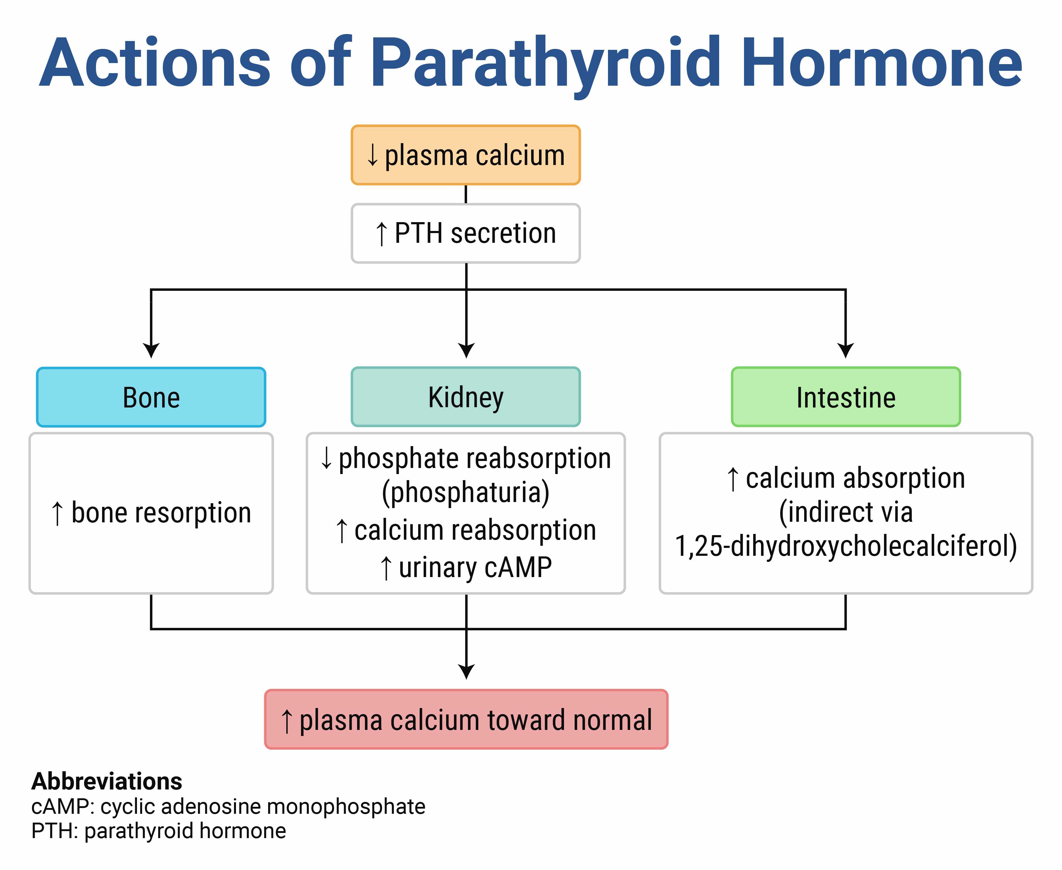
Patients with hypoparathyroidism will usually have a low or normal alkaline phosphatase measurement (A). It is typically well above standard reference ranges in children due to bone growth and can be three times the normal value without suggesting a pathologic process. Most patients with hypoparathyroidism will have a normal calcifediol measurement (C) but a low calcitriol (1,25-hydroxy vitamin D) level. Uncommonly, patients will have a very high calcitriol level, but this is much less likely than an elevated phosphorus level. Patients with hypoparathyroidism will have a normal magnesium measurement (D), but it is good to keep in mind that there are two rare genetic syndromes described that present as tetany and seizures in infants from hypocalcemia caused by low magnesium levels. It is unclear how they are related but in these rare cases, treatment with magnesium restores both magnesium and calcium levels.
300
Which of the following is the most reliable indication of an Achilles tendon rupture?
- A. Diminished Achilles reflex
- B. Inability to ambulate
- C. Inability to plantar flex the foot
- D. Pain along the posterior ankle
- E. Positive calf squeeze test
E. Positive Calf Squeeze Test
Patients often present with a history of sudden pain and a popping sensation in the posterior ankle during physical activity. On examination, there may be edema and ecchymosis over the Achilles tendon, and a palpable defect may be appreciated. The calf squeeze test, or Thompson test, is the most reliable indicator of an Achilles tendon rupture, with a sensitivity of 96%. With the patient prone or kneeling on a chair, the posterior calf is squeezed at its widest point. The absence of plantar flexion indicates a rupture of the tendon. Management includes immobilization with a short leg cast with the ankle in slight plantar flexion, known as the equinus position, and orthopedic consultation.
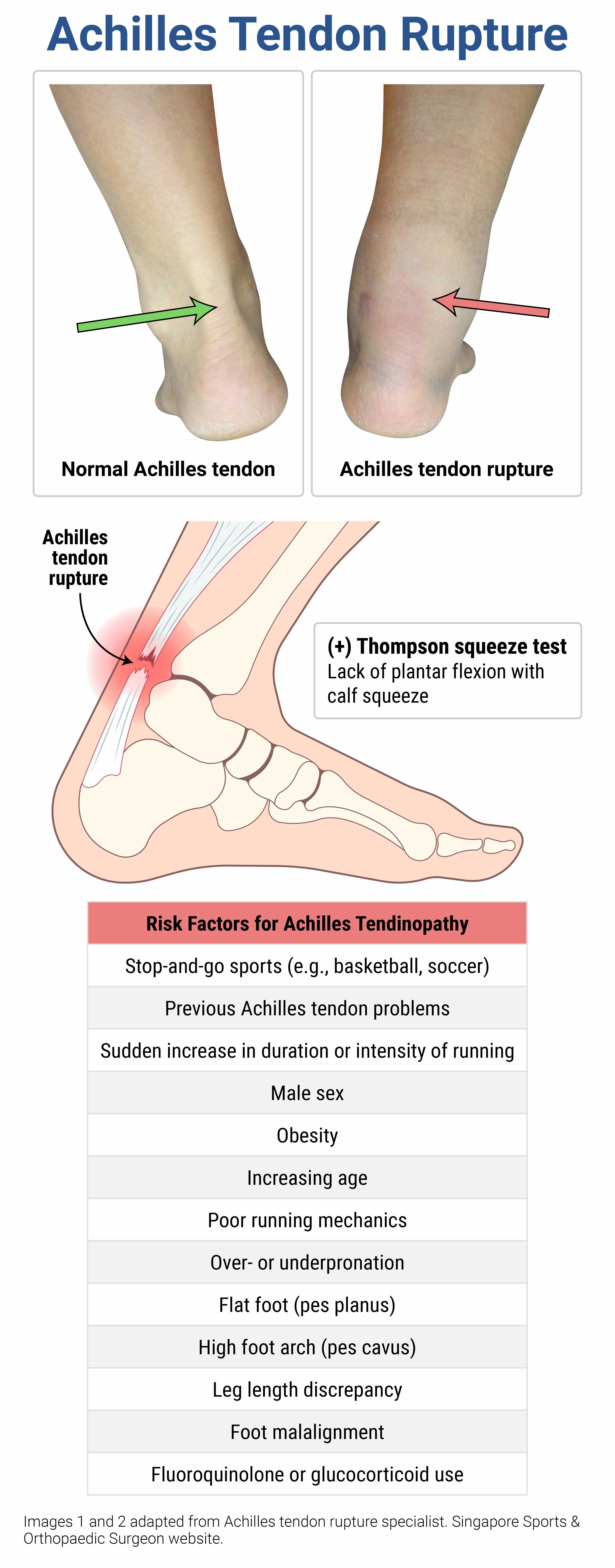
A tendon rupture can result in a decreased Achilles reflex due to the structural integrity of the tendon being impaired. However, the Achilles reflex (A) primarily assesses the S1 and S2 nerve roots. A diminished reflex is not a specific or reliable indicator for an Achilles tendon rupture and can be seen in other conditions, including peripheral nerve disorders such as neuropathy and Guillain-Barré syndrome.
Because of the multiple muscles involved in plantar flexion of the foot that remain intact and functional even with an Achilles tendon rupture, the ability or inability to ambulate (B) and plantar flex the foot (C) does not reliably exclude or diagnose an Achilles tendon rupture.
Pain along the posterior ankle (D) is typical. However, one study showed that up to one-third of patients with tendon rupture report no pain.
300
Which of the following views in the FAST exam will demonstrate free fluid in the peritoneal cavity first in a supine adolescent or adult patient?
- A. Pelvic (retrovesicular) view
- B. Pericardial (cardiac) view
- C. Perihepatic (right upper quadrant) view
- D. Perisplenic (left upper quadrant) view
C. Perihepatic View
In the supine patient, the right upper quadrant (Morison pouch) view on a FAST exam is most likely to reveal free fluid in the peritoneal space before the other four views in adolescents and adults. The posterior peritoneum attaches in such a way that free fluid from any injury anywhere will travel to the right upper quadrant in a supine patient (it is the most dependent region in the supramesocolic region). The right upper quadrant should be divided into three zones: above/below the diaphragm; Morison pouch (hepato-renal recess); and paracolic gutter (around the left hepatic edge/inferior pole of the kidney).
The pelvic (A) and perisplenic (D) views are not the earliest views to demonstrate the presence of free fluid in a supine patient. It was thought that the pelvic view, being the most anatomically dependent, would be the most sensitive view, but studies have only demonstrated this to be true in younger children.
The pericardial view (B) is used to visualize pericardial effusion, not peritoneal free fluid.
300
A 3-month-old boy is brought to the ED by his parent for an episode of not breathing and turning blue and floppy for a few seconds after crying. He recovered after his parent gave him assisted breaths. She reports her son was diagnosed with a tracheoesophageal fistula that was repaired at birth with no complications. He recovered uneventfully and was discharged home after 2 weeks. About 1 week ago, the boy developed a brassy, barking cough and noisy breathing. He also has a small amount of spit-up after feeding but no back arching. He has otherwise been doing well, breastfeeding, and gaining weight appropriately. In the ED, his vital signs are within normal limits. On exam, he is comfortable but has inspiratory and expiratory stridor, consistent with what his parent described as noisy breathing. Which of the following is the most likely cause of these symptoms?
- A. Gastroesophageal reflux disease
- B. Leak at the anastomosis site of the tracheoesophageal fistula repair
- C. Recurrence of the tracheoesophageal fistula
- D. Tracheomalacia
D. Tracheomalacia
A brassy, barking cough and biphasic stridor in an infant who has had tracheoesophageal fistula repair is best explained by tracheomalacia. Tracheomalacia can present as an isolated congenital lesion, but it is also a frequent complication of surgical repair of esophageal atresia and tracheoesophageal fistula. Tracheomalacia can be found in pathology specimens in 75% of patients who have esophageal atresia and tracheoesophageal fistula and typically is found at or above the level of the original site of the lesion. Most infants have only the brassy, barking cough, but severe tracheomalacia may manifest with stridor at rest, biphasic stridor, dyspnea with feeding, or expiratory wheezing with respiratory infections. Some infants who have esophageal atresia and tracheoesophageal fistula with severe tracheomalacia can also develop death spells that begin after 2–3 months of age, as described in this patient. Such spells are associated with feeding, crying, or coughing and are characterized by cyanosis, apnea, bradycardia, and hypotonia that require resuscitation. Many patients with tracheomalacia can be cared for with close observation, but patients with recurrent death spells may require more invasive intervention such as nasal continuous positive airway pressure temporarily and aortopexy or tracheostomy for long-term relief
300
A 9-year-old boy presents to the emergency department for inability to walk. He has had 2 days of leg pain and has been irritable for the past day. The morning of presentation, he refused to get out of bed or bear weight. On his initial examination, he has 1/5 strength in his lower extremities and 4/5 strength in his upper extremities. Patellar reflexes cannot be elicited. Which of the following is the most likely CSF finding?
- A. WBC count of 1,100/µL, glucose of 20 mg/dL, protein of 135 mg/dL
- B. WBC count of 1,100/µL, glucose of 20 mg/dL, protein of 80 mg/dL
- C. WBC count of 120/µL, glucose of 65 mg/dL, protein of 80 mg/dL
- D. WBC count of 2/µL, glucose of 65 mg/dL, protein of 135 mg/dL
- E. WBC count of 2/µL, glucose of 65 mg/dL, protein of 40 mg/dL
Analysis of the CSF helps make the diagnosis of Guillain-Barré. CSF studies will show an elevated protein level, typically twice the upper limit of normal. The WBC count in the CSF is within normal limits. This dissociation between high CSF protein and normal cell counts (albuminocytologic dissociation) is diagnostic for Guillain-Barré syndrome.
Bacterial meningitis may have cloudy or purulent CSF. However, it could still be clear. The opening pressure is usually elevated (> 25 cm H2O), as is the WBC count, which is usually > 1,000/µL (A, B). The glucose will be low (< 40% of the serum glucose), and the protein will be high (> 50 mg/dL).
Aseptic or viral meningitis may have an elevated opening pressure with clear CSF. The glucose level is within normal limits, but the protein will be elevated (> 50 mg/dL). The WBC count ranges from 10–500/µL (C) and will not be within the reference range.
Normal CSF has a low WBC count, a glucose level > 60% of the serum glucose level, and a protein level < 45 mg/dL (E). The fluid should appear clear, and the opening pressure should not be elevated.
400
A 7-month-old boy presents with pallor and decreased weight gain over the past month. His hemoglobin level is 4 g/dL. What is the most likely cause of his anemia?
- A. Alpha thalassemia intermedia
- B. Beta thalassemia major
- C. Iron deficiency anemia
- D. Sickle cell anemia
B. Beta Thal Major
Beta thalassemia major is the severe form of homozygous beta thalassemia, in which little or no beta-globin chains are produced. Children can start to develop anemia at 2–3 months of age because the fetal gamma-globin chains disappear in the first few months of life. Generally, children present between 6 and 24 months of age with pallor, decreased growth, and hepatosplenomegaly.
Loss of two to three of the four alpha-globin genes leads to alpha thalassemia intermedia (A), which is associated with chronic anemia and hemolysis. Like sickle cell anemia, these patients have chronically low hemoglobin but usually only have problems with acute anemia with concomitant infection or illness, which was not mentioned in this patient. Alpha thalassemia major is a complete loss of the four alpha-globin genes resulting in hydrops fetalis.
Iron deficiency anemia (C) presents in children between 1 and 2 years old as patients begin to drink cow milk. Patients have microcytic, hypochromic anemia without jaundice or hepatosplenomegaly. Generally, iron deficiency does not cause such a low hemoglobin level or require transfusion.
Sickle cell anemia (D) is tested during newborn screening. Patients generally have chronically low hemoglobin ranging from 7–12 g/dL. Patients with sickle cell disease are at increased risk of sepsis due to splenic infarction and functional asplenia.
400
A 10-month-old boy with arginine vasopressin deficiency and developmental delay presents to the emergency department with fussiness and decreased oral intake. His foster parents report he was recently discharged after a brain injury from nonaccidental trauma and is currently receiving subcutaneous desmopressin in addition to a low-solute diet and furosemide. He has some obvious motor and speech delays, is fussy but consolable, and has an otherwise unremarkable exam, including vital signs within normal limits for his age. His serum sodium is 126 mEq/L. His weight is 400 g more than when he was discharged 2 days ago. Which of the following is the most appropriate next step in management?
- A. Administer intravenous dextrose in sterile water
- B. Decrease the desmopressin dose
- C. Increase solutes in the diet
- D. Restrict water intake
B. Decrease the Desmopressin Dose
Arginine vasopressin deficiency (central diabetes insipidus) is the decreased release of antidiuretic hormone (ADH), causing an inability to concentrate urine and a significant loss of total body water. It is usually permanent when idiopathic but is often transient after neurosurgery or cerebral trauma. Symptoms consist of polyuria and nocturia but can result in severe hypernatremia when patients lack a thirst mechanism, cannot express their thirst, or lack access to water (as with an infant or toddler).
Treatment of infants with arginine vasopressin deficiency also includes a low-solute diet and a thiazide diuretic that together ultimately reduce overall urine output by inducing mild hypovolemia and reducing water delivery to sites that are sensitive to ADH. Infants taking desmopressin are at high risk of sodium derangements, as in this baby who has signs of volume overload, and they need to have their sodium levels checked frequently. Initial treatment in infants with arginine vasopressin deficiency and signs of volume overload is to reduce the dose of desmopressin and recheck the serum sodium within 24 hours.
400
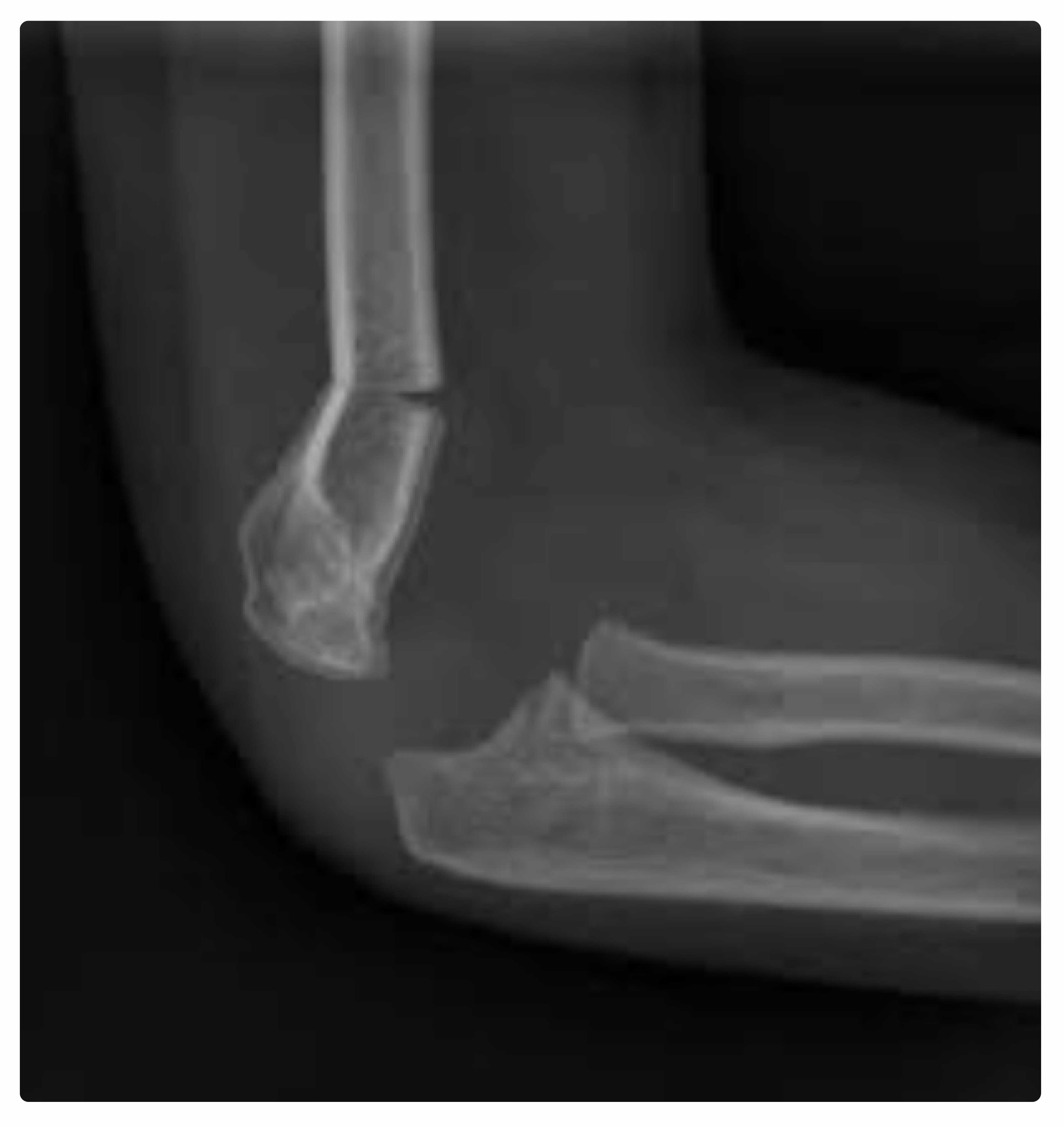
A 9-year-old girl presents to the ED with an elbow injury after falling off of a trampoline. She has a strong radial pulse, normal sensation, and although anxious, she is still able to show normal motion of her hand. The above X-ray is obtained. Reduction and splinting in the emergency department will most likely result in which one of the following outcomes?
- A. Anterior interosseous nerve injury
- B. Cubitus varus deformity
- C. Limb length discrepancy
- D. Volkmann contracture
- B. Cubitus varus deformity
Supracondylar fractures make up the vast majority of elbow fractures in children. Unlike other areas, the distal humerus has very little remodeling potential, so near anatomic alignment is required for a good cosmetic and functional outcome. The Gartland classification is commonly used for supracondylar fractures, describing them as type I, type II, or type III. Type I fractures are usually only seen by a posterior fat pad without cortex disruption. These are nondisplaced, stable, and heal well with splinting alone. A posterior long-arm splint should be used with the arm in pronation or neutral position at 90 degrees at the elbow. Type II supracondylar fractures have anterior cortex disruption, but the posterior cortex is intact. If nondisplaced, these can be treated like a Gartland type I supracondylar fracture, but this is uncommon. Type III fractures have disruption of both the anterior and posterior cortex.
Displaced Gartland type II fractures and all type III fractures require admission for operative closed reduction with internal fixation. Patients with a displaced supracondylar fracture who undergo reduction in the ED and splinting rather than operative reduction and fixation are at risk of developing a functionally significant cubitus varus deformity where the elbow is permanently deviated while extended with the forearm bent toward the body.

The most common nerve injury from a supracondylar fracture is an anterior interosseous nerve injury (A), which is typically due to the fracture itself but can be worsened during fracture reduction. Nerve injuries can have long-term sequelae, but most are neuropraxias that resolve within 2–3 months.
A limb length discrepancy (C) is not described as a complication of a supracondylar fracture.
A Volkmann contracture (D) is an uncommon complication of both proper and improper supracondylar management that results in a compartment injury with muscle ischemia, permanently fixed flexion of the elbow and wrist, and forearm pronation.
400
A 4-year-old 20 kg boy presents to the emergency department with fever, cough, and difficulty breathing. He is toxic-appearing and unresponsive to attempts at IV placement. His temperature is 41.3°C, heart rate is 180 bpm, respiratory rate is 82/min, blood pressure is 64/30 mm Hg, and oxygen saturation is 71%. He has no breath sounds on the right, and his tachypnea is quiet and shallow. His capillary refill is 6 sec. You and your team push an IV fluid bolus and provide bag-mask ventilation while preparing for intubation. Which of the following is the most appropriate induction agent for this patient?
- A. Etomidate 6 mg
- B. Ketamine 30 mg
- C. Midazolam 4 mg
- D. Propofol 70 mg
B. Ketamine 30 mg
Each induction agent for rapid sequence intubation has its own characteristics that make it more or less useful in certain situations. Ketamine is a preferred agent for patients with hypotension, as it is typically associated with tachycardia and increased blood pressure. It can also allow the patient to maintain airway reflexes until paralysis takes effect. Patients should be aware that it is not always associated with an increase in blood pressure, as there are reported cases of hypotension presumed to occur when the body reduces endogenous stress hormone release during its use. The appropriate dose of ketamine as an induction agent is 1.5 mg/kg until adult doses are reached.
Etomidate (A) is not recommended as it can cause transient adrenal insufficiency, which is not ideal in the setting of septic shock. Ketamine would be a better choice. The appropriate dose of etomidate is 0.2–0.4 mg/kg.
Midazolam (C) is uncommonly used for induction, as it typically does not cause enough anesthesia unless used at high doses. High doses of midazolam also cause hypotension. The appropriate dose of midazolam is 0.1–0.3 mg/kg with a maximum of 4 mg.
Propofol (D) is also uncommonly used in rapid sequence intubation, as it is also associated with hypotension. The recommended dose of propofol for induction is 2–3 mg/kg.
400
A 2-year-old vaccinated boy presents to the emergency department in the afternoon with a sudden onset of noisy and difficult breathing. He woke in his usual state of health this morning and was his normal active self. He has not had any fevers, nasal congestion, or rhinorrhea. On exam, he is afebrile and awake but anxious. He is tachypneic with intercostal retractions and has audible inspiratory stridor at rest. He is drooling and has a high-pitched seal-like bark when he coughs. After a dose of nebulized racemic epinephrine, he continues to have stridor at rest. Which of the following is the most appropriate next step?
- A. Intramuscular dexamethasone
- B. Nebulized racemic epinephrine
- C. Oxygen by high-flow nasal cannula
- D. Soft tissue neck and chest X-ray
D. Soft tissue neck and chest xray
By far, the most common cause of stridor at rest in children is laryngotracheitis (i.e., croup). This commonly presents between 6 months and 3 years of age with inspiratory stridor at rest after about 12–48 hours of symptoms of an upper respiratory infection with cough, nasal congestion, rhinorrhea, and fevers. Croup is typically worse at night and frequently presents at that time. When the onset of stridor is sudden, the patient is outside of the typical age range, or the patient lacks a viral respiratory prodrome, additional workup and evaluation are necessary. Other causes of stridor include bacterial tracheitis, which usually presents in older, more toxic-appearing patients with dysphagia and drooling. Epiglottitis is now uncommon in children due to the Haemophilus influenzae vaccination, but it historically manifested as an ill patient with a fever, drooling, and soft stridor while in a tripoding position. Patients with a retropharyngeal abscess can present with stridor but more commonly with fever and limitation in neck range of motion. Noninfectious causes of acute stridor include foreign body aspiration, epiglottis, laryngeal injury, angioneurotic edema (i.e., anaphylaxis), and spasmodic croup. Of these, a toddler with sudden-onset stridor without fever or upper respiratory symptoms and a poor response to racemic epinephrine most likely has a tracheal airway foreign body, which can be evaluated with a soft tissue neck and chest X-ray.
Steroids such as intramuscular dexamethasone (A) are appropriate for treating laryngotracheitis, but most children can tolerate the medication orally. Agitating a patient with a tracheal foreign body by giving an intramuscular injection can turn a partial airway obstruction into a complete airway obstruction.
A second dose of nebulized racemic epinephrine (B) is appropriate for treating a patient with continued stridor at rest when their history and exam are consistent with laryngotracheitis.
Oxygen by high-flow nasal cannula (C) is commonly used to reduce the work of breathing in patients with bronchiolitis but will not help in a patient with an airway foreign body.
400
A young adult patient presents with altered mental status. Vital signs reveal a temperature of 106.4°F (41.3°C) and an irregular heart rate of 163 bpm. His blood pressure is 110/73 mm Hg. An ECG shows rapid atrial fibrillation. What treatment should be immediately initiated?
- A. Aggressive cooling with ice water immersion
- B. Calcium channel blocker
- C. Cold intravenous fluids
- D. Synchronized cardioversion
- A. Aggressive cooling with ice water immersion
This patient presents with heat stroke, and the focus of management should be on immediate, rapid cooling. Heat illness exists on a spectrum from minor heat illness (heat cramps, heat edema, heat syncope) through heat exhaustion to heat stroke. Heat stroke is a life-threatening disease characterized by elevated temperature (usually > 104°F [40°C]) and multisystem organ dysfunction. Additional complications of heat stroke include rhabdomyolysis, liver failure, dysrhythmias, pulmonary edema, acute kidney failure, and disseminated intravascular coagulation. Management begins with rapid cooling. This is typically accomplished by either cool mist with fans or ice water immersion (the most rapid approach). Patients should also receive intravenous hydration.
Administration of a calcium channel blocker (B) for rate control is not indicated. Most tachydysrhythmias will resolve with aggressive cooling in the setting of heat stroke. Any delay in cooling should be avoided.
Cold intravenous fluids (C) can be used as an adjunct to cooling but should not be the first or primary method.
Synchronized cardioversion (D) for the patient’s atrial fibrillation is contraindicated, as the duration of this rhythm is unknown, and it will further delay cooling since the patient must be dry to be cardioverted (i.e., you cannot perform either major method of cooling).
500
Which of the following characteristics suggests transfusion-related acute lung injury instead of transfusion-associated circulatory overload?
- A. High fever
- B. Hypertension
- C. Hypoxemia
- D. Jugular venous distention
- E. Pulmonary edema
Transfusion-related acute lung injury (TRALI) complicates transfusions in 1 of every 5,000 units of packed red blood cells. Although rare, it is associated with high mortality. The pathophysiology is thought to be due to an acute neutrophilic response that leads to endothelial damage and massive capillary leak in the pulmonary vasculature.
The diagnosis is clinical, with patients presenting with signs and symptoms of acute lung injury or acute respiratory distress syndrome. TRALI is characterized by hypoxemia (oxygen saturation < 90% on room air) and bilateral pulmonary infiltrates presenting within 6 hours of blood transfusion. There should not be any evidence of pulmonary artery hypertension or generalized fluid overload. Hypotension, tachycardia, and fever are common. Fever is not common in transfusion-associated circulatory overload (TACO).
TRALI is generally characterized by hypotension, not hypertension (B).
Similar to TRALI, TACO also presents with hypoxemia (C), respiratory distress, and pulmonary edema (E) on chest X-ray. However, hypertension is typical, and high fever is unlikely.
TACO is also associated with distended neck veins (D) and other markers of generalized volume overload. Treatment of TACO consists of aggressive diuresis and respiratory support.
500
A 12-year-old girl presents to the emergency department with a firm anterior neck mass. Her parent first noticed it this morning, but the patient says it has been slowly growing over the last few weeks. The patient is short for her age but reports no weight changes, palpitations, constipation, diarrhea, or difficulty with temperature changes. Her exam is only remarkable for an enlarged and firm but nontender goiter. Laboratory results show normal free T4 and thyroid-stimulating hormone values. Which of the following is the most likely diagnosis?
- A. Chronic autoimmune thyroiditis
- B. Graves disease
- C. Nontoxic nodular goiterYour Answer
- D. Subacute granulomatous thyroiditis
A. Chronic autoimmune thyroiditis
Chronic autoimmune thyroiditis is so common that it is also the most common cause of hypothyroidism in children, even though most cases are euthyroid. Treatment should be directed by a pediatric endocrinologist, but there is evidence that treatment with thyroid hormone decreases goiter size even in euthyroid children with the disease.
Graves disease (B) is the most common cause of goiter with hyperthyroidism in children and is not shown in this patient.
A nontoxic nodular goiter (C) is the most common cause of euthyroid goiter in adults but not children.
Subacute granulomatous thyroiditis (D) is a rare cause of goiter in children but should be suspected in children with a painful goiter, which is not shown in this patient.
500
A 2-year-old boy is brought to to the emergency department with bilious emesis that began 3 hours prior to arrival. His parent seems distracted by the three other children they have with them in the department. On physical exam, you note that the boy has bruises in multiple stages of healing, including on his torso. His parent says the boy has not eaten for 2 days and reported abdominal pain 1 day ago. You are concerned he may have a bowel obstruction, but what other injury should be suspected?
- A. Kidney contusion
- B. Liver laceration
- C. Pancreatic injury
- D. Splenic rupture
In the setting of suspected bowel obstruction and possible nonaccidental trauma or child abuse, the diagnosis of a duodenal hematoma should be considered. In all cases of diagnosed duodenal hematoma, pancreatic injury should be considered due to the anatomic location of the pancreas in relation to the duodenum and the force needed to cause the primary duodenal injury. This occurs when a child is struck forcefully with a small-diameter object. Duodenal hematomas develop slowly over time, and as the hematoma develops, this can cause gastric distention and gastric outlet obstruction. Once obstruction occurs, the patient presents with signs of obstruction and bilious emesis.

Kidney contusions (A) are rare, even in the setting of child abuse, and also do not cause bilious emesis.
Liver laceration (B) and splenic rupture (D) are the most common injuries in pediatric blunt abdominal trauma, but neither of these causes a patient to have bilious emesis. These are also the most common injuries seen in individuals who have nonaccidental trauma with blunt abdominal trauma.
500
An 8-year-old boy presents to the emergency department after suddenly collapsing at school. He arrives with emergency medical service personnel, who are performing chest compressions and ventilations through a supraglottic device, and he is apneic and pulseless. The patient has good chest rise and bilateral breath sounds with the supraglottic device. Which of the following is the most appropriate initial ventilatory rate during the resuscitation of this patient?
- A. 1 ventilation every 2–3 seconds
- B. 1 ventilation every 6–8 seconds
- C. 2 ventilations for every 15 compressions
- D. 2 ventilations for every 30 compressions
A. 1 ventilation every 2-3 seconds
Patients in cardiopulmonary arrest with an advanced airway should receive asynchronous ventilation every 2–3 seconds according to the latest American Heart Association guidelines. An endotracheal tube and a supraglottic device are considered advanced airways, through which breaths should not be coordinated with chest compressions to minimize interruptions. One breath every 2–3 seconds (20–30 breaths per minute) can seem slow during a resuscitation, and most clinicians find it difficult to avoid overventilation of patients during a stressful resuscitation. Remember that each positive pressure breath increases intrathoracic pressure, causing a drop in venous return, pulmonary perfusion, and cardiac output, thereby reducing the success of defibrillation and overall survival.
One ventilation every 6–8 seconds (A) is the recommended rate of ventilation for rescue breathing in adult patients.
Two ventilations every 15 compressions (C) is appropriate for two rescuers in a prepubertal child without an advanced airway.
Two ventilations every 30 compressions (D) is appropriate for one rescuer of any age patient, or two rescuers in postpubertal patients who do not have an advanced airway.
500
A 2-month-old term male infant presents to the ED with trouble feeding. His mother reports it is taking longer for him to feed and he often seems like he is choking. She reports it began at birth and has been slowly worsening, and while the infant frequently spits up, it has been similar to her other two children. He has had no fevers, vomiting, or other ill symptoms. On exam the infant is well appearing with vital signs within normal limits for age. A complete exam, including of the oropharynx and oral mucosa, is normal. The patient is gaining weight well but has frequent coughing episodes on observed breastfeeding. Which of the following is the most appropriate next step?
- A. Fiberoptic endoscopic evaluation of swallowing
- B. Limited famotidine trial
- C. Reassurance and discharge
- D. Upper gastrointestinal series
D. Upper GI Series
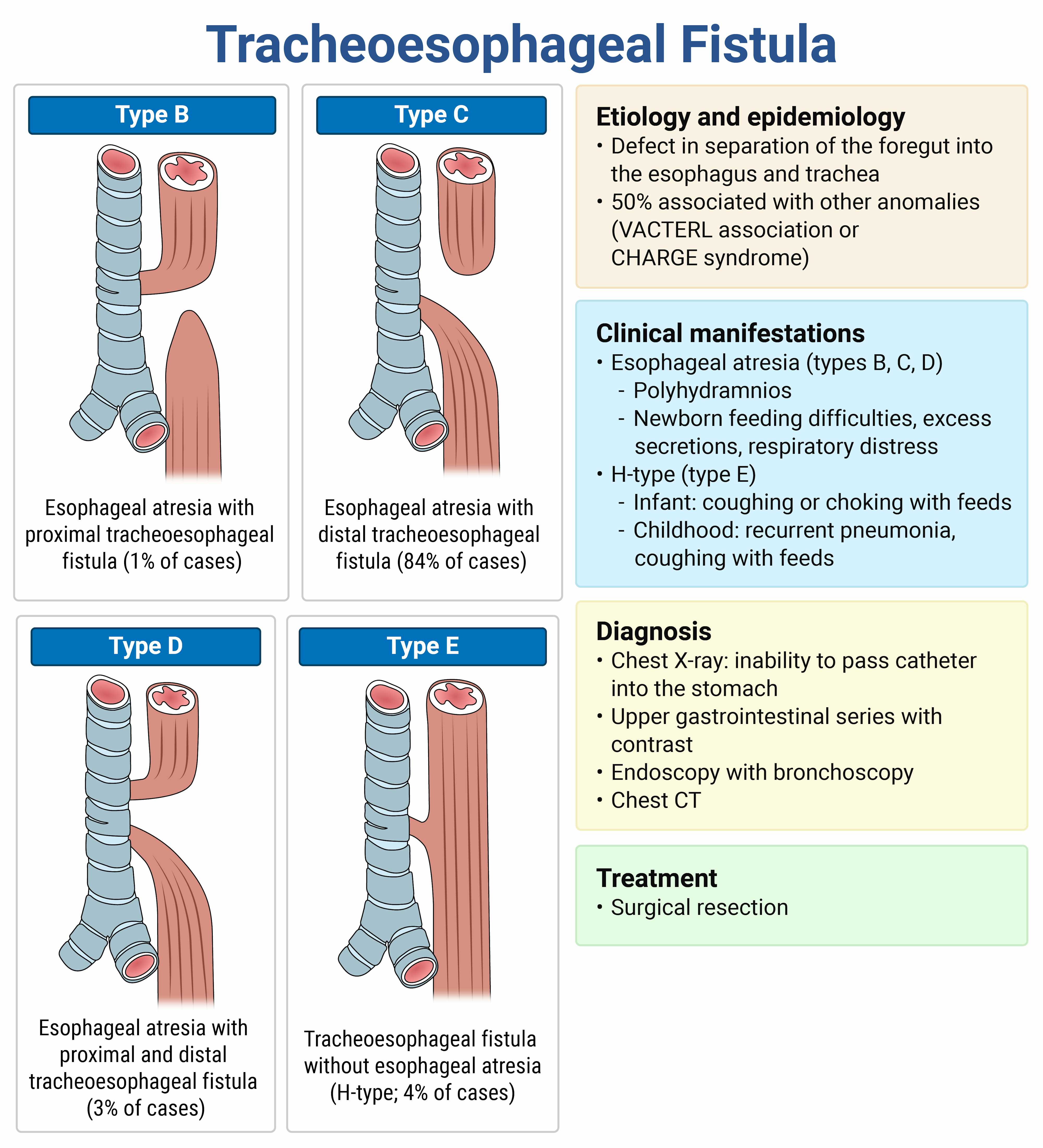
Dysphagia is described as both difficulty with or abnormality with swallowing. Most pediatric patients with dysphagia have an associated infection, such as stomatitis, pharyngitis, or a peritonsillar abscess. When unrelated to infection, dysphagia is an uncommon problem in children and, unlike many other pediatric concerns, is usually associated with serious illness. A complete history and physical examination will suggest a diagnosis in most patients. Causes of dysphagia in infants and children include birth trauma from intubation or meconium aspiration, congenital anomalies (e.g., tracheoesophageal fistula, vascular ring, or choanal stenosis), extreme cases of gastroesophageal reflux, foreign body, anaphylaxis, airway obstruction, and botulism.
In this patient, a history of worsening dysphagia since birth suggests a congenital anomaly. When dysphagia presents at birth, passing an 8 F catheter through the nares and into the stomach can evaluate for choanal stenosis or esophageal obstruction such as esophageal atresia. This is often done with an X-ray to confirm placement in the stomach, followed by an upper gastrointestinal series with catheter removal to evaluate for an H-type tracheoesophageal fistula. An upper gastrointestinal series will also evaluate the esophagus for stenosis or an esophageal web.
A fiberoptic endoscopic evaluation of swallowing (A) involves threading an endoscopic camera through the nose to evaluate swallowing in real time. This is one method for screening for pre-esophageal disorders, but it does not detect congenital esophageal anomalies.
A trial of acid suppression, such as a limited famotidine trial (B), is reserved for infants with gastroesophageal reflux with significant symptoms (e.g., failure to thrive) and who do not improve with conservative therapy. Infants with gastroesophageal reflux typically present with frequent spit-ups, fussiness, and occasionally failure to thrive, not with dysphagia.
Children with dysphagia require further evaluation to determine a cause and to arrange definitive treatment, so reassurance and discharge (C) is inappropriate in this patient.
500
A 4-month-old girl presents to the emergency department with vomiting for the last 3 days. Lab studies show a pH < 6.9, PCO2 of 12 mm Hg, lactic acid of 0.4 mmol/L, glucose of 54 mg/dL, sodium of 145 mEq/L, chloride of 110 mEq/L, and bicarbonate of 10 mEq/L, and there are marked ketones in her urine. Which of the following is the most likely diagnosis?
- A. Fatty acid oxidation defect
- B. Gastroenteritis
- C. Glycogen storage disease
- D. Organic acidemia
D. Organic Acidemia
Gauthami Says: Metabolic acidosis (maybe more than is expcted from gastro), marked ketones (rules out FAO disorder)
This patient has a profound metabolic acidosis and cannot compensate even by reducing carbon dioxide with tachypnea. Such a profound metabolic acidosis is commonly the presentation of an undiagnosed inborn error of metabolism. Inborn errors of metabolism most likely to present with a severe anion gap metabolic acidosis with hypoglycemia and elevated ketones are organic acidemias, such as isovaleric acidemia and holocarboxylase synthetase deficiency.
Fluid resuscitation and dextrose administration are used to treat a child with severe metabolic acidosis due to an undiagnosed inborn error of metabolism. Making the child NPO and infusing dextrose effectively stops catabolism and prevents the further creation of toxic metabolites. This is typically done with 10% dextrose in water at twice the maintenance rate for an infant, which can be administered peripherally. The ideal calculated rate should deliver 8–10 mg/kg/min.
Patients with a fatty acid oxidation defect (A) typically present with hypoglycemia and hepatic encephalopathy without metabolic acidosis due to the lack of ketone production.
Patients with a metabolic acidosis due to gastroenteritis (B) usually have a nonanion gap metabolic acidosis.
Patients with glycogen storage disease (C) present with hypoglycemia, seizures, and hepatomegaly with metabolic acidosis and an elevated lactate, many of which are not present in this case. The patient's lactate level is normal, and there is no mention of hepatomegaly.
1000
A patient is sent home with a prescription for an antibiotic after being diagnosed with a bacterial upper respiratory infection. He returns 2 days later because his eyes are yellow. Vital signs include a blood pressure of 129/71mm Hg, heart rate of 83 bpm, respiratory rate of 17/min, oxygen saturation of 97% on room air, and temperature of 98.3°F. Laboratory studies show a hemoglobin of 10 g/dL, hematocrit of 31%, and reticulocyte count of 2.1%. Peripheral smear shows small, round inclusions in the red blood cells. Which of the following antibiotics was most likely initially prescribed?
- A. Amoxicillin
- B. Azithromycin
- C. Cephalexin
- D. Ciprofloxacin
- E. Doxycycline
Glucose-6-phosphate dehydrogenase (G6PD) deficiency is a genetic disorder resulting in a deficiency of the G6PD enzyme, which is crucial for red blood cell function. The condition follows an X-linked recessive inheritance pattern. Pathophysiologically, the deficiency leads to a reduced ability to handle oxidative stress, causing red blood cells to hemolyze prematurely. It is often triggered by infections, certain medications, or foods like fava beans. Symptoms may include jaundice, fatigue, pallor, and dark urine. The physical exam may reveal pallor and jaundice.
Laboratory findings typically show anemia, elevated reticulocyte count, and the presence of Heinz bodies (small, round inclusions) in red blood cells on peripheral smear. Management involves avoiding oxidative stressors, treating acute hemolysis supportively with hydration, and if necessary, giving blood transfusions. Education on trigger avoidance is key in managing the condition. Ciprofloxacin can cause oxidative stress that leads to hemolytic anemia. Other medications associated with hemolysis in G6PD deficiency include those shown below.
1000
A 14-year-old girl presents to the emergency department with palpitations. Her parents are concerned she may be using illicit drugs since she is having difficulty concentrating at school, fatigue, and weight loss despite an increase in appetite over the last few months. On review of systems, the patient also reports diarrhea, infrequent periods, and heat intolerance. You see pronounced exophthalmos on your exam. Which of the following additional findings might you expect on physical exam?
- A. Anhidrosis
- B. Atrial fibrillation
- C. Decreased deep tendon reflexes
- D. Tongue fasciculations
D. Tongue Fasciculations
Exam findings of hyperthyroidism include tremors, tongue fasciculations, lid lag, hyperactive reflexes, proximal muscle weakness, warm skin, and sweating. Most patients with Graves disease will have a diffuse goiter. Proptosis with hyperthyroidism is specific to Graves disease.
Anhidrosis (A) is part of the anticholinergic toxidrome that can also present with tachycardia, flushed skin, and behavior disturbances. Such patients tend to be more acute in onset, have dilated pupils, and are associated with an ingestion not described above.
Atrial fibrillation (B) is seen in 10–20% of adults with hyperthyroidism but is extremely uncommon in children with hyperthyroidism.
Most patients with hyperthyroidism will have increased deep tendon reflexes, not decreased deep tendon reflexes (C).
1000
A 5-year-old boy was sledding and hit a fence. He is brought to the ED via EMS with a report of anterior neck pain and shortness of breath. On exam, you note crepitus, a flattened thyroid prominence, and tenderness on palpation of the anterior neck. The nurse alerts you that the patient's oxygen saturation is 89% on room air. Definitive airway management should include which of the following?
- A. Continuous positive airway pressure via noninvasive mask
- B. Cricothyrotomy
- C. Endotracheal intubation
- D. Surgical consult for tracheostomy
- D. Surgical consult for tracheostomy
This patient should be evaluated for emergent tracheostomy. The patient likely has a laryngotracheal fracture or disruption. This most commonly presents after blunt anterior neck trauma with findings of crepitus over the larynx, laryngeal or tracheal tenderness, a flattened thyroid prominence, anterior neck deformity, and severe shortness of breath. Patients will typically have abnormal neck plain film radiography showing air in the peritracheal tissue. Attempts at endotracheal intubation may cause increased laryngotracheal trauma and a total loss of the airway. Cricothyrotomy is generally not utilized for emergent airway access in children < 8 years. Moreover, cricothyrotomy in children with direct laryngeal trauma may result in retrotracheal placement of the airway. Tracheostomy is the preferred method of obtaining a definitive airway.
Continuous positive airway pressure (A) could worsen the subcutaneous emphysema.
Cricothyrotomy (B) is contraindicated in children < 8 years of age and can result in posterior placement of the artificial airway.
Endotracheal intubation (C) may separate tissues and worsen the tenuous airway.
1000
A 3-year-old girl presents to the emergency department after being found apneic and pulseless at the bottom of a pool. The patient is intubated using high-quality, advanced life support skills and achieves a return of spontaneous circulation. The patient remains comatose without sedatives. Which of the following core temperatures should the team target for this patient?
- A. 30.0°C
- B. 35.5°C
- C. 37.0°C
- D. 38.5°C
C. 37
While mild hypothermia after an out-of-hospital cardiac arrest is shown to improve neurologic outcomes in some adults, these findings have not been duplicated in large pediatric studies. Despite this, the current recommendation for children who have a return of spontaneous circulation after cardiac arrest but remain comatose is targeted temperature management of sustained normothermia between 36 and 37.5°C or hypothermia between 32 and 34°C. Normothermia is typically chosen and should be maintained for 5 days. If hypothermia is chosen, it should only be maintained for 2 days, followed by 3 days of normothermia. There are some cases where hypothermia has shown a significant improvement in neurologic outcomes in children, specifically for asphyxiated newborns ≥ 36 weeks gestation when started within 6 hours. All such infants with moderate to severe hypoxic-ischemic encephalopathy should be offered therapeutic hypothermia. Fevers must be aggressively treated after a return of spontaneous circulation, as there is good evidence a fever can result in long-term neurologic complications in these patients.
1000
A 9-year-old girl with a history of prematurity at 25 weeks gestational age, cerebral palsy, gastrostomy tube dependence, and tracheostomy and ventilator dependence presents to the ED with a fever. Her parents state that for the last 2 days, they have noticed thick, malodorous secretions around her tracheostomy site, and today, they increased her oxygen supplementation. On examination, she is ill-appearing with a delayed capillary refill. Her temperature is 102.2°F (39°C). Her laboratory workup demonstrates leukocytosis with a left shift. Upon chart review, you notice that she has been infected previously with pan-sensitive gram-negative bacilli. Which of the following represents the best next step in management?
- A. Admission with intravenous ceftriaxone
- B. Admission with intravenous piperacillin-tazobactam
- C. Discharge home with cephalexin
- D. Discharge home with ciprofloxacin
This patient is presenting with signs and symptoms that are concerning for bacterial tracheitis. Given advances in neonatology and critical care, many children with extreme prematurity are able to survive. Chronic respiratory support may be necessary secondary to neurologic and neuromuscular disorders, central hypoventilation syndromes, obstructive apnea, or congenital facial and airway anomalies.
Admission with ceftriaxone (A) will not provide adequate coverage against Pseudomonas species.
Discharging home with cephalexin (C) or ciprofloxacin (D) is not appropriate for this patient.
1000
A 4-year-old boy presents to the emergency department unresponsive following an unknown ingestion. His arterial blood gas reveals a pH of 7.20, PaCO2 of 20 mm Hg, and bicarbonate of 12 mEq/L. Which of the following is likely the cause of his symptoms?
- A. Carbon monoxide
- B. Ethylene glycol
- C. Oil of wintergreen
- D. Volatile hydrocarbons
C. Oil of wintergreen
Salicylate poisoning is a common occurrence due to the widespread availability of prescription and over-the-counter preparations. In addition to oral preparations, multiple forms of salicylates are available as oral preparations and topical medications (oil of wintergreen) and flavoring agents. These products contain methyl salicylates and acetylsalicylates, both of which rapidly convert to salicylate once ingested. Oil of wintergreen is a highly potent and concentrated form of methyl salicylate (5 mL of the oil contains 7–9 grams of aspirin) and is deadly to a toddler.
Carbon monoxide (A) poisoning can cause syncope and coma in severe poisoning and will also cause a metabolic lactic acidosis, but carbon monoxide poisoning is not known to cause a concomitant respiratory alkalosis, which is present in this case.
Ethylene glycol (B) is a toxic alcohol that causes CNS depression, metabolic lactic acidosis, and kidney failure due to calcium oxalate crystal formation. A concomitant respiratory alkalosis does not occur with ethylene glycol poisoning.
Volatile hydrocarbons (D) are one of the most commonly used drugs among adolescents. Products used include glue, spray paint, electronics cleaners, and others. Volatile hydrocarbons cause neurologic depression and cardiac toxicity, which can lead to “sudden sniffing death".