Hernias
Hernias
Pharm and Genetics
Rando
Review
100
These are the boundaries of the inferior lumbar triangle (Petit Triangle)
Superior to Iliac crest
Lateral edge Lat Dorsi
Posterior edge External oblique
Cause: Weakness in the lumbodorsal fascia
Best repaired with mesh anchored to the fascia over the bone.
100
These are the Boundaries of the Superior lumbar triangle (Grynfeltt triangle)
12th rib
Paraspinal muscles (lat edge of quadratus lumborum)
Posteruir edge of Internal oblique
Lumbar hernias are more common in this space than in the Petit Triangle.
Cause: Weakness in the lumbodorsal fascia
Best repaired with mesh anchored to the fascia over the b
100
During a total thyroidectomy, the left inferior pole is being dissected out, and the artery is encountered. What is the vessel and its origin?
Inferior thyroid artery from the thyrocervical trunk
100
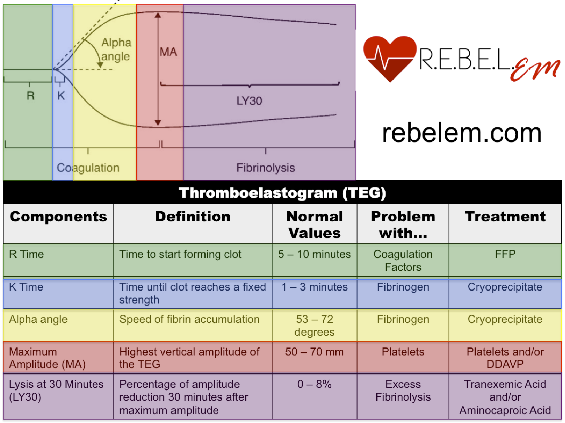
What product/s do you give a patient with elevated MA on TEG?
200
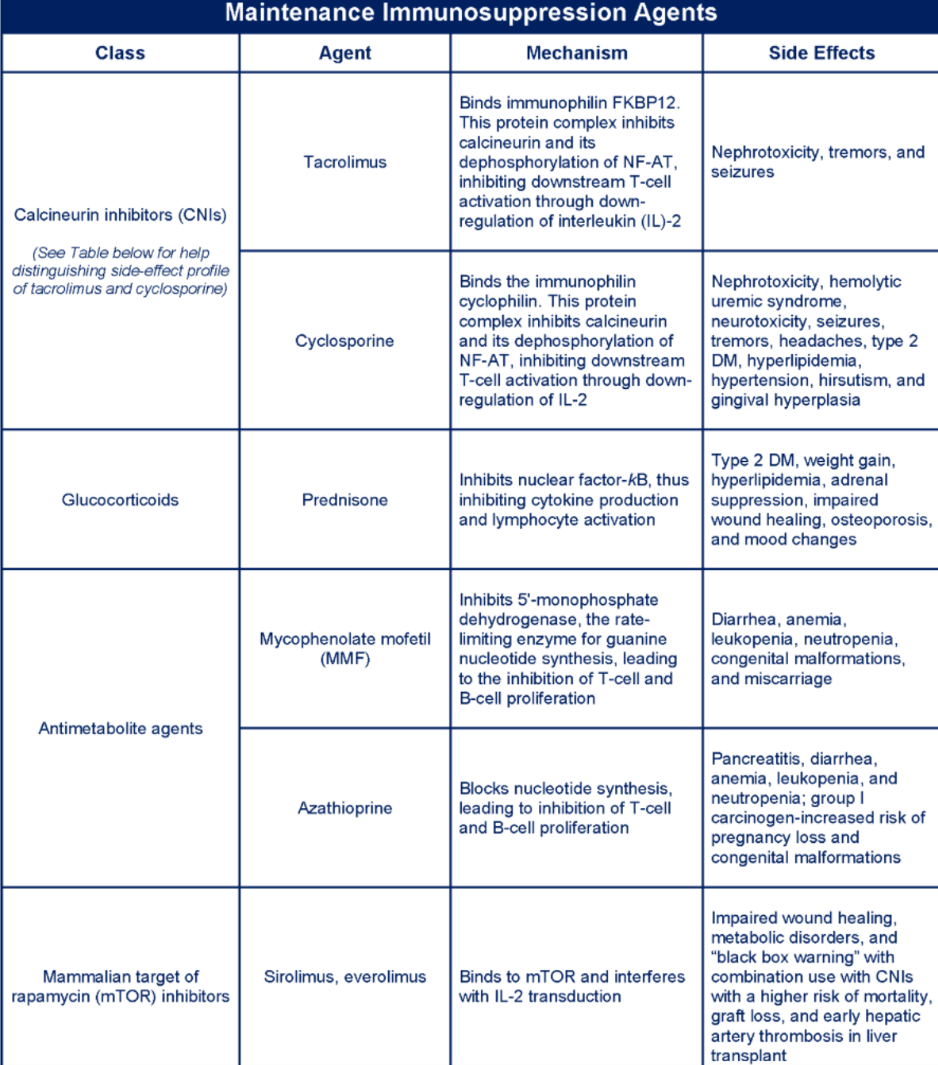
What class of transplant immunology medications causes nephrotoxicity? Give one example.
200
Name at least 6 guidelines for surgical intervention in asymptomatic primary hyperparathyroidism? (As in, when would you operate? There are 8 answers)
- Age younger than 50
- Calcium higher than 1mg/dl obove normal (8.88-10.4)
- Nephrolithiasis
- Nephrocalcinosis
- Osteoporosis w. T-score less than -2.5
- Compression fracture
- Urinary Ca greater than 250ug per 24 hours (Women) or 300up (men)
- Neuropsychiatric symptoms
300
This is the genetic mutation in a patient presenting with midline neck swelling and numerous painless nodules on the lips or tongue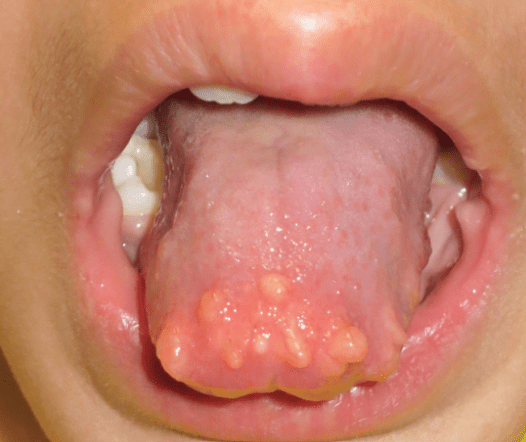
RET
MEN2 is an AD disorder.
MEN2A: Medullary thyroid carcinoma, pheochromocytoma, and hyperparathyroidism
MEN2B: Medullary thyroid carcinoma, pheochromocytoma, Mucosal neuromas, Marfanoid habitus
300
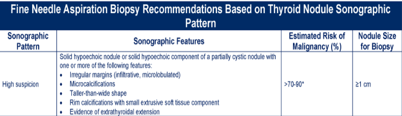
What are the high-risk/suspicious sonographic features seen on imaging for a thyroid nodule? (6)
400
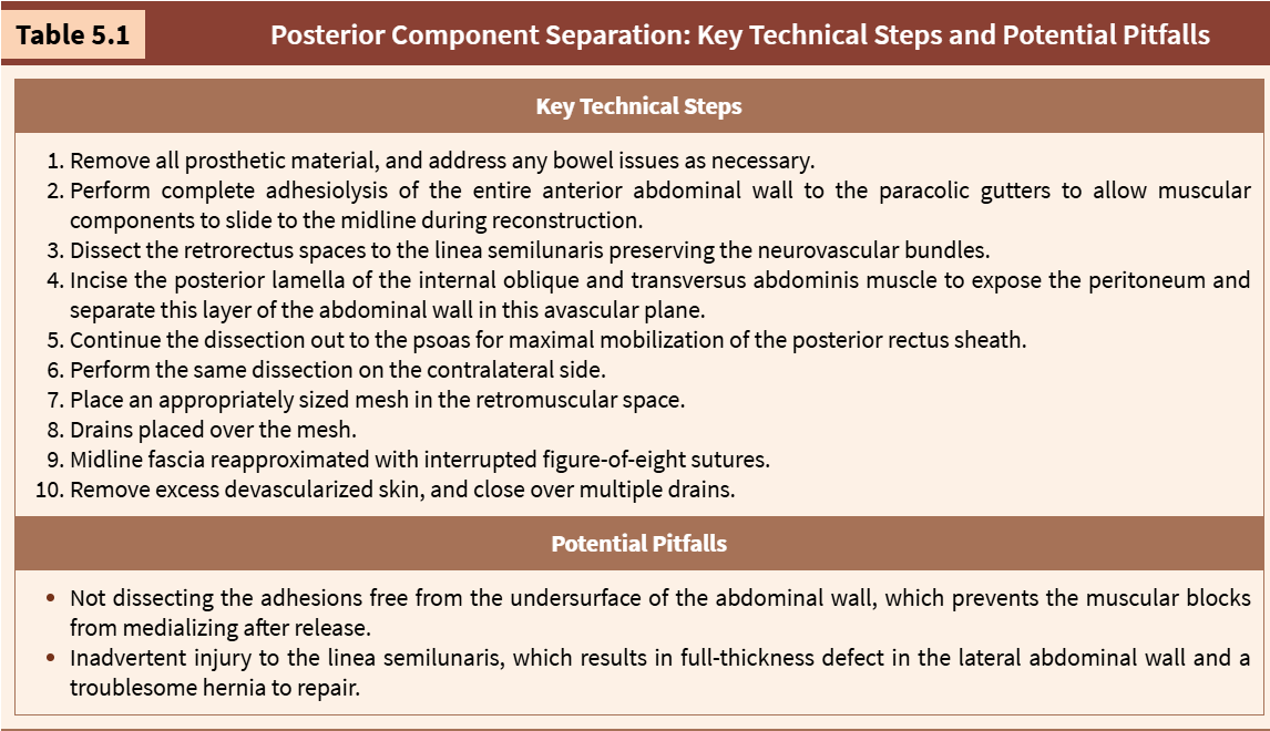
You are performing a posterior component separation to close a complex recurrent hernia that was previously repaired with mesh. You have removed all prosthetic material, and you have performed a complete adhesiolysis of the anterior abdominal wall to allow components to slide to the midline. What is your next step?
400
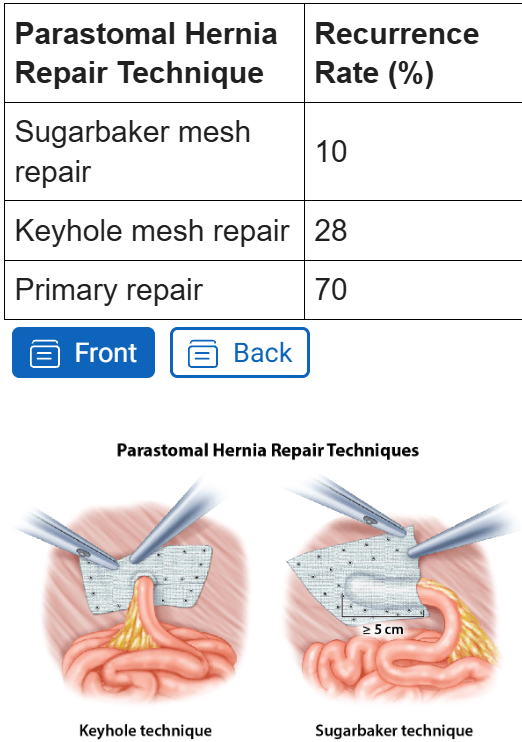
A 61-year-old man with a BMI of 38 kg/m², poorly controlled type II diabetes (HbA1c 9.2%), and an active 20-pack-year smoking history underwent an abdominoperineal resection with end colostomy for rectal adenocarcinoma four years ago. He presents to the surgery clinic with a gradually enlarging bulge around his colostomy and increasing difficulty maintaining an appliance seal, with intermittent leakage and skin excoriation. He denies obstructive symptoms. On examination there is a large, easily reducible bulge encircling the stoma that enlarges with Valsalva. A CT scan confirms a loop of colon and omentum herniating through the parastomal fascial defect, without evidence of obstruction or strangulation. He is highly motivated to proceed with definitive repair.
What is the most appropriate management for this patient?
A. Primary suture (fascial) repair of the parastomal defect
B. Open onlay biologic mesh repair
C. Stoma relocation to the contralateral abdominal wall without mesh
D. Minimally invasive (laparoscopic or robotic) mesh repair using a Sugarbaker technique
E. Continued nonoperative management with a hernia support belt indefinitely
D. Minimally invasive (laparoscopic or robotic) mesh repair using a Sugarbaker technique
Patients with a symptomatic parastomal hernia and risk factors for wound complications and recurrence (obesity, diabetes, smoking) are preferentially managed with a minimally invasive, mesh-reinforced repair. Mesh reinforcement markedly reduces recurrence versus primary suture repair, and the minimally invasive approach reduces wound-related morbidity compared with open repair in high-risk patients.
400
A 69-year-old woman presents with several months of gradual abdominal distention and a 30-lb weight gain. Her history is notable for 15 years of work in a shipyard, coronary artery disease, prior tobacco use, and stage II breast cancer treated with surgery and chemotherapy 12 years ago. A screening colonoscopy 3 years ago was normal. She has no changes in bowel function. On examination, she has a distended, mildly tender abdomen with a fluid wave; there is no palpable pelvic or breast mass and no lymphadenopathy. CT of the chest, abdomen, and pelvis shows moderate ascites, diffuse smooth and nodular peritoneal thickening with an "omental cake," mesenteric infiltration, small bilateral pleural plaques, and scattered sub-centimeter bilateral lung nodules.
What is the most likely diagnosis?
A. Peritoneal carcinomatosis from metastatic lung cancer
B. Peritoneal carcinomatosis from breast cancer
C. Peritoneal carcinomatosis from metastatic colon cancer
D. Peritoneal carcinomatosis from gastric cancer
E. Primary peritoneal mesothelioma
E. Primary peritoneal mesothelioma
In a patient with a documented history of asbestos exposure who presents with isolated peritoneal disease — ascites, peritoneal and mesenteric thickening, omental caking, and accompanying pleural plaques — without an identifiable dominant primary tumor, primary peritoneal mesothelioma should be strongly suspected. Sx typically include abd pain, weight gain, and increased abd girth.
A. Metastatic lung cancer — Peritoneal metastasis from lung cancer is uncommon and almost always occurs in the setting of a dominant lung primary and widespread systemic (brain, bone, liver, adrenal) disease rather than isolated peritoneal disease.B. Breast cancer — Peritoneal spread from breast cancer typically occurs late, in the context of established metastatic disease elsewhere (bone, liver, nodes), and most often with lobular histology. Isolated peritoneal recurrence 12 years out from stage II disease with no locoregional or systemic recurrence would be distinctly unusual
C. Colon cancer — A normal colonoscopy 3 years ago and the absence of GI symptoms, obstruction, or a colonic mass make colorectal carcinomatosis unlikely.
D. Gastric cancer — Gastric carcinomatosis usually presents with GI symptoms (early satiety, weight loss, obstruction) and often gastric wall thickening/linitis plastica; none are present here
.